

Main decision tree
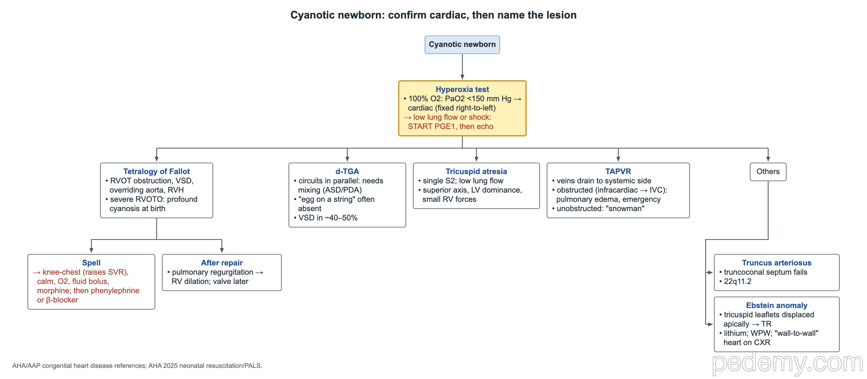
Cyanotic newborn: confirm cardiac, then name the lesion
Key points
- Hyperoxia test: PaO2 below about 150 mm Hg on 100% oxygen points to cyanotic heart disease.
- Cyanotic newborn with low lung blood flow or shock: start prostaglandin E1, then get an echo.
- CCHD pulse-oximetry screen at 24–48 hours; screening earlier than 24 hours gives more false positives.
- Tetralogy spell: knee-chest position and calming, then oxygen, fluid bolus and morphine; phenylephrine or a β-blocker if needed.
- Large VSD causes heart failure at 4–8 weeks, as pulmonary resistance falls.
- ASD: wide, fixed split S2 with a pulmonary flow murmur.
- Coarctation: weak, delayed femoral pulses and arm BP above leg BP; about half also have a bicuspid aortic valve.
The cyanotic newborn: hyperoxia test, PGE1 and CCHD screening
On 100% oxygen, lung disease usually lets the PaO2 rise well above 150 mm Hg. A fixed right-to-left shunt in the heart keeps it low. A cyanotic newborn whose PaO2 stays around 40 mm Hg on 100% oxygen, with reduced lung blood flow on X-ray, needs prostaglandin E1 to keep the ductus open, then an echocardiogram.
- Duct-dependent obstruction of the systemic outflow can cause shock in the first days; heart failure from a large left-to-right shunt usually appears over the following weeks.
- Pink arms with blue legs: right-to-left flow across the ductus.
- CCHD pulse-oximetry screening is done at 24–48 hours of age. Screening before 24 hours gives more false-positive results.
- The 2025 AAP algorithm passes a baby with saturation ≥95% in both the right hand and a foot, and allows only 1 repeat screen.
- A planned home birth still needs CCHD screening at 24–48 hours.
Tetralogy of Fallot
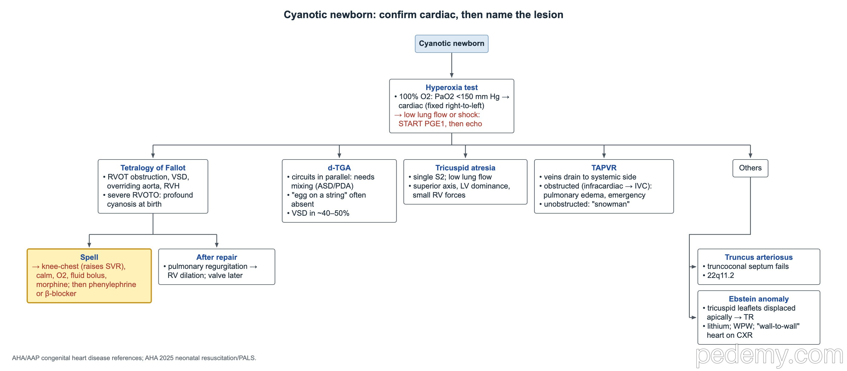
Tetralogy of Fallot has 4 parts: right ventricular outflow tract obstruction, a VSD, an overriding aorta and right ventricular hypertrophy. A cyanotic infant with a harsh murmur at the left upper sternal border (from the outflow obstruction), a single S2 and a boot-shaped heart fits.
In a hypercyanotic spell, squatting or the knee-chest position raises systemic vascular resistance, so less blood shunts right to left across the VSD.
- Spell treatment: knee-chest position and calming, then oxygen, an IV fluid bolus and morphine; if needed, phenylephrine or a β-blocker.
- Most common long-term problem after repair: pulmonary regurgitation from the outflow-tract patch, which dilates the right ventricle. Many need a pulmonary valve replacement later.
d-TGA and other cyanotic lesions
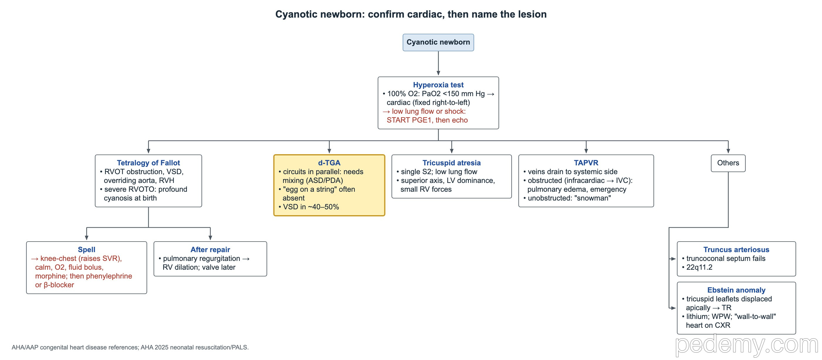
In d-transposition of the great arteries (d-TGA), the systemic and pulmonary circuits run in parallel, so the baby survives only through mixing at an ASD, VSD or PDA. Cyanosis is profound soon after birth and barely improves on 100% oxygen. A blue newborn with a loud single S2, a quiet precordium and an egg-on-a-string heart fits; the X-ray sign is often absent, so get an urgent echo. A VSD is present in about 40–50%. A 3-day-old with profound cyanosis and shock needs prostaglandin E1 first.
| Lesion | Key clues |
|---|---|
| Tricuspid atresia | Cyanosis, single S2, low lung blood flow; ECG with a superior (leftward) axis, LV dominance and small RV forces |
| TAPVR | Pulmonary veins drain to the systemic side. Unobstructed supracardiac type: late "snowman" heart. Obstructed (usually infracardiac): pulmonary edema and an emergency |
| Truncus arteriosus | The truncoconal (aorticopulmonary) septum fails to divide the common trunk; linked to 22q11.2 deletion |
| Ebstein anomaly | Septal and posterior tricuspid leaflets displaced toward the apex, part of the RV atrialized, tricuspid regurgitation. Massive "wall-to-wall" heart on X-ray; WPW and SVT. Linked to first-trimester lithium (small absolute risk) |
Left-to-right shunts: VSD, ASD, AV canal and PDA
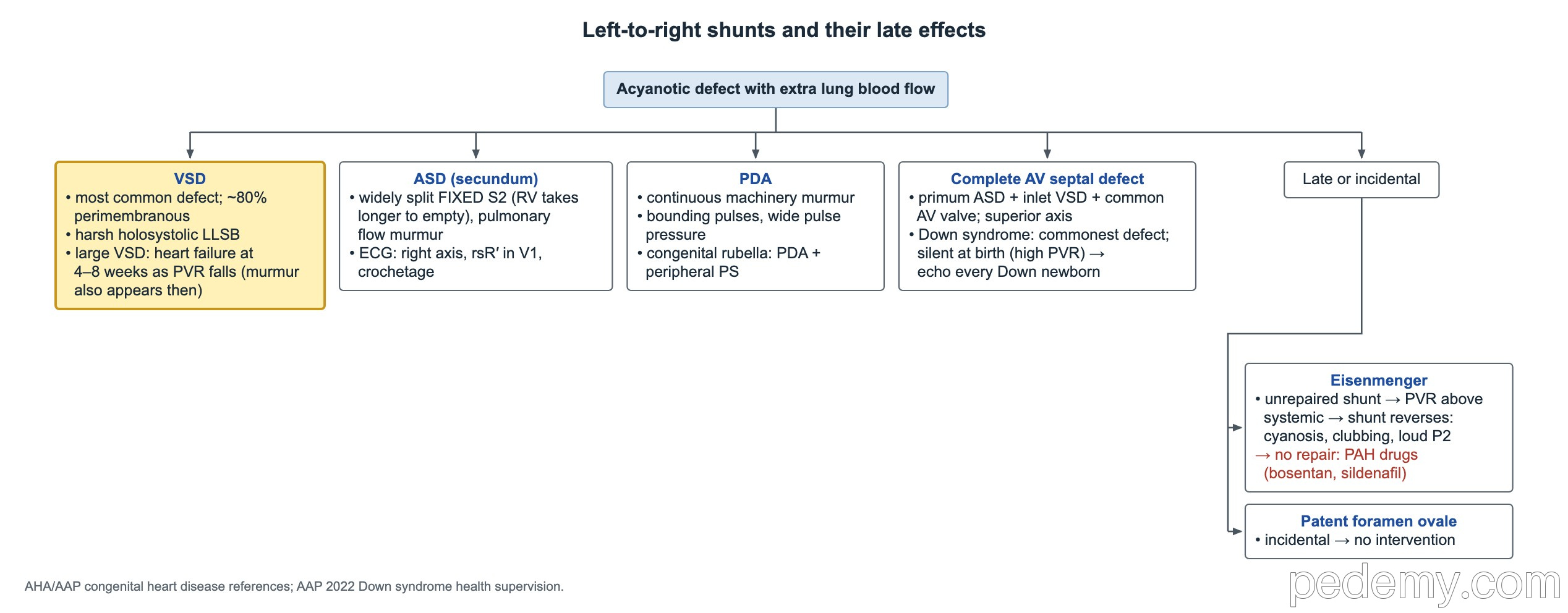
The VSD is the most common congenital heart defect recognized in infancy. Heart failure from a large VSD appears weeks after birth, not at birth: as pulmonary vascular resistance falls over the first 4–8 weeks, more blood floods the lungs, and the murmur appears then too.
| Lesion | What to know |
|---|---|
| VSD | About 80% are perimembranous, just below the aortic valve; aortic cusp prolapse can cause aortic regurgitation. Harsh holosystolic murmur at the left lower sternal border |
| Secundum ASD | Pulmonary flow murmur with a wide, fixed split S2: the RV takes longer to empty its extra volume and the shunt evens out breathing effects. ECG: right axis, rsR′ in V1, notched inferior R waves (crochetage) |
| Complete AV septal defect | Primum ASD, inlet VSD and a common AV valve; superior axis; most common lesion in Down syndrome |
| PDA | Continuous machinery murmur with bounding pulses and a wide pulse pressure (runoff into the pulmonary artery) |
| Congenital rubella | PDA with cataracts, deafness and blueberry-muffin spots: the mother was not immune to rubella before pregnancy |
| Patent foramen ovale | Incidental finding in a healthy child; no intervention or follow-up needed |
Eisenmenger syndrome and long-term cyanosis
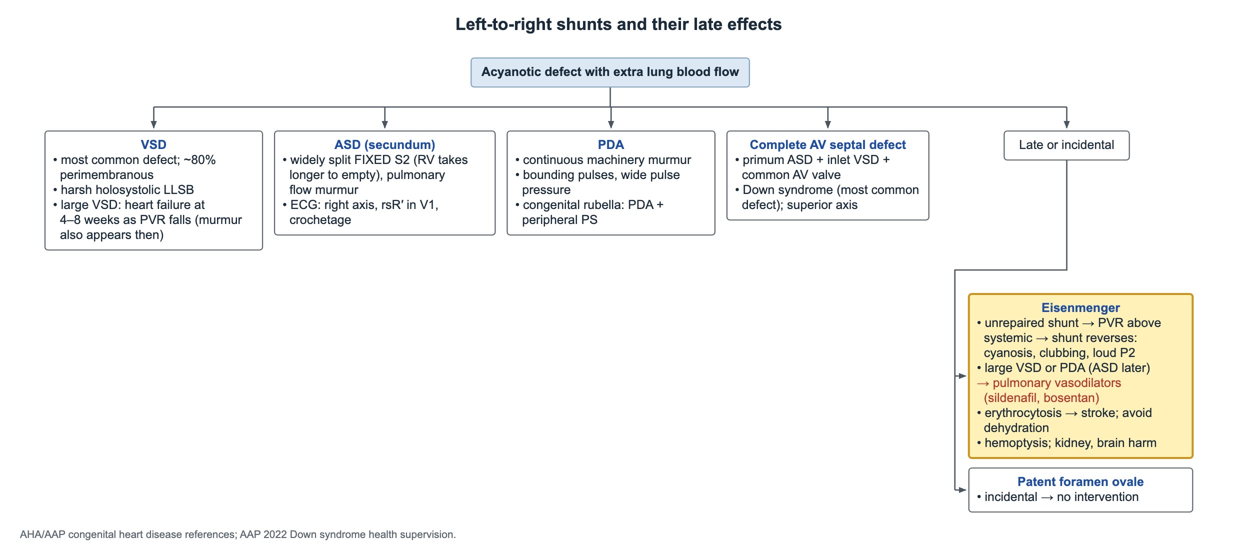
A large shunt left unrepaired, usually a large VSD or PDA (ASD later), damages the lung vessels. Pulmonary resistance rises above systemic, and the shunt reverses to right-to-left. A teen with an unrepaired VSD who is now cyanotic, with clubbing and a loud P2, has Eisenmenger syndrome.
- If repair is not possible, pulmonary vasodilators (bosentan, sildenafil, prostanoids) can help.
- Long-standing cyanosis causes secondary erythrocytosis; the thick blood raises the risk of stroke and TIA, and dehydration is a key trigger.
- Massive hemoptysis can kill quickly.
- Other organs harmed: kidneys, blood (bleeding and thrombosis) and brain.
Coarctation, aortic stenosis and bicuspid aortic valve
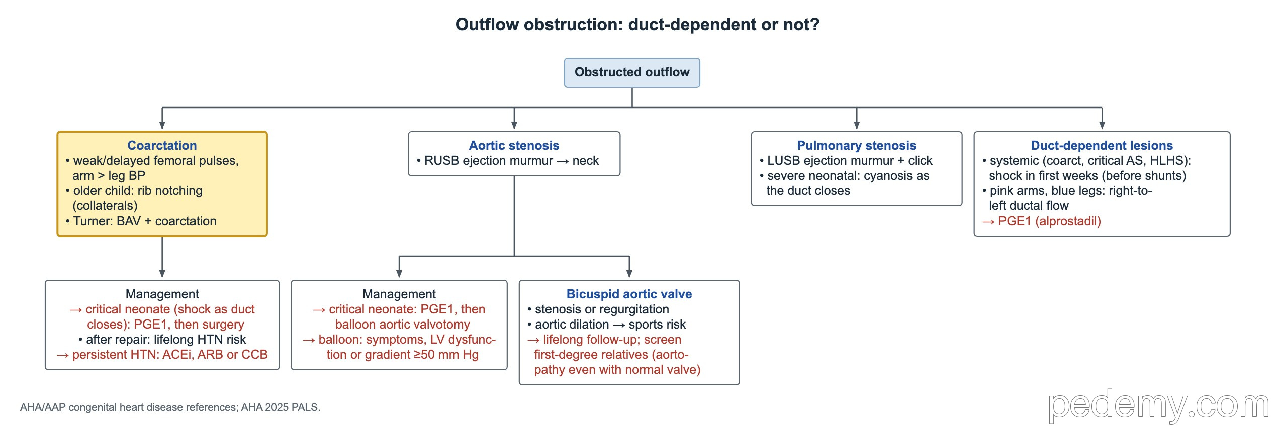
- Coarctation: weak or delayed femoral pulses compared with the right arm, and arm BP above leg BP. The commonest coexisting defect is a bicuspid aortic valve, in about half.
- Older child: inferior rib notching on X-ray, from enlarged intercostal collateral arteries.
- Critical coarctation in a neonate (shock as the duct closes): prostaglandin E1 to reopen the ductus, then surgery. School-age hypertension from coarctation: surgical or catheter repair.
- After repair, hypertension is a lifelong risk, including masked hypertension; follow with ambulatory BP monitoring. If BP is high, first rule out recoarctation (arm-leg gradient, echo, CT or MRI). Persistent stage 1 hypertension with no gradient: an ACE inhibitor, ARB or calcium-channel blocker.
- Critical valvar aortic stenosis in a neonate with poor LV function: prostaglandin E1, then balloon aortic valvotomy. Balloon valvotomy in older children for symptoms, LV dysfunction or a gradient ≥50 mm Hg.
- Bicuspid aortic valve: lifelong follow-up even without symptoms, because aortic stenosis, regurgitation and aortic dilation can develop. Screen first-degree relatives with echo; a relative with a normal three-leaflet valve can still have a dilated aorta.
- Bicuspid valve and sports: first update the cardiology assessment of the valve and aortic size; with aortic dilation, avoid collision and contact sports.
ALCAPA and vascular rings
In ALCAPA, the left coronary artery arises from the pulmonary artery instead of the aorta. The baby is well at birth, while pulmonary pressure is high. At 2–3 months, pulmonary pressure falls, left coronary perfusion becomes inadequate and the heart becomes ischemic: the infant sweats or cries with feeds, the LV is weak and the ECG shows an infarct pattern with deep Q waves in I and aVL. A febrile illness can tip the infant into heart failure because it raises oxygen demand. After stabilization, the treatment is urgent surgery.
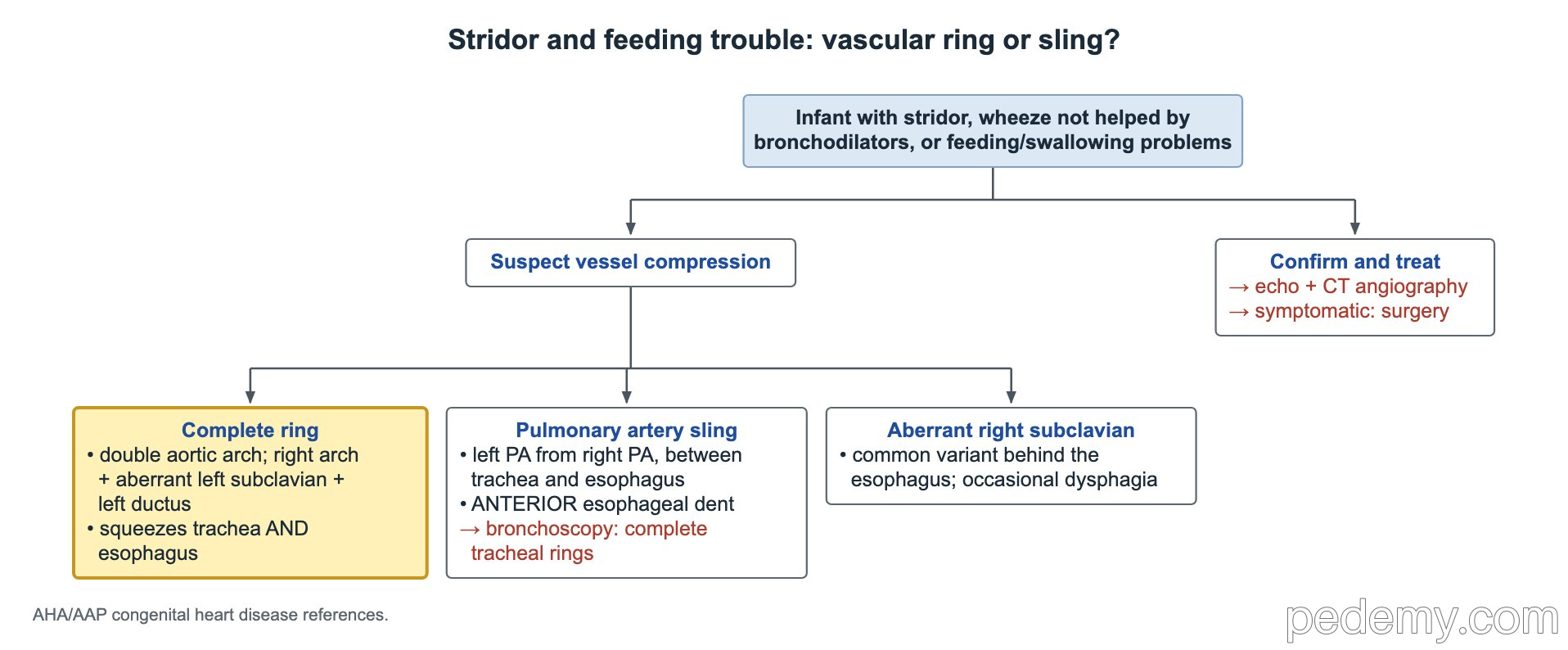
- Infant with wheeze that does not respond to albuterol, stridor or feeding problems: think of a vascular ring or sling compressing the airway and esophagus.
- Most common complete rings: double aortic arch, and right aortic arch with an aberrant left subclavian artery and a left-sided ductus. They can squeeze both the trachea and the esophagus.
- Pulmonary artery sling: the left pulmonary artery arises from the right one and passes behind the trachea and in front of the esophagus, giving an anterior indentation on esophagram. Examine the airway with bronchoscopy, because complete tracheal rings and tracheal stenosis are common.
- Aberrant right subclavian artery: a common variant that passes behind the esophagus and occasionally causes dysphagia.
- Imaging that maps the vessels: echocardiography plus CT angiography. Persistent symptomatic compression: surgery.
Common questions
What does a failed hyperoxia test mean?
A PaO2 that stays below about 150 mm Hg on 100% oxygen points to a fixed right-to-left shunt from cyanotic heart disease rather than lung disease.
When is newborn pulse-oximetry screening for CCHD done?
At 24–48 hours of age. Screening before 24 hours gives more false-positive results. The 2025 AAP algorithm passes a baby with saturations of 95% or more in both the right hand and a foot.
How do you treat a tet spell?
Place the child in the knee-chest position and calm them, then give oxygen, an IV fluid bolus and morphine. If the spell continues, give phenylephrine or a β-blocker.
Study this topic
Sources
- AAP 2025: Newborn screening for critical congenital heart disease, new algorithm (Pediatrics)
- ACC/AHA 2025: Guideline for the management of adults with congenital heart disease (Circulation)
- AAP 2017: Clinical practice guideline for high blood pressure in children and adolescents (Pediatrics)
Written for Pedemy in our own words from the sources above; checked November 2026. For education only, not medical advice; check doses and local protocols before treating a patient. Pedemy is independent and not affiliated with or endorsed by the organizations cited.