

Main decision tree
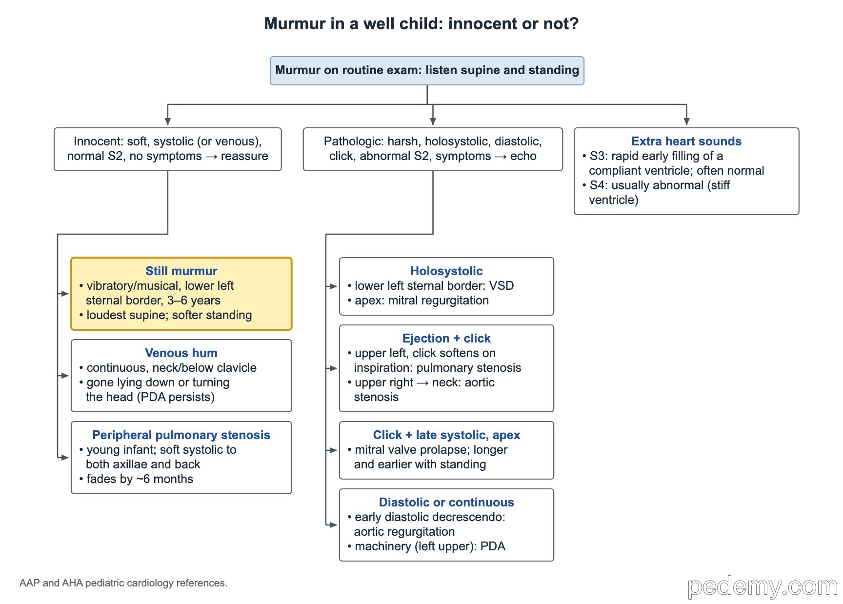
Murmur in a well child: innocent or not?
Key points
- Innocent murmurs are soft (grade 1–2/6), systolic or venous, with a normal S2 and no symptoms: no echo needed.
- Holosystolic, diastolic, harsh or grade ≥3/6 murmurs, a click, an abnormal S2 or symptoms → echo and cardiology.
- Still murmur: musical or vibratory at the lower left sternal border, loudest supine, softer standing; peaks at 3–6 years.
- Venous hum: continuous below the clavicles, gone lying down or turning the head; a PDA murmur persists supine.
- A small VSD murmur often appears 2–6 weeks after birth, as pulmonary vascular resistance falls.
- Standing or Valsalva makes the HCM murmur louder and the AS murmur softer; squatting does the reverse.
- S3 can be normal in a healthy child; S4 is usually abnormal (stiff ventricle).
Innocent or pathologic?
Listen with the child supine and then standing. An innocent murmur is soft, short and systolic (or venous), the second heart sound splits normally, and the child is well. Explain to the family that the heart is normal; no test is needed.
The Still murmur is the most common innocent murmur. It is a low-pitched, musical or vibratory ("twanging string") midsystolic sound at the lower left sternal border, usually in children aged 3–6 years. It is loudest supine and softens when the child sits or stands, and it gets louder with fever or exercise.
- Peripheral pulmonary flow murmur (PPS): a young infant with a soft systolic murmur heard in both axillae and the back; it fades by about 6 months.
- Venous hum: continuous, under the clavicles or at the base of the neck, gone when supine (see below).
- Pulmonary flow murmur: soft ejection murmur at the upper left sternal border in older children and teens.
| Red flag | Why it matters |
|---|---|
| Holosystolic or diastolic timing | Diastolic murmurs are never innocent |
| Grade 3/6 or louder, or a thrill | A thrill means grade 4/6 |
| Harsh quality | Turbulent flow through a narrow valve or defect |
| Ejection click | Valvar aortic or pulmonary stenosis, or bicuspid aortic valve |
| Abnormal S2 (fixed split, single, loud) | ASD, pulmonary hypertension or outflow disease |
| Louder on standing | Hypertrophic cardiomyopathy |
| Symptoms or abnormal pulses | Poor growth, sweating with feeds, exertional chest pain or syncope, weak femoral pulses |
Venous hum or PDA?
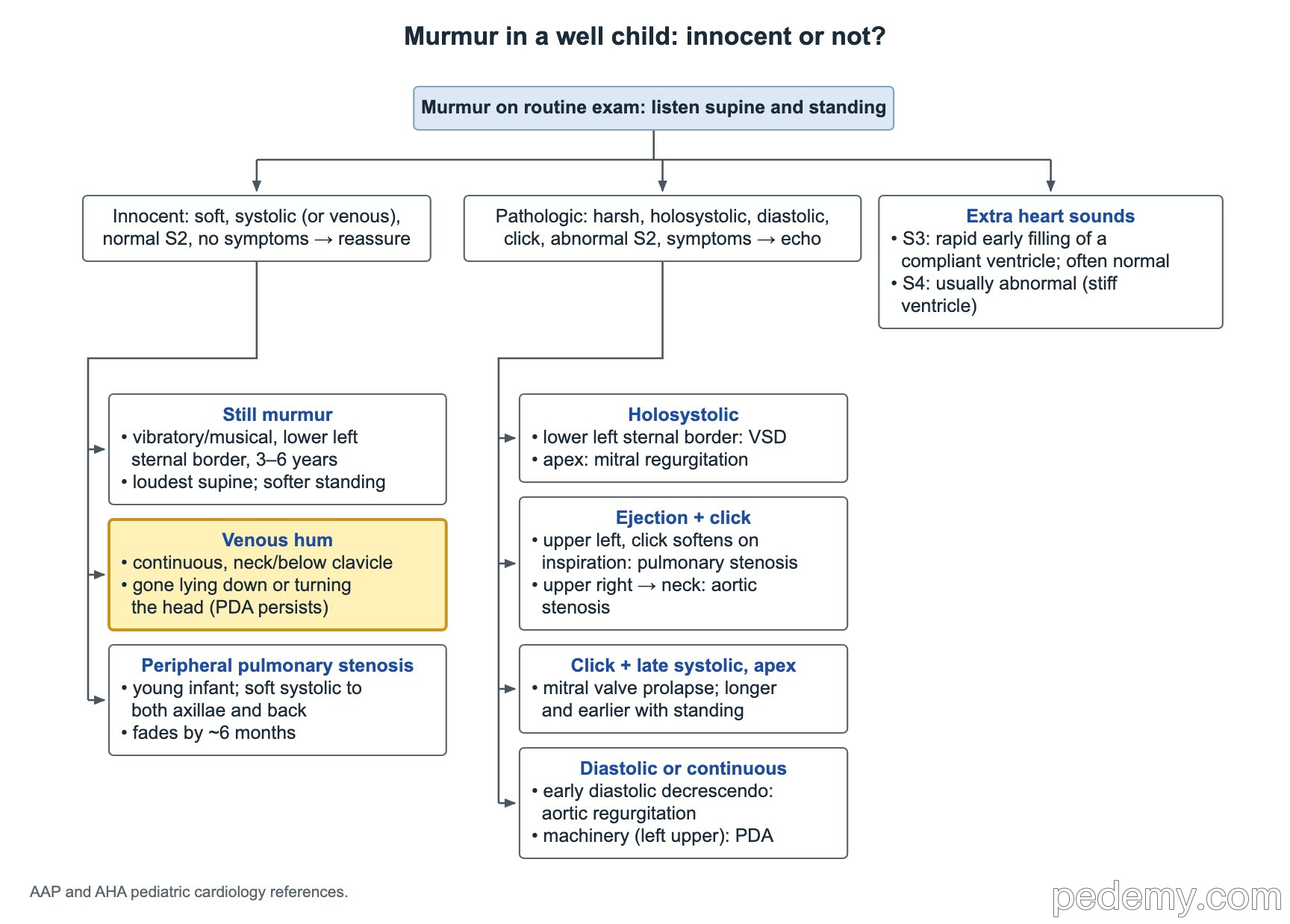
Both are continuous murmurs heard below the clavicle, so position is the test. A venous hum comes from blood returning through the jugular veins; it is common in toddlers and preschool children. It disappears when the child lies flat, turns the head or has light pressure placed over the jugular vein.
A PDA murmur is a continuous "machinery" murmur at the left infraclavicular area that persists supine. Look for bounding pulses and a wide pulse pressure. A venous hum needs no testing; a suspected PDA needs an echo.
Holosystolic murmurs: VSD and mitral regurgitation
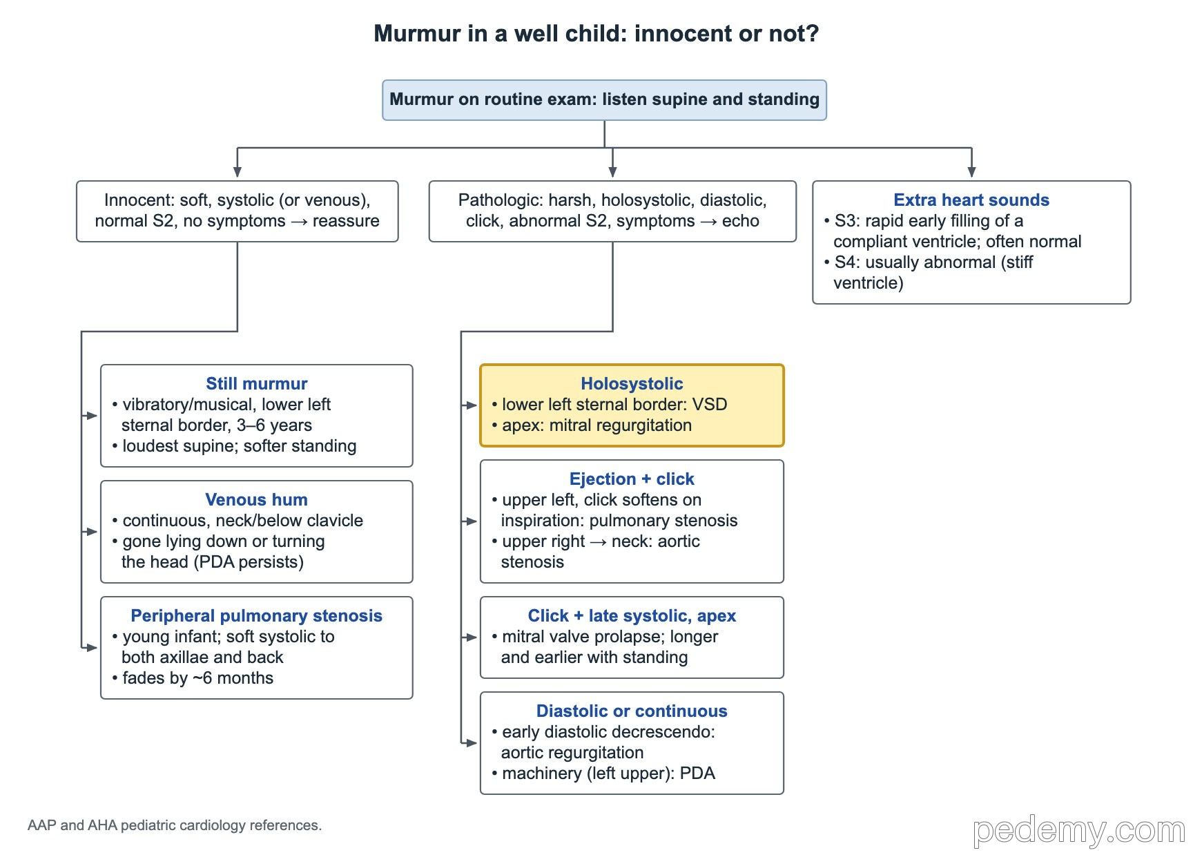
- VSD: harsh holosystolic murmur at the lower left sternal border. Small defects are often the loudest.
- Mitral regurgitation: holosystolic (blowing) murmur at the apex that radiates to the left axilla.
- A small VSD may be silent at birth. Pulmonary vascular resistance is high in the first days; as it falls over 2–6 weeks, the left-to-right pressure gradient rises and the murmur appears.
- A large VSD can have a soft murmur and present at 1–2 months with heart failure: fast breathing, sweating with feeds and poor weight gain.
Ejection murmurs with a click: aortic and pulmonary stenosis
Critical pulmonary stenosis in a newborn is duct-dependent: the infant becomes more cyanotic as the ductus arteriosus closes. Start prostaglandin E1 (alprostadil) to reopen the duct, then the valve is opened with a balloon.
Critical aortic stenosis is also duct-dependent and presents with shock in the first weeks as the duct closes.
| Lesion | Where and what |
|---|---|
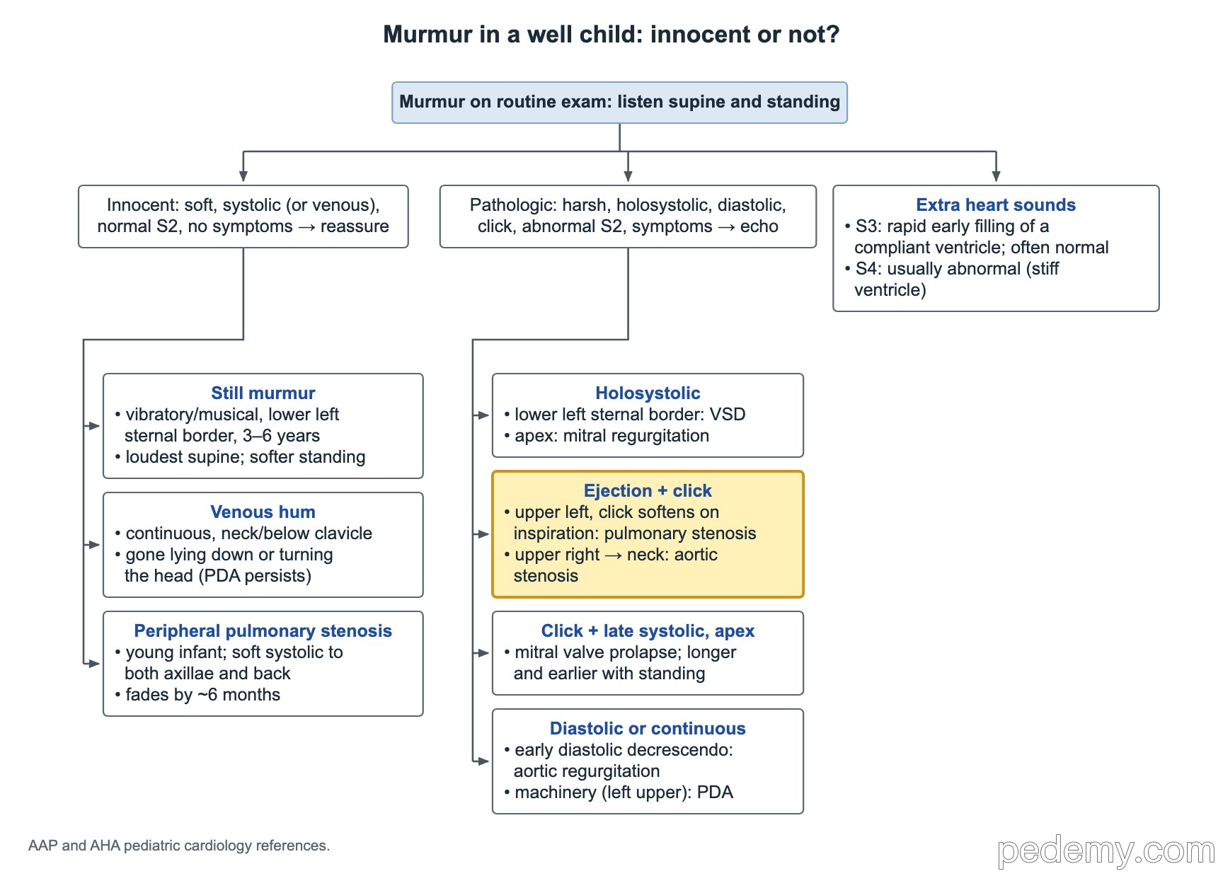
| Valvar pulmonary stenosis | Ejection murmur at the upper left sternal border; ejection click that softens or disappears on inspiration |
| Valvar aortic stenosis | Harsh ejection murmur at the upper right sternal border radiating to the neck; apical click that does not vary with breathing |
| Bicuspid aortic valve | Ejection click; may develop stenosis, regurgitation or aortic dilation over time |
Maneuvers: HCM, aortic stenosis and mitral valve prolapse
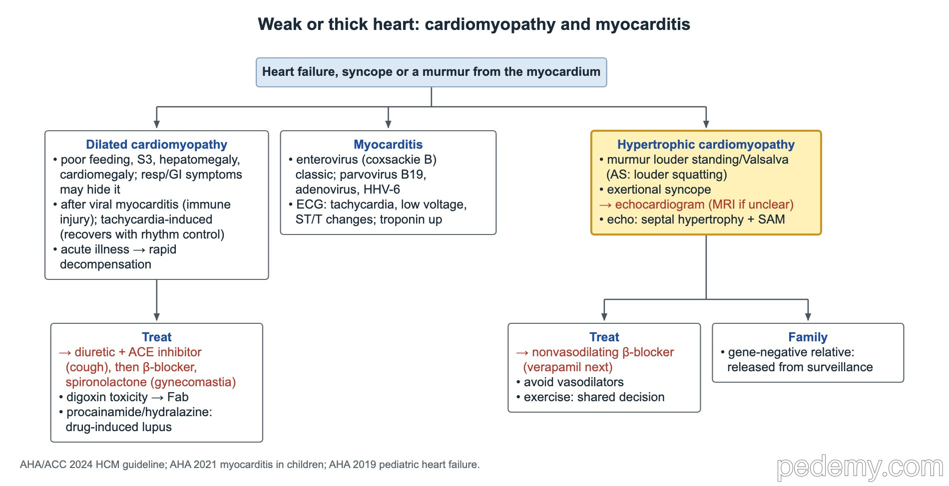
Standing and the Valsalva strain lower the volume in the left ventricle. Squatting raises venous return and afterload. These changes separate fixed obstruction from dynamic obstruction.
- Mitral valve prolapse: midsystolic click followed by a late systolic murmur at the apex.
- HCM: the murmur, exertional syncope or a family history leads to an echo (cardiac MRI if the echo is unclear); the echo shows septal hypertrophy, often with systolic anterior motion of the mitral valve.
- HCM treatment starts with a non-vasodilating β-blocker (verapamil if β-blockers fail or are not tolerated); avoid vasodilators and dehydration.
- Screen first-degree relatives; a relative who tests negative for the family's disease-causing gene variant can stop surveillance.
| Lesion | Standing or Valsalva | Squatting |
|---|---|---|
| Hypertrophic cardiomyopathy (dynamic outflow obstruction) | Louder | Softer |
| Valvar aortic stenosis (fixed obstruction) | Softer | Louder |
| Mitral valve prolapse | Click earlier, murmur longer | Click later, murmur shorter |
| Still murmur | Softer | — |
Diastolic and continuous murmurs
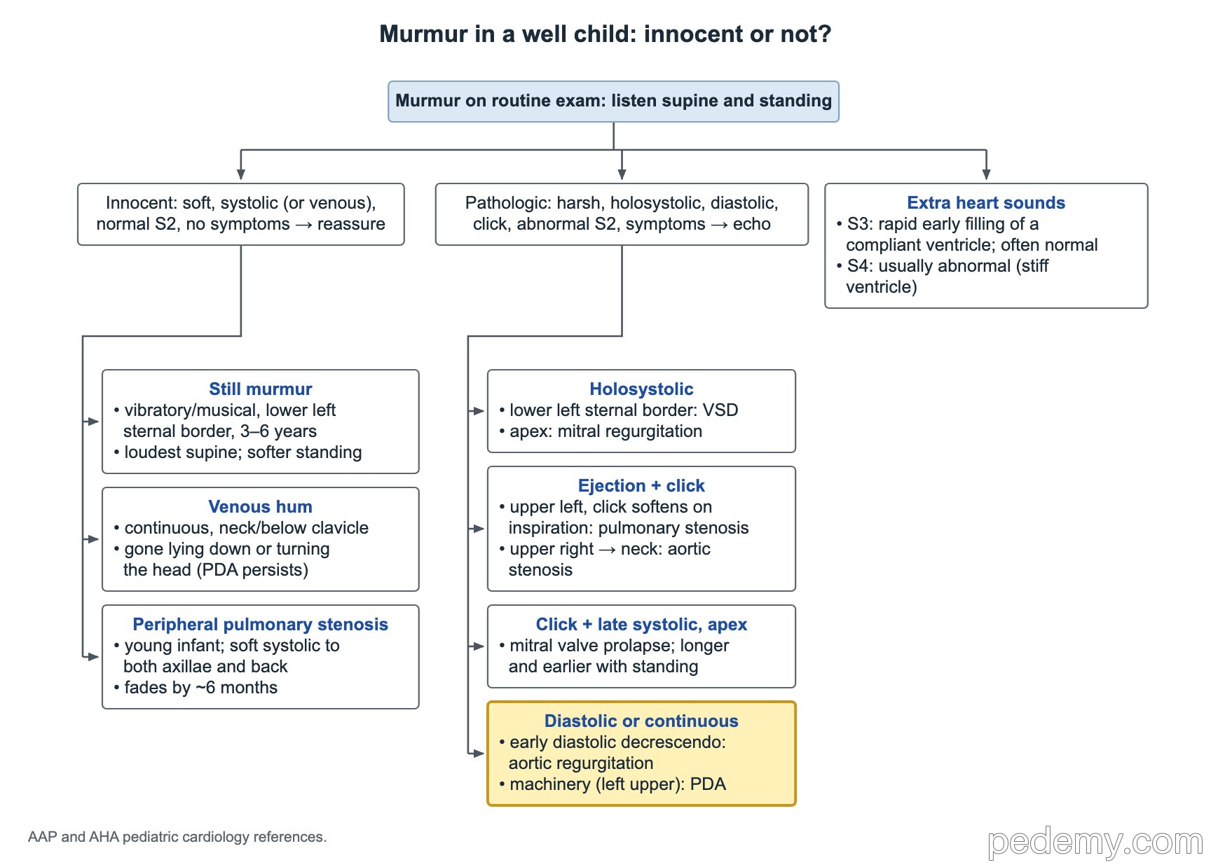
- Aortic regurgitation: high-pitched, early diastolic decrescendo murmur at the left sternal border, best heard with the patient sitting forward in full expiration. Causes in children include bicuspid aortic valve and a VSD with aortic cusp prolapse.
- PDA: continuous machinery murmur at the left infraclavicular area.
- Every diastolic murmur needs an echo; the only common benign continuous murmur is the venous hum.
S2, S3 and S4
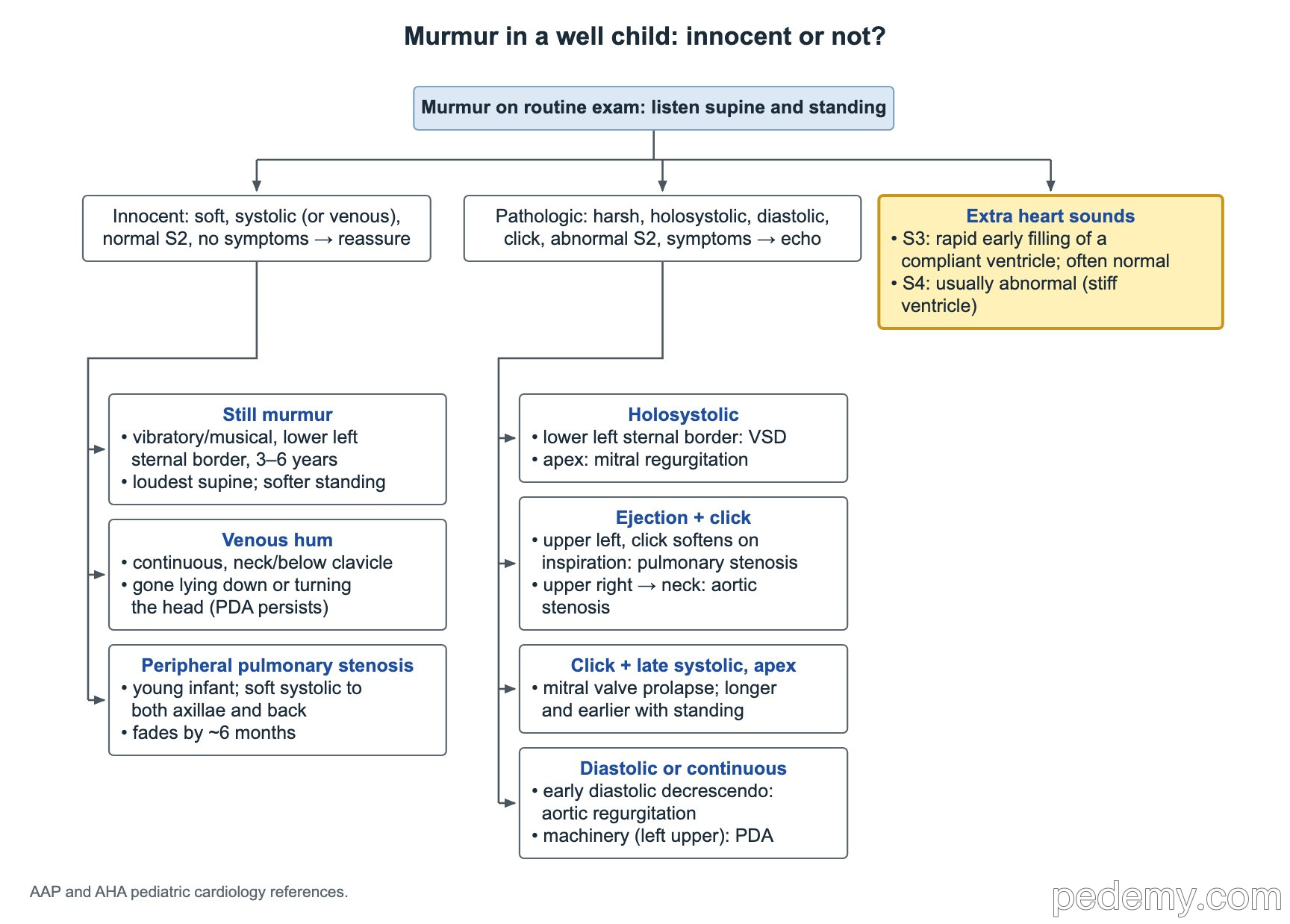
- S2 normally splits on inspiration and closes on expiration. A wide, fixed split suggests an ASD; a single loud S2 suggests pulmonary hypertension or an absent pulmonary valve sound.
- S3: early diastole, produced by rapid early filling of a compliant ventricle with a large volume of blood. It is often normal in children, teens and athletes, but also occurs with volume overload and heart failure.
- S4: late diastole, from atrial contraction into a stiff ventricle. In a child it is usually abnormal (for example, HCM or severe hypertension).
Common questions
Does an innocent heart murmur need an echocardiogram?
No. A soft systolic or venous murmur with a normal S2, normal pulses and a well child needs no echo. Get an echo when there are red flags such as a diastolic or holosystolic murmur, a click, grade 3/6 or louder, or symptoms.
Do innocent murmurs go away?
Many fade with age. The peripheral pulmonary flow murmur usually goes by about 6 months, and the Still murmur and venous hum often fade by adolescence. They can sound louder during fever.
How do you tell a venous hum from a PDA?
Lay the child down. A venous hum disappears supine or when the head is turned; a PDA murmur persists.
Study this topic
Sources
- ACC/AAP/AHA 2014: Appropriate use criteria for initial transthoracic echocardiography in outpatient pediatric cardiology (JACC)
- AHA/ACC/PACES 2024: Guideline for the management of hypertrophic cardiomyopathy (JACC)
- ACC/AHA 2020: Guideline for the management of valvular heart disease, executive summary (JACC)
Written for Pedemy in our own words from the sources above; checked November 2026. For education only, not medical advice; check doses and local protocols before treating a patient. Pedemy is independent and not affiliated with or endorsed by the organizations cited.