

Main decision tree
Slow, blocked or unusual ECG in a child
Key points
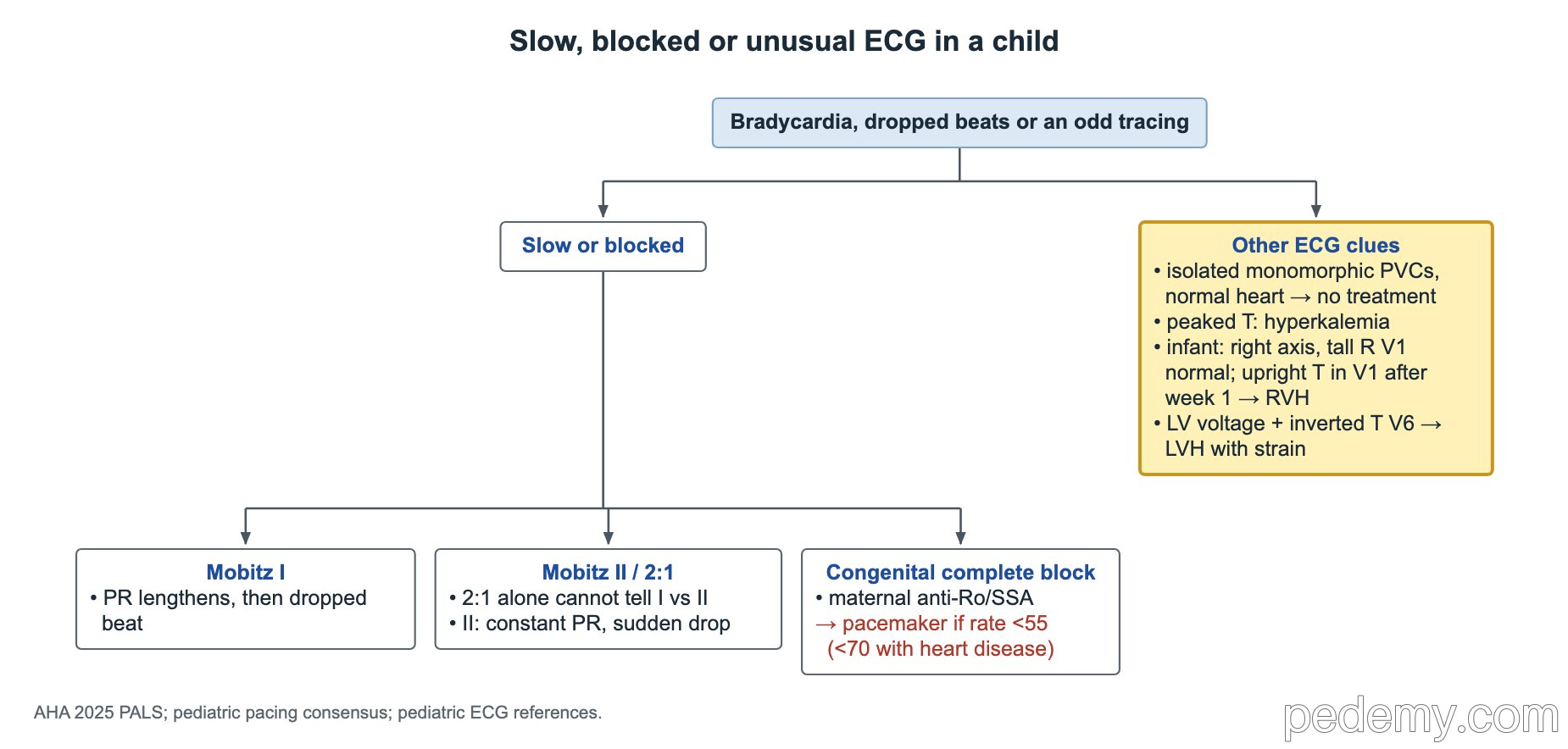
- Right axis and a tall R in V1 are normal in infancy; an upright T in V1 after the first week (until adolescence) suggests RVH.
- Mobitz I: PR lengthens before a dropped beat, usually benign; Mobitz II: constant PR with a sudden drop, needs cardiology.
- Congenital complete heart block is linked to maternal anti-Ro/SSA antibodies, often in a mother without symptoms.
- SVT: abrupt, fixed rate, often ≥220/min in infants or ≥180/min in children; most have a structurally normal heart.
- Stable SVT: vagal maneuvers, then adenosine 0.1 mg/kg (max 6 mg), then 0.2 mg/kg (max 12 mg); unstable: synchronized cardioversion 0.5–1 J/kg, then 2 J/kg.
- WPW: short PR, delta wave, wide QRS; recurrent SVT with WPW → catheter ablation.
- QTc above 450 ms is prolonged; torsades → IV magnesium; long-term LQTS → nadolol or propranolol.
The normal ECG by age, and other clues
Before birth the right ventricle does most of the work, so a newborn's ECG shows right axis deviation and a tall R wave in V1. This right ventricular dominance is normal at 2 months and fades over childhood.
- The T wave in V1 is upright in the first days of life, then turns negative by about 1 week and stays negative until adolescence.
- An upright T wave in V1 between 1 week and adolescence suggests right ventricular hypertrophy.
- Excess left-sided R-wave voltage with inverted T waves in V6 in an infant is left ventricular hypertrophy with a strain (repolarization) pattern: get an echo.
- Isolated premature ventricular complexes that are infrequent and monomorphic, in a child with a normal heart, no symptoms and no worrying family history, need no antiarrhythmic treatment.
- Hyperkalemia: tall peaked T waves first, then a longer PR, flattened or lost P waves and a widening QRS. Very high levels (roughly above 8 mEq/L) lead to a sine-wave pattern, ventricular fibrillation or asystole.
- Hyperkalemia with ECG changes is an emergency: give IV calcium to stabilize the heart, then shift potassium into cells (insulin with glucose, inhaled albuterol) and remove it from the body.
AV block: which degree?
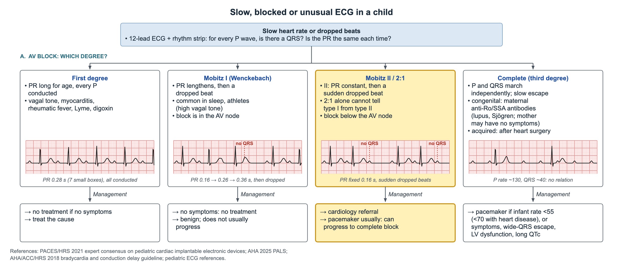
Use a 12-lead ECG and a long rhythm strip. Ask two questions for every P wave: is there a QRS after it, and is the PR the same each time?
| Block | ECG | Meaning and management |
|---|---|---|
| First degree | PR long for age; every P conducted | High vagal tone, myocarditis, rheumatic fever, Lyme disease, digoxin; no treatment if no symptoms, treat the cause |
| Second degree, Mobitz I (Wenckebach) | PR lengthens beat by beat until a P wave is not conducted; the cycle repeats | Block in the AV node; common in sleep and athletes; benign and rarely progresses |
| Second degree, Mobitz II | Constant PR in conducted beats, then a sudden non-conducted P wave | Block below the AV node; can progress to complete block; refer to cardiology, a pacemaker is usually needed |
| 2:1 AV block | Every second P wave not conducted; conducted PRs constant | Cannot be called type I or type II from this pattern alone; look for other conduction ratios on a longer strip |
Complete (third-degree) heart block
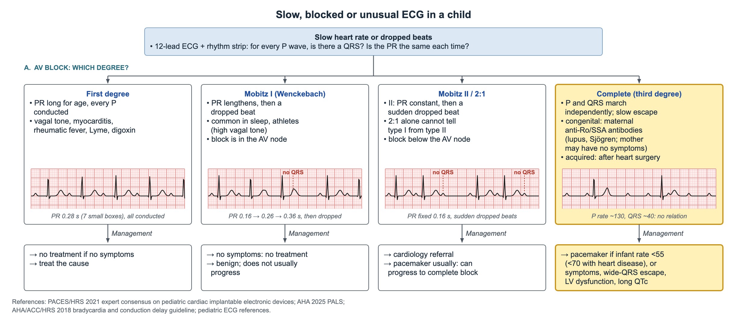
P waves and QRS complexes march independently, with a slow escape rhythm. Congenital complete heart block is usually caused by maternal anti-Ro/SSA (and anti-La/SSB) antibodies crossing the placenta and injuring the fetal conduction system. The mother may have lupus or Sjögren syndrome, or no symptoms at all. Acquired complete block in children most often follows heart surgery.
- Permanent pacemaker for an infant with congenital complete heart block when the mean ventricular rate is 50/min or less.
- Also pace for a wide-QRS escape rhythm, ventricular dysfunction, or heart failure or low-output symptoms, at any rate.
- Infants of mothers with known anti-Ro/SSA antibodies are followed with fetal heart monitoring during pregnancy.
Narrow regular tachycardia: SVT
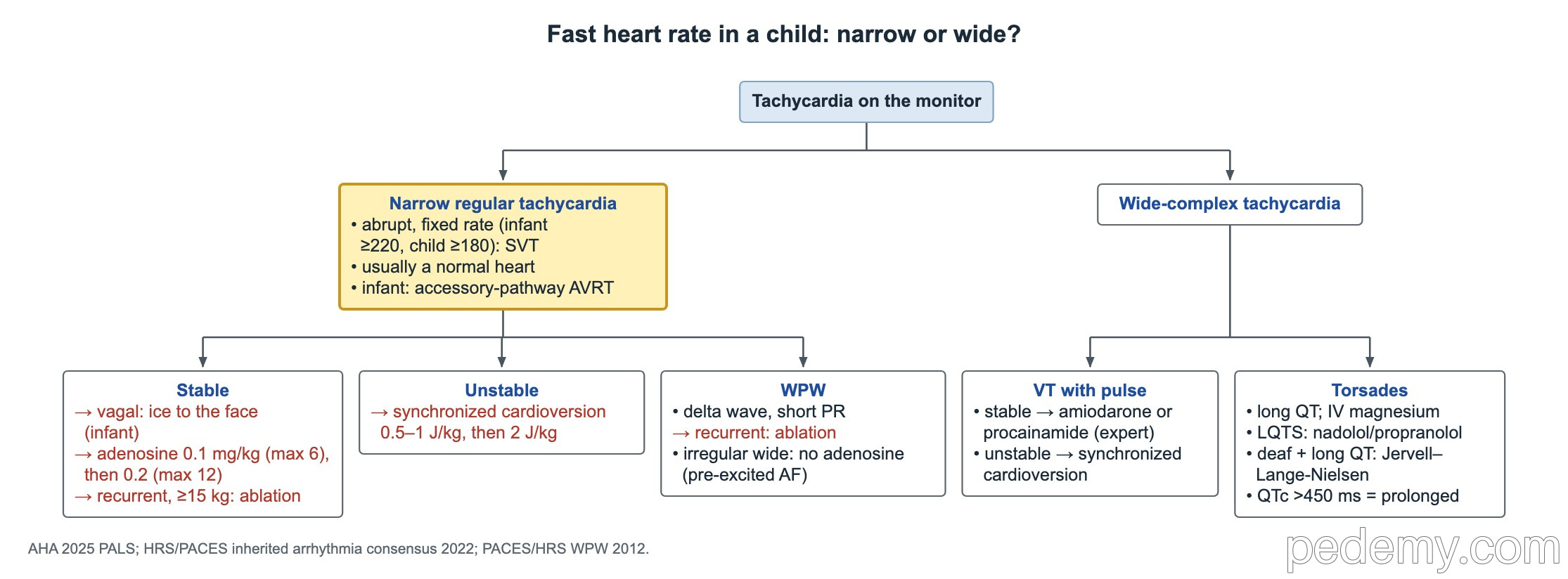
SVT starts and stops abruptly and the rate is fixed, without beat-to-beat change. Rates are often 220/min or more in infants and 180/min or more in children, and P waves are absent or abnormal. Sinus tachycardia has a variable rate, normal P waves and a cause such as fever, pain or dehydration.
Most children with SVT have a structurally normal heart. In infants the usual mechanism is reentry through an accessory pathway (AVRT); AV nodal reentry (AVNRT) becomes more common in adolescents.
- Stable: vagal maneuvers first (ice to the face in an infant; Valsalva in an older child).
- Then adenosine 0.1 mg/kg (max 6 mg) as a rapid IV push with a flush; second dose 0.2 mg/kg (max 12 mg).
- Unstable (hypotension, poor perfusion, altered mental status): synchronized cardioversion 0.5–1 J/kg, then 2 J/kg.
- Recurrent SVT needing repeated ED adenosine in a child of 15 kg or more, with no delta wave: catheter ablation is the definitive treatment.
Wolff-Parkinson-White (WPW)
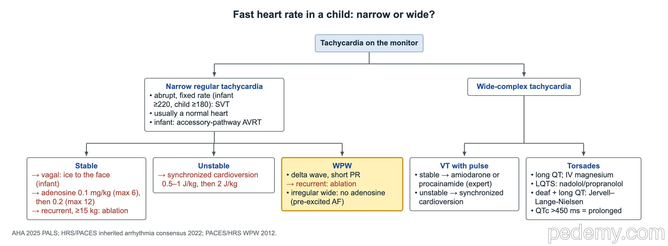
In WPW an accessory pathway conducts from atria to ventricles and bypasses the AV node. The early activation gives a short PR interval, a slurred upstroke of the QRS (delta wave) and a wide QRS.
- A teen with WPW and recurrent SVT: radiofrequency catheter ablation of the accessory pathway is the definitive treatment.
- Atrial fibrillation conducted down the pathway gives an irregular, wide tachycardia that can turn into ventricular fibrillation.
- For irregular wide tachycardia with pre-excitation, do not give adenosine or other AV-node blockers; use synchronized cardioversion if unstable and get expert help.
Wide-complex tachycardia
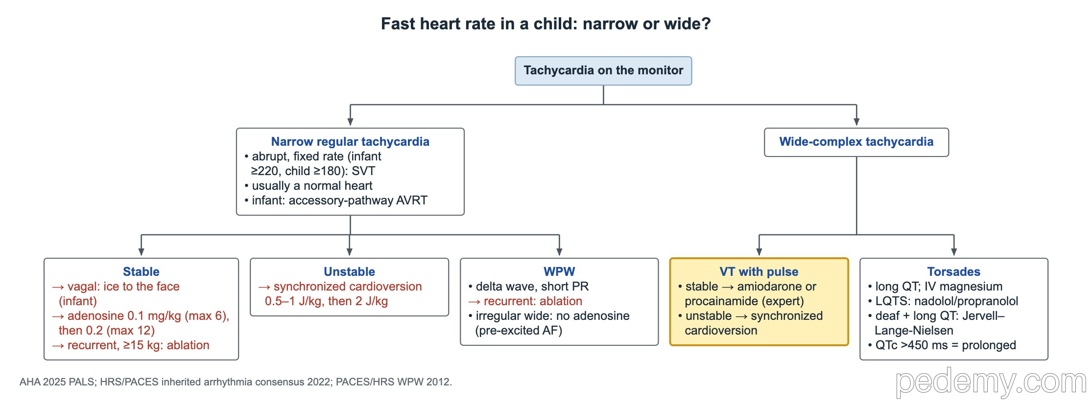
Treat a wide-complex tachycardia in a child as ventricular tachycardia until proven otherwise. Check for a pulse and perfusion first.
- Pulse with good perfusion: if regular and monomorphic, adenosine may be tried; otherwise amiodarone or procainamide with expert consultation (not both together).
- Pulse with hypotension or poor perfusion: synchronized cardioversion 0.5–1 J/kg, then 2 J/kg. Do not delay it for fluids or drugs.
- No pulse: start CPR and defibrillate as for pulseless VT or VF.
Long QT syndrome and torsades
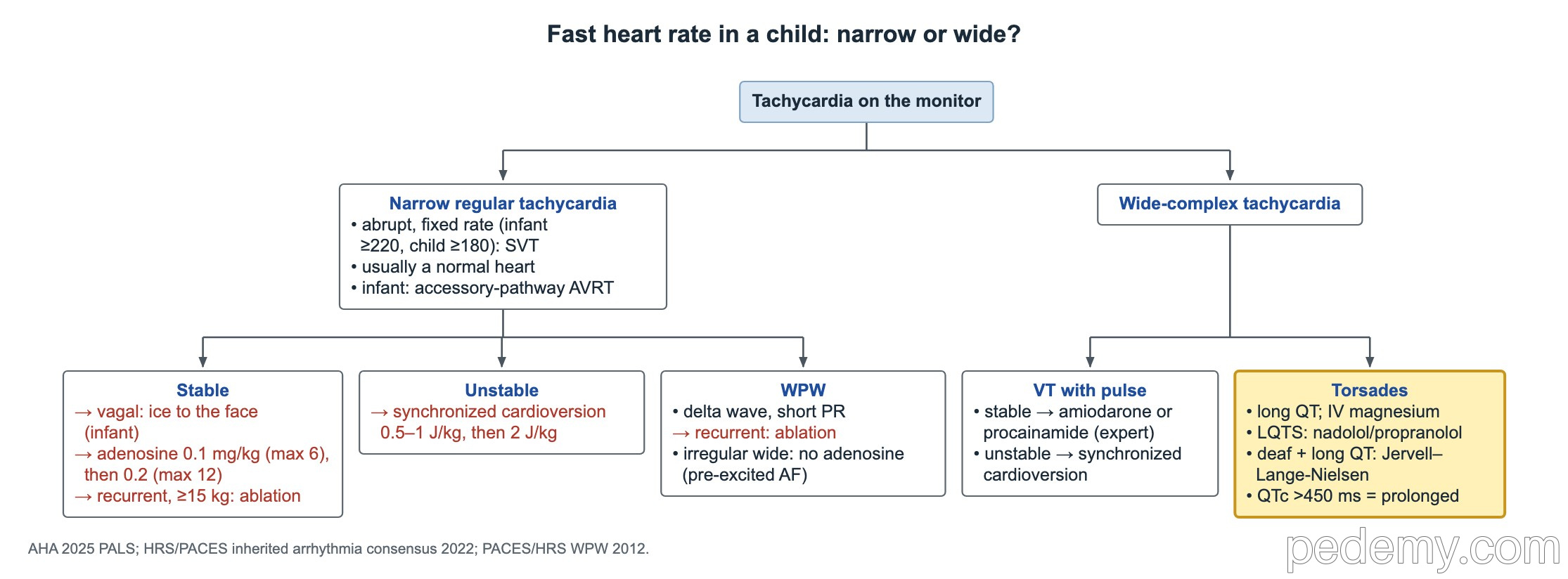
Torsades de pointes is a polymorphic VT in which the QRS axis twists around the baseline. It points to a long QT interval. Give IV magnesium sulfate; defibrillate if the child is pulseless.
- A corrected QT (QTc) above 450 ms (0.45 s) is prolonged in a child.
- Congenital LQTS with syncope: a β-blocker is first-line long-term treatment, nadolol or propranolol (propranolol in infants).
- Congenital deafness plus a long QT suggests Jervell and Lange-Nielsen syndrome.
- Avoid QT-prolonging medicines and correct low potassium and magnesium.
- Screen first-degree relatives with an ECG.
Common questions
What heart rate is SVT in a baby?
SVT in infants is usually 220 beats per minute or more, with a fixed rate that starts and stops suddenly. In older children it is usually 180 or more.
What is the adenosine dose for SVT in children?
0.1 mg/kg (max 6 mg) as a rapid IV push followed by a saline flush. If it fails, give 0.2 mg/kg (max 12 mg).
What QTc is prolonged in children?
A QTc above 450 ms is prolonged. Long QT with fainting, seizures with exercise or startle, or a family history of sudden death needs cardiology review.
Study this topic
Sources
- AHA/AAP 2025: Part 8, Pediatric advanced life support (Circulation)
- PACES 2021: Expert consensus on cardiovascular implantable electronic devices in pediatric patients (JACC Clin Electrophysiol)
- PACES/HRS 2016: Expert consensus on catheter ablation in children and patients with congenital heart disease (Heart Rhythm)
Written for Pedemy in our own words from the sources above; checked November 2026. For education only, not medical advice; check doses and local protocols before treating a patient. Pedemy is independent and not affiliated with or endorsed by the organizations cited.