

Main decision tree
Head injury: who needs CT? (PECARN)
Key points
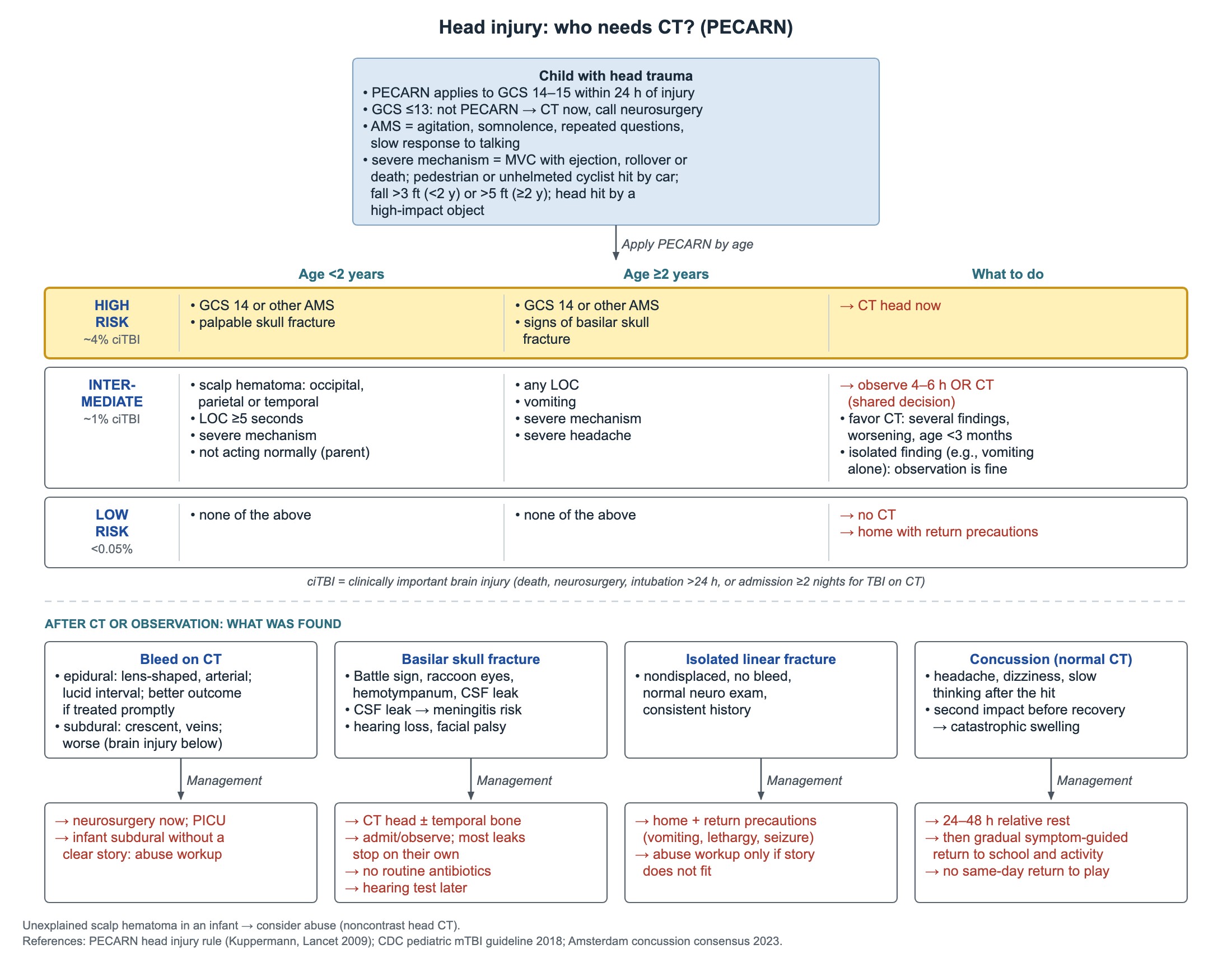
- PECARN high risk (GCS 14 or altered mental status, palpable or basilar skull fracture): about 4% risk of clinically important brain injury → CT now.
- PECARN intermediate risk (about 1%): observe 4–6 hours or CT, a shared decision; vomiting alone does not mandate CT.
- Isolated nondisplaced linear skull fracture, normal exam, story fits → home with return precautions.
- Tension pneumothorax in a child: needle at the 2nd intercostal space, midclavicular line, then chest tube; no X-ray first.
- Neurogenic shock = hypotension with bradycardia → fluids to euvolemia, then norepinephrine.
- Salter–Harris II (physis + metaphysis) is the most common; III and higher often need surgery and carry the most growth arrest.
- Nursemaid's elbow, 6 months–5 years: no X-ray if classic; hyperpronation is the preferred first reduction.
Head injury: who needs CT (PECARN)
PECARN applies to children with GCS 14–15 seen within 24 hours of head trauma. A GCS of 13 or less is outside the rule: get a CT now and call neurosurgery. A "mild" injury (GCS 13–15) does not by itself mean no CT; apply the rule.
Altered mental status means agitation, somnolence, repeated questions or slow responses to speech.
- Severe mechanism: crash with ejection, rollover or a death; pedestrian or unhelmeted cyclist struck by a car; fall over 3 ft (under 2 years) or over 5 ft (2 years and older); head struck by a high-impact object.
- In the intermediate group, lean toward CT with several findings, worsening symptoms, or age under 3 months.
- One isolated finding, such as vomiting alone in an alert child with GCS 15: observation is reasonable.
- ciTBI means death, neurosurgery, intubation over 24 hours, or admission for 2 nights or more for brain injury on CT.
- A 2024 multicenter study again confirmed both age-based PECARN head rules.
| Risk group | Age under 2 years | Age 2 years and older | Plan |
|---|---|---|---|
| High (about 4% ciTBI) | GCS 14 or other altered mental status; palpable skull fracture | GCS 14 or other altered mental status; signs of basilar skull fracture | CT head now |
| Intermediate (about 1%) | Occipital, parietal or temporal scalp hematoma; LOC 5 seconds or more; severe mechanism; not acting normally per parent | Any LOC; vomiting; severe mechanism; severe headache | Observe 4–6 hours or CT, decided with parents |
| Low (under 0.05%) | None of the above | None of the above | No CT; home with return precautions |
Skull fractures, bleeds and concussion
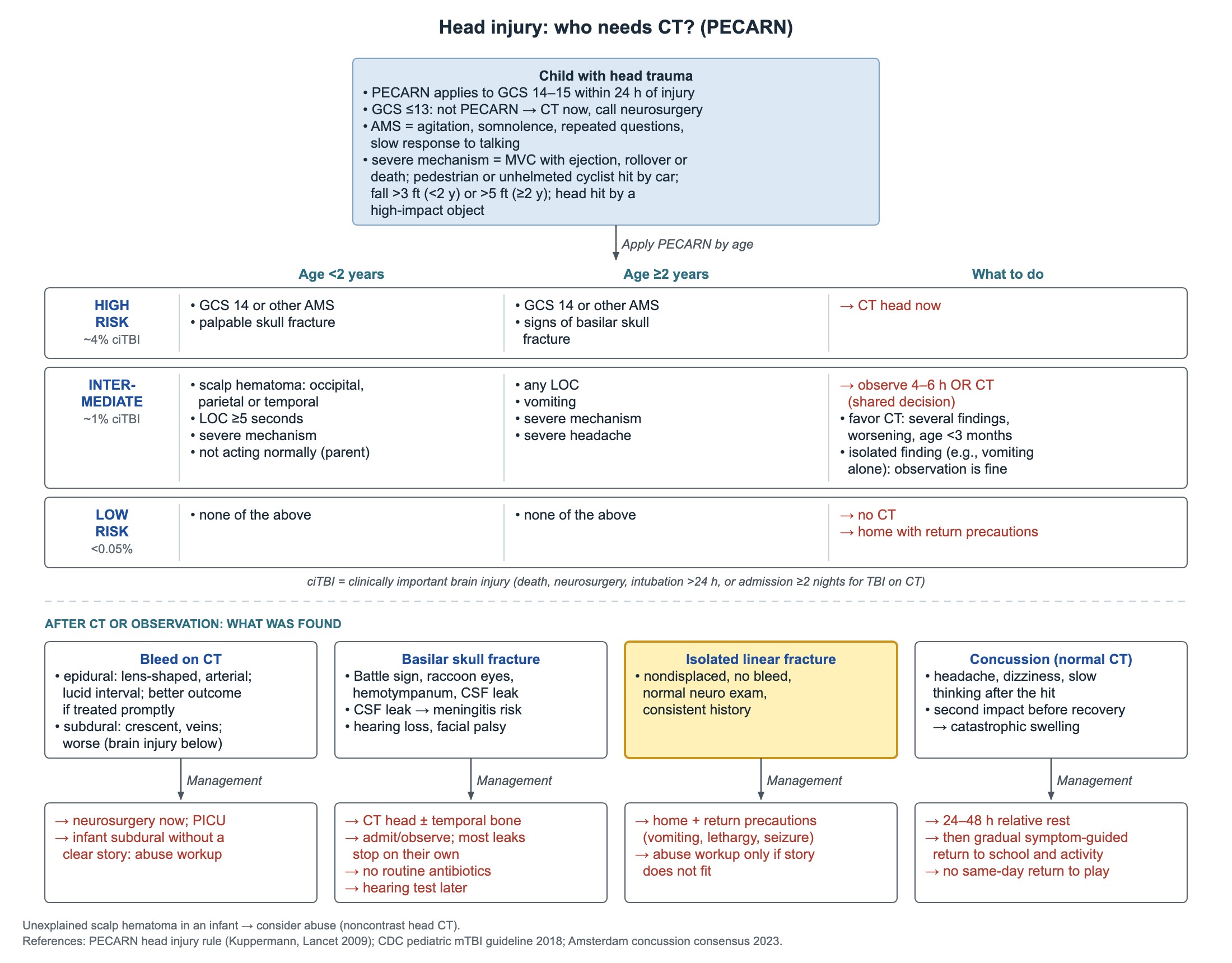
- Delayed bleeding after an isolated linear fracture with a normal CT is rare, so a well child does not need a routine overnight stay.
- Return precautions for infants: repeated vomiting, drowsiness, inconsolable crying, weakness or seizure.
- Complex, depressed or multiple skull fractures, or a story that does not fit, raise concern for abuse.
- If CT shows only a linear fracture, add brain MRI when abuse is suspected or neurologic findings are unexplained.
- Infant with a large unexplained scalp hematoma and abuse concern: first imaging for brain injury is a noncontrast head CT.
- GCS 12 or lower (or an abnormal CT): emergency neurosurgery consult. GCS 8 or lower: intubate.
- CSF leaks usually stop on their own; a persistent leak raises meningitis risk. Hearing loss and facial palsy can follow temporal-bone fractures.
- Second-impact syndrome: a new hit before recovery from the first concussion may cause catastrophic brain swelling.
| Finding | Key features | Management |
|---|---|---|
| Epidural hematoma | Lens-shaped, arterial; may have a lucid interval | Neurosurgery now; better outcome than subdural if treated promptly |
| Subdural hematoma | Crescent-shaped, venous | Neurosurgery; worse prognosis because the brain below is more injured; infant with no clear story → abuse evaluation |
| Basilar skull fracture | Battle sign, raccoon eyes, hemotympanum, CSF leak | CT head with temporal-bone imaging when needed; observe; no prophylactic antibiotics; watch for meningitis; hearing test later |
| Isolated linear fracture | Nondisplaced, no bleed, normal neuro exam, history fits | Home with return precautions; routine admission not needed |
| Concussion (normal CT) | Headache, dizziness, slow thinking after the hit | 24–48 hours relative rest, then gradual symptom-guided return; no same-day return to play |
Chest, abdomen and spinal cord injury
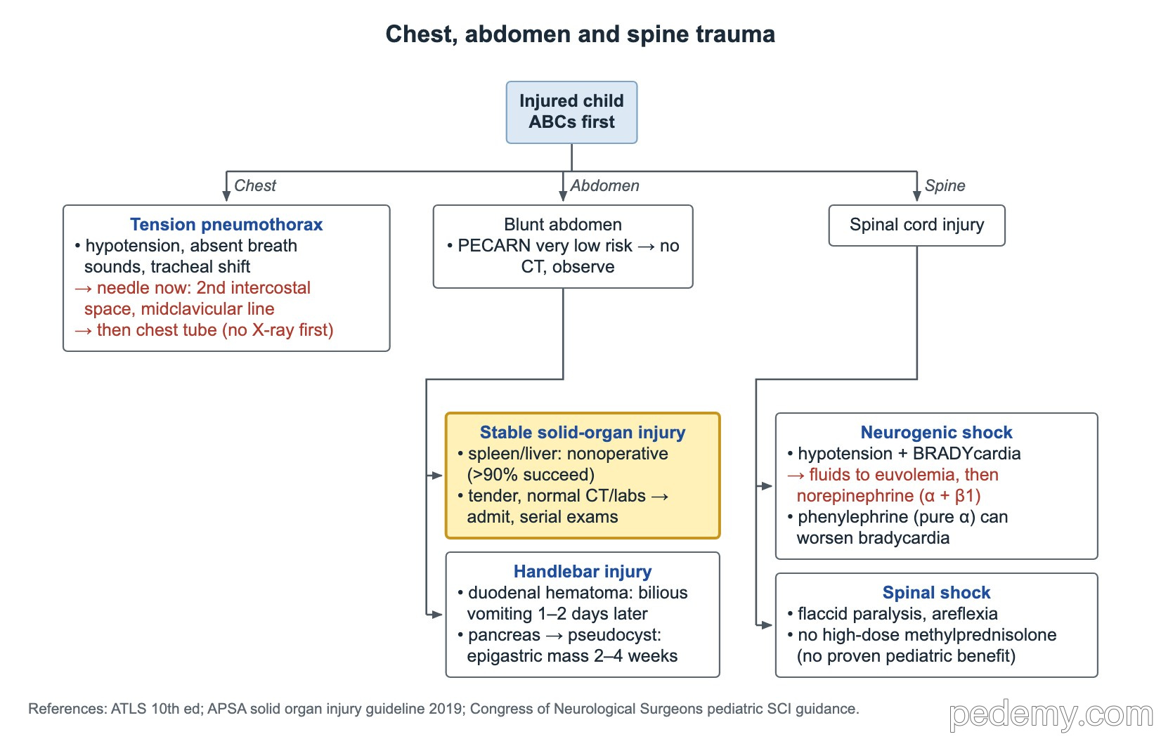
Start with the ABCs. Tension pneumothorax is a clinical diagnosis: hypotension, absent breath sounds on one side and tracheal shift. Decompress at once with a needle in the 2nd intercostal space, midclavicular line, then place a chest tube. Do not wait for an X-ray.
- PECARN abdominal rule: normal GCS, no abdominal tenderness, no seat-belt sign, no chest wall injury or breath-sound change, and no vomiting → very low risk; observe without CT.
- Stable spleen or liver injury: nonoperative care with monitoring succeeds in over 90%.
- Abdominal tenderness with normal vitals, CT and labs: admit for serial abdominal exams.
- Handlebar injury, bilious vomiting and upper abdominal pain 1–2 days later: duodenal hematoma.
- Handlebar injury, rising lipase and an epigastric mass 2–4 weeks later: pancreatic pseudocyst.
| Spinal cord injury | Defining feature | Management |
|---|---|---|
| Neurogenic shock | Hypotension with bradycardia (lost sympathetic tone) | Fluids to euvolemia, avoid repeated boluses; then norepinephrine (alpha-1 vasoconstriction plus beta-1 heart-rate support). Phenylephrine is pure alpha and may worsen bradycardia. |
| Spinal shock | Flaccid paralysis and areflexia below the injury | Supportive care; routine high-dose methylprednisolone is not recommended (no proven benefit, more complications) |
Fracture patterns in children
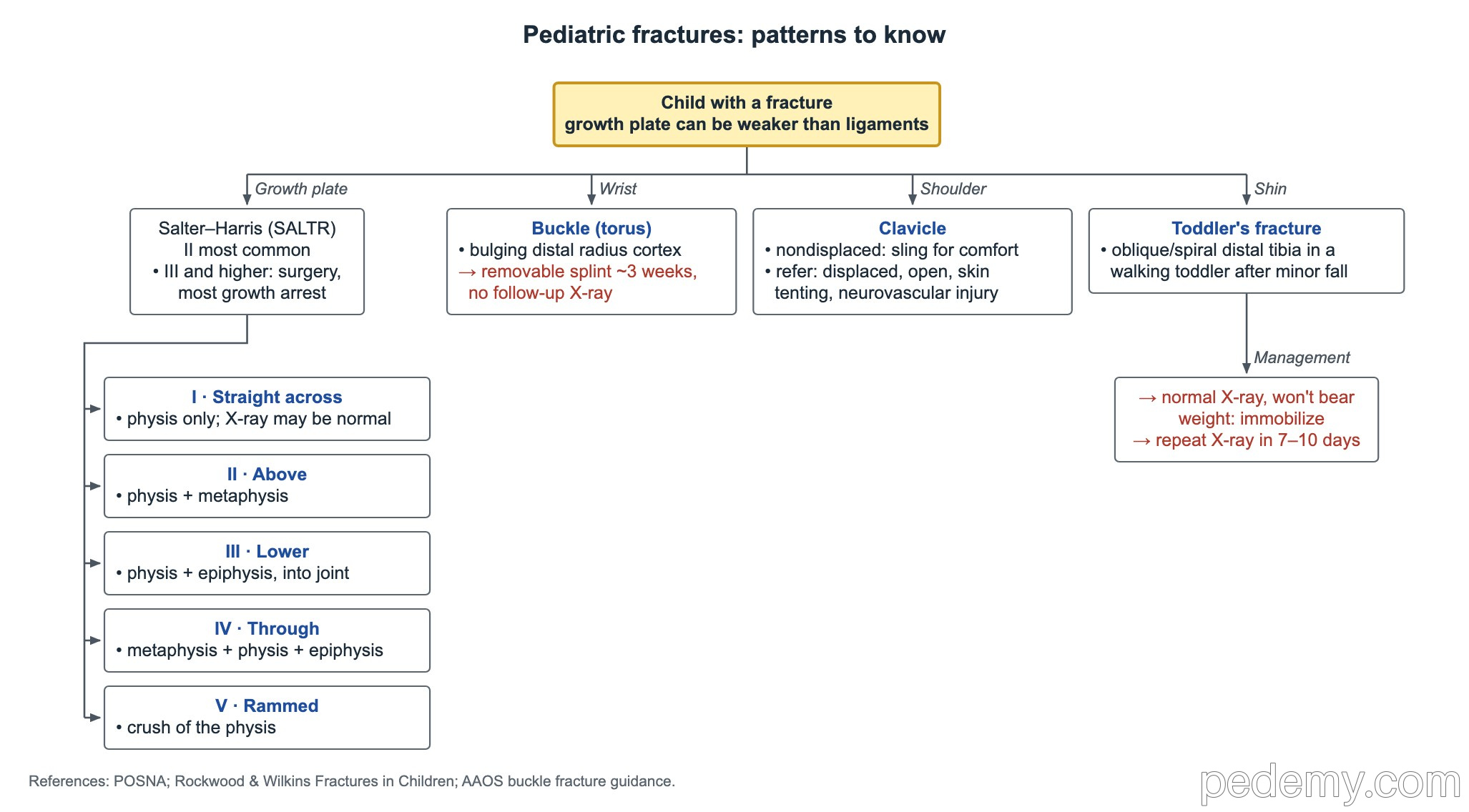
The growth plate (physis) can be weaker than the nearby ligaments, so a child who "sprains" a joint may instead have a physeal fracture. Salter–Harris types follow the SALTR memory aid.
- Types III and higher more often need surgery and carry the highest risk of growth arrest.
- Buckle (torus) fracture of the distal radius: bulging cortex, no fracture line → removable splint for about 3 weeks, no follow-up X-ray.
- Clavicle fracture, nondisplaced: sling for comfort. Refer if displaced, open, tenting the skin or with neurovascular injury.
- Toddler's fracture: oblique or spiral distal tibia fracture in a walking toddler after a minor fall.
- Suspected toddler's fracture with a normal X-ray and refusal to bear weight: immobilize and repeat the X-ray in 7–10 days.
- Ottawa ankle rule: X-ray the ankle with malleolar pain plus bony tenderness at the posterior distal 6 cm or tip of either malleolus, or inability to take 4 steps both right after injury and in the ED.
- Ottawa foot rule: X-ray the foot with midfoot pain plus navicular or 5th-metatarsal-base tenderness, or inability to take 4 steps.
- The Ottawa rules are highly sensitive, but they do not rule out every fracture.
| Salter–Harris type | SALTR | What it involves |
|---|---|---|
| I | Straight across | Physis only; X-ray may look normal |
| II | Above | Physis + metaphysis; the most common type |
| III | Lower | Physis + epiphysis, into the joint |
| IV | Through | Metaphysis + physis + epiphysis |
| V | Rammed | Crush of the physis |
Supracondylar fracture and compartment syndrome
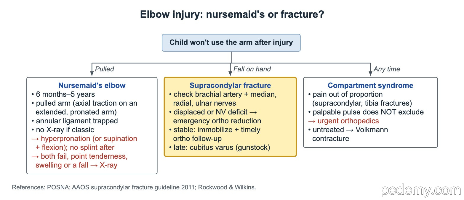
A supracondylar humerus fracture usually follows a fall on an outstretched hand. Always check the brachial artery and the median, radial and ulnar nerves.
- Significantly displaced fracture or a neurovascular deficit: emergency orthopedic reduction.
- Stable, nondisplaced fracture with intact neurovascular status: immobilize with timely orthopedic follow-up.
- Most common late deformity after malunion: cubitus varus (gunstock deformity).
- Compartment syndrome risk is highest with supracondylar and tibial fractures.
- Key sign: pain out of proportion to the injury. A palpable distal pulse does not exclude it.
- Suspected compartment syndrome: urgent orthopedic consultation.
- Untreated forearm compartment syndrome leads to Volkmann ischemic contracture.
Nursemaid's elbow (radial head subluxation)
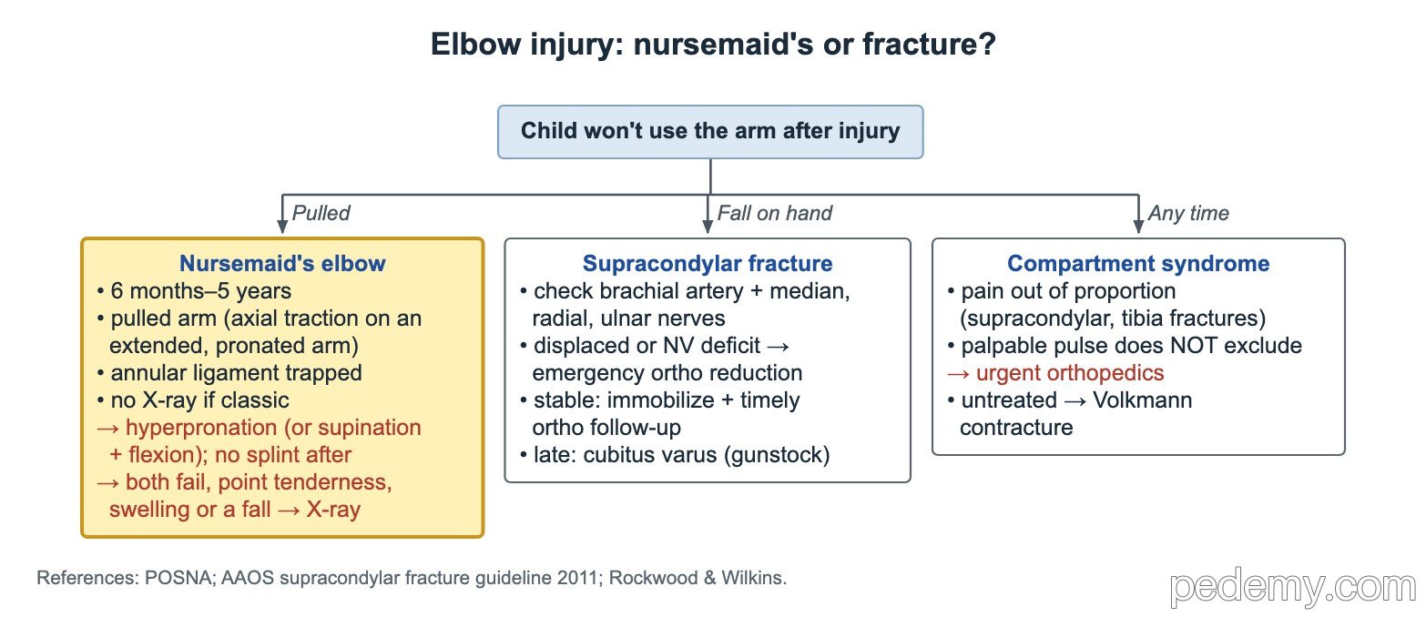
A child aged 6 months to 5 years suddenly stops using one arm after it was pulled, with little swelling or pain. Axial traction on an extended, pronated arm lets the radial head slip and the annular ligament becomes trapped in the joint.
- Classic story and exam: no X-ray needed before reduction.
- X-ray first if there was a fall, focal bony tenderness or swelling.
- Two techniques: hyperpronation (preferred first, thumb on the radial head) or supination with flexion.
- After a successful reduction the child uses the arm normally within minutes; no splint is needed.
- If two different reduction attempts fail, get elbow X-rays to look for a fracture.
Straddle injuries and knocked-out teeth
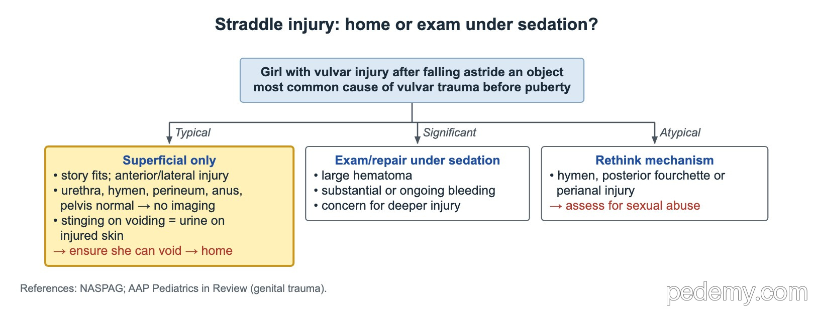
Falling astride an object, such as a bike crossbar, is the most common cause of vulvar injury before puberty. The injury is usually anterior or lateral.
- Superficial injury with normal urethra, hymen, perineum, anus and pelvis on a full exam: no imaging; confirm she can void, then discharge.
- Dysuria after a superficial abrasion comes from urine stinging the raw skin, not from urethral injury.
- Large hematoma, substantial or ongoing bleeding, or concern for deeper injury: exam or repair under sedation.
- Injury to the hymen, posterior fourchette or anus is atypical for a straddle fall: assess for penetrating injury and sexual abuse.
| Avulsed tooth | What to do |
|---|---|
| Primary (baby) tooth | Never replant: it can damage the permanent tooth bud |
| Permanent tooth, can replant | Replant at the scene: hold the crown, brief rinse, do not scrub the root; urgent dentist |
| Permanent tooth, cannot replant | Store in HBSS (Save-A-Tooth) if available, otherwise cold milk; see a dentist urgently |
| Dry time over 60 minutes | Root ligament cells are unlikely to survive; still see a dentist urgently |
Common questions
Does a child with a head injury and vomiting need a CT scan?
Not always. Under PECARN, vomiting alone in an alert child aged 2 or older with GCS 15 is intermediate risk; observation for 4–6 hours is a reasonable choice, decided with the parents.
Do children with a linear skull fracture need to be admitted?
No, if the fracture is nondisplaced, there is no bleed, the neuro exam is normal and the story fits. They can go home with return precautions.
How do you reduce a nursemaid's elbow?
Hyperpronation of the forearm with a thumb over the radial head is the preferred first method; supination with flexion is the alternative. No splint is needed after a successful reduction.
What should a knocked-out permanent tooth be stored in?
Replant it right away if possible. If not, store it in HBSS (Save-A-Tooth) or cold milk and see a dentist urgently. Never replant a baby tooth.
Study this topic
Sources
- Kuppermann et al. PECARN head injury prediction rules (Lancet 2009)
- Holmes et al. PECARN abdominal and head trauma rules: prospective validation (Lancet Child Adolesc Health 2024)
- CDC guideline on diagnosis and management of mild TBI among children (JAMA Pediatrics 2018)
- Consensus statement on concussion in sport, Amsterdam 2022 (Br J Sports Med 2023)
- IADT guidelines: avulsion of permanent teeth (Dent Traumatol 2020)
Written for Pedemy in our own words from the sources above; checked November 2026. For education only, not medical advice; check doses and local protocols before treating a patient. Pedemy is independent and not affiliated with or endorsed by the organizations cited.