

Main decision tree
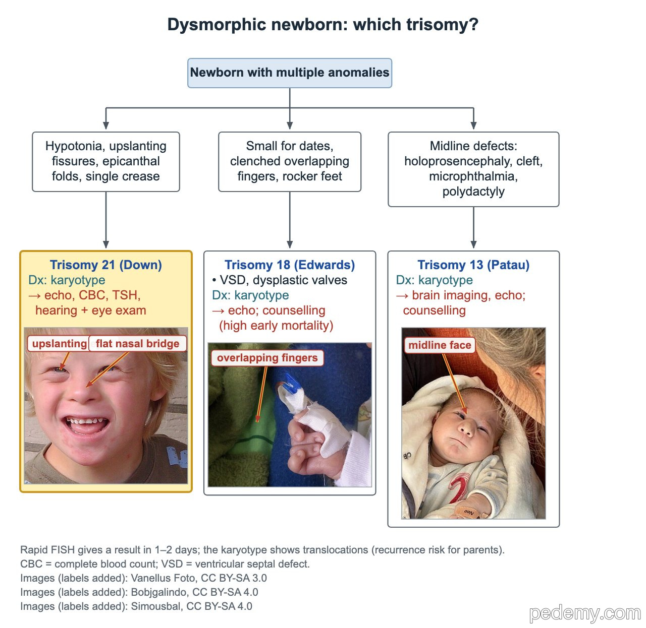
Dysmorphic newborn: which trisomy?
Key points
- Down syndrome: about 95% free trisomy 21 from meiotic nondisjunction; recurrence about 1% (or the maternal-age risk if higher).
- Robertsonian translocation (about 3–4%): karyotype both parents; a 21;21 carrier parent → essentially 100% of liveborn children.
- Every newborn with Down syndrome needs an echo, even without a murmur; TSH at 6 months, 12 months, then yearly.
- Turner syndrome: 45,X or mosaic, about 1 in 2,000–2,500 girls; not linked to maternal age; karyotype confirms.
- 22q11.2 deletion: about 90% de novo; an affected parent passes it to 50% of children; schizophrenia risk later.
- Prader–Willi = loss of paternal 15q11–q13; Angelman = loss of maternal UBE3A; methylation analysis is the first test.
- Fragile X: CGG expansion in FMR1; test with CGG repeat analysis (PCR with Southern blot), not microarray.
Which trisomy? Trisomies 21, 18 and 13
Each common trisomy has a recognizable newborn pattern. All three are confirmed by karyotype; rapid FISH can give a result in 1–2 days, but only the karyotype shows a translocation.
| Trisomy | Newborn clues | Key associations |
|---|---|---|
| 21 (Down) | Hypotonia, upslanting palpebral fissures, epicanthal folds, flat nasal bridge, single palmar crease, Brushfield spots | AV canal, VSD, ASD, tetralogy; duodenal atresia; leukemia; hypothyroidism |
| 18 (Edwards) | Growth restriction, clenched hands with overlapping fingers, hypoplastic nails, rocker-bottom feet | VSD (most common) and multiple dysplastic valves; high early mortality |
| 13 (Patau) | Midline defects: holoprosencephaly, cleft lip and palate, microphthalmia; polydactyly | Brain imaging and echo; high early mortality |
Down syndrome: mechanism and recurrence
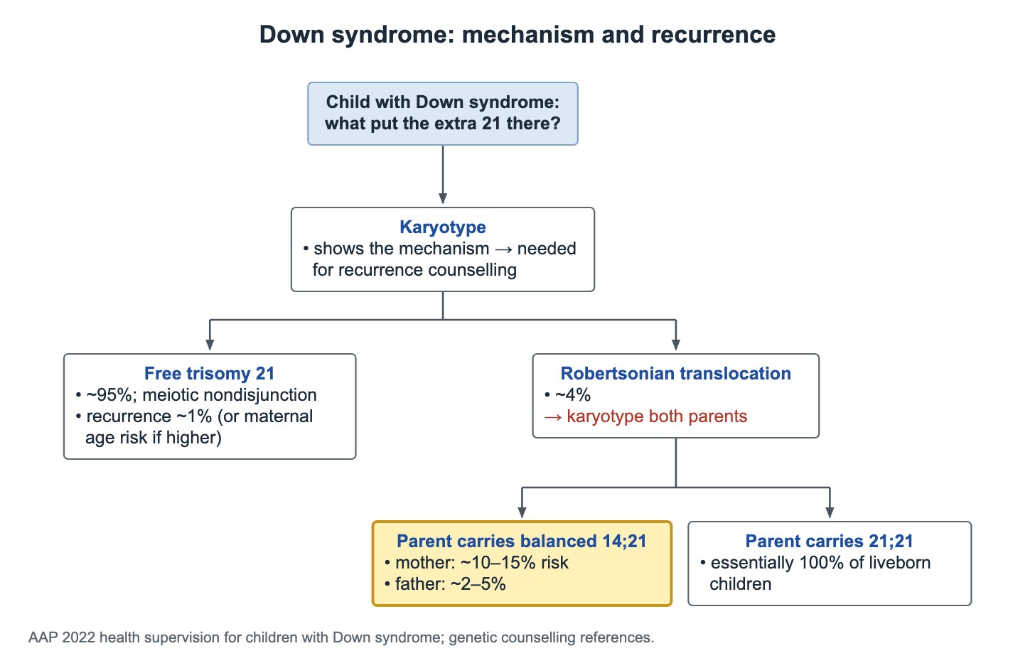
A karyotype (chromosome analysis) is needed before recurrence counseling, because it shows how the extra chromosome 21 material got there. Microarray and FISH can show extra 21 material but cannot show a balanced translocation.
Down syndrome can come from free trisomy 21, a translocation carrying extra chromosome 21 material, or mosaicism.
- Free trisomy 21: about 95%, from meiotic nondisjunction. Parents' karyotypes are normal; recurrence is about 1%, or the maternal-age risk if that is higher.
- Robertsonian translocation: about 3–4% of cases. Karyotype both parents to find a balanced carrier and set the recurrence risk.
- Mother carries a balanced 14;21 translocation: about 10–15% risk.
- Father carries a balanced 14;21 translocation: much lower, a few percent.
- Either parent carries a balanced 21;21 translocation: essentially 100% of liveborn children have Down syndrome.
Down syndrome: what to check and when
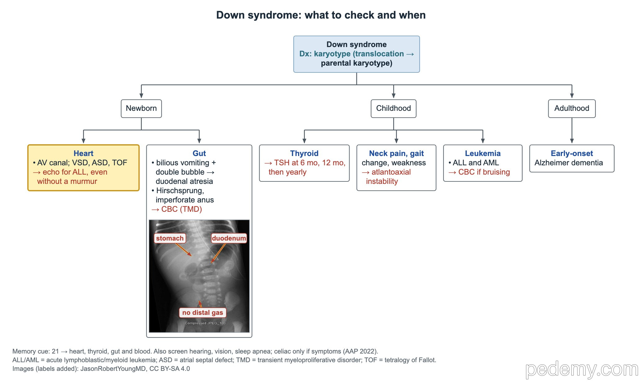
About half of children with Down syndrome have a heart defect, and many have no murmur at first, so every newborn needs an echocardiogram read by a pediatric cardiologist. The AV canal defect is the classic lesion; VSD, ASD and tetralogy of Fallot also occur.
- Gut: duodenal atresia (bilious vomiting, double-bubble X-ray), Hirschsprung disease, imperforate anus, reflux, and later celiac disease.
- Celiac disease: test with tTG-IgA plus total IgA when symptoms such as poor weight gain or loose stools appear (AAP 2022 no longer advises routine screening without symptoms).
- Blood: CBC in the newborn. Transient abnormal myelopoiesis (blasts in blood, pustular rash, large liver) usually resolves by about 3 months but raises the later risk of acute megakaryoblastic leukemia.
- Leukemia risk is raised; ALL is the most common leukemia overall, and AML is especially common in early childhood.
- Thyroid: newborn screen, then TSH at 6 months, 12 months and every year, mainly for hypothyroidism.
- Neck: atlantoaxial instability. No routine neck X-rays if well; neck pain, gait change or weakness → stop activity and get urgent neutral C-spine X-rays, then MRI.
- Also check hearing, vision and sleep apnea; early-onset Alzheimer disease is a later-life risk.
Heart defect and calcium: Williams or 22q11.2 deletion
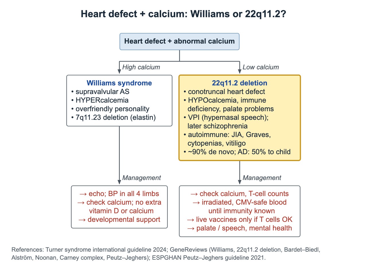
22q11.2 deletion is autosomal dominant: about 90% of deletions are new (de novo), and an affected parent passes it to 50% of children.
| Feature | Williams syndrome | 22q11.2 deletion |
|---|---|---|
| Deletion | 7q11.23, includes ELN (elastin) | 22q11.2 |
| Calcium | High | Low |
| Heart | Supravalvular aortic stenosis | Conotruncal defects |
| Other clues | Periorbital fullness, stellate irises, overly friendly personality | Immune deficiency, palate problems, velopharyngeal insufficiency (nasal speech) |
| Later risks | Hypertension | Schizophrenia; autoimmunity: juvenile arthritis, Graves disease, cytopenias, vitiligo |
| Management | Echo; blood pressure in all 4 limbs; avoid extra vitamin D or calcium | Check calcium and T-cell counts; irradiated, CMV-safe blood until immunity is known; live vaccines only if T cells are adequate |
Prader–Willi, Angelman and fragile X
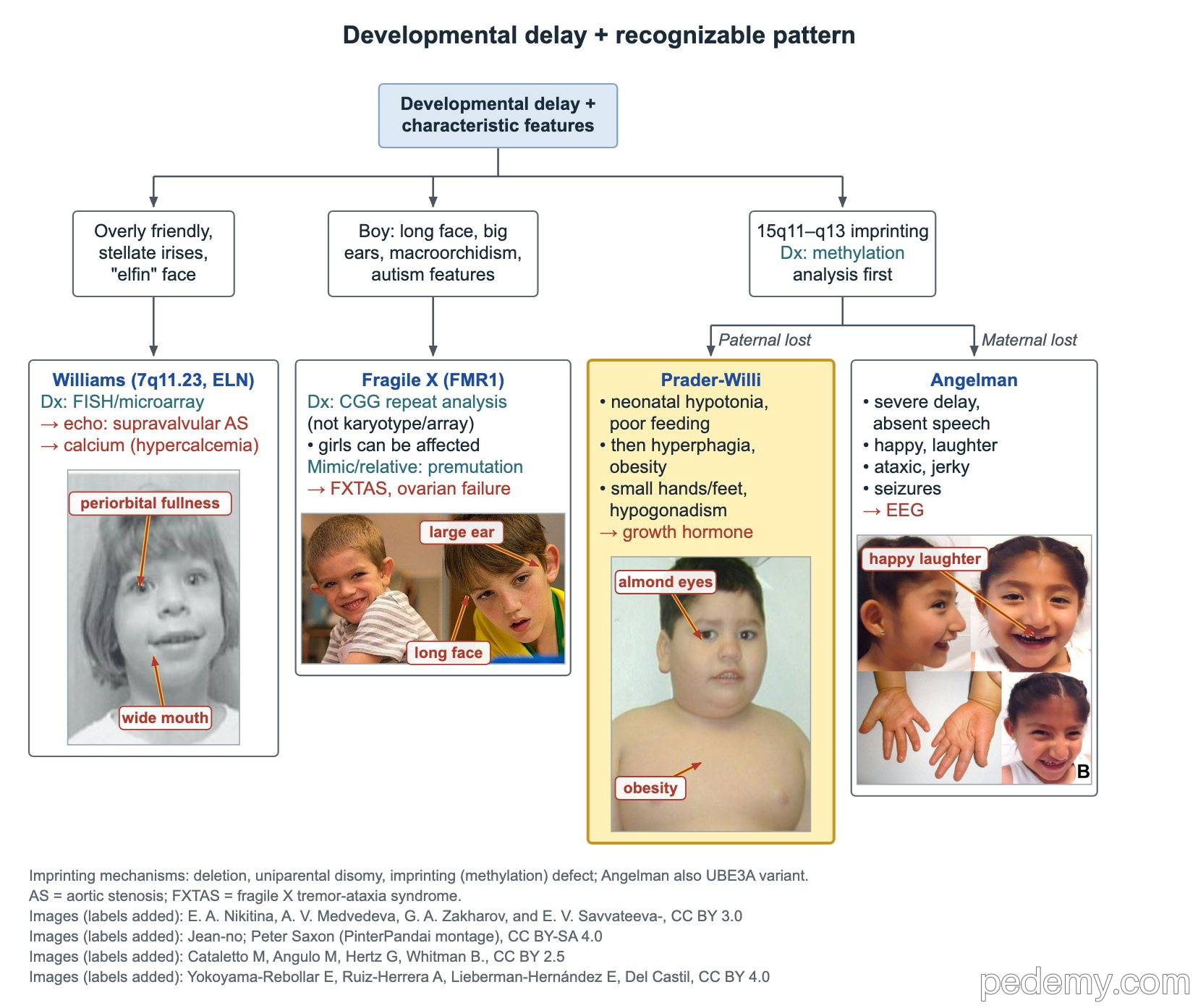
Prader–Willi and Angelman syndromes both involve the imprinted 15q11–q13 region. Which parent's copy is lost decides the syndrome. Methylation analysis is the first test because it detects deletion, uniparental disomy and imprinting defects.
- Fragile X: CGG repeat expansion in FMR1; the full mutation hypermethylates and silences the gene.
- Boys: intellectual disability, long face, large ears, joint hypermobility, large testes after puberty; mitral valve prolapse may cause a click.
- Girls can also be affected.
- Premutation carriers: mothers risk primary ovarian insufficiency; older carriers risk fragile X tremor-ataxia syndrome (FXTAS).
- Diagnosis: FMR1 CGG repeat analysis by PCR with Southern blot.
| Feature | Prader–Willi | Angelman |
|---|---|---|
| Lost copy | Paternal 15q11–q13 | Maternal 15q11–q13 (UBE3A) |
| Mechanisms | Deletion, maternal uniparental disomy, imprinting defect | Deletion, paternal uniparental disomy, imprinting defect, UBE3A variant |
| Infancy | Severe hypotonia, poor suck and feeding | Developmental delay |
| Later | Hypothalamic hyperphagia and obesity, small hands and feet, hypogonadism, almond-shaped eyes | Severe disability, absent speech, happy demeanor with frequent laughter, jerky ataxic movements, seizures |
| Treatment point | Growth hormone | EEG and seizure care |
Turner syndrome and other sex-chromosome aneuploidy
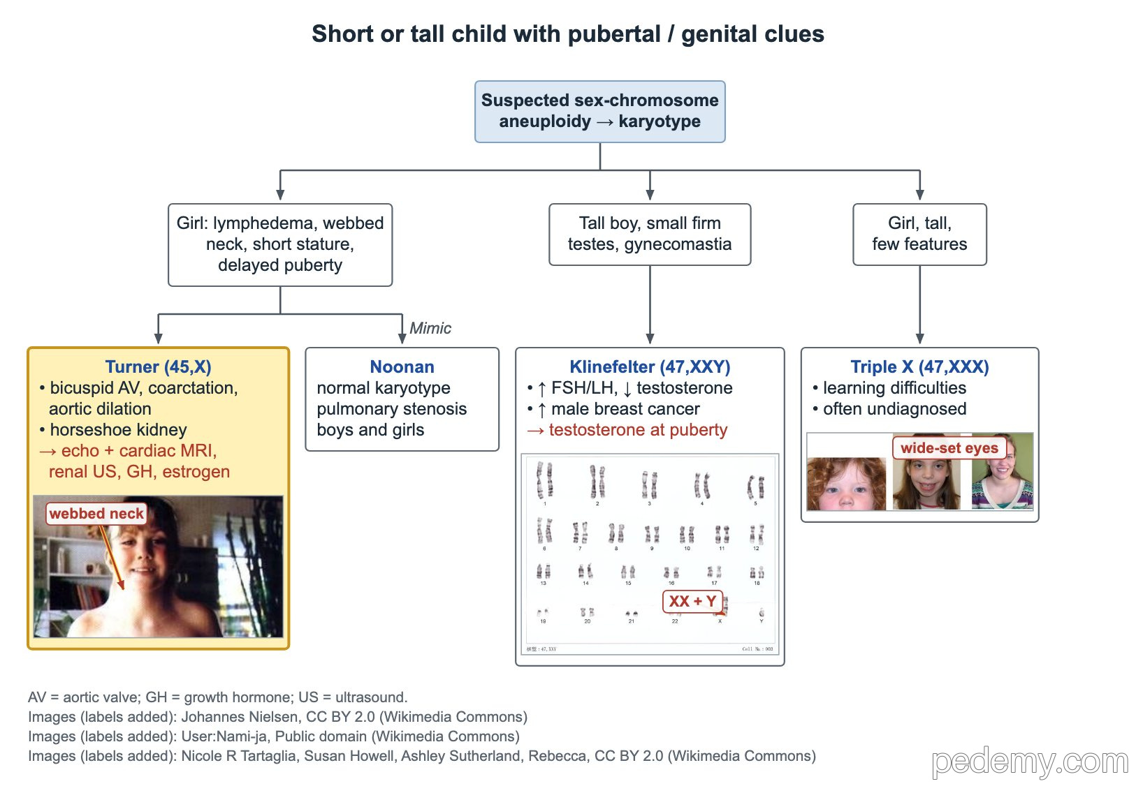
Turner syndrome (45,X or mosaic) affects about 1 in 2,000–2,500 liveborn girls. It usually comes from meiotic nondisjunction, the missing sex chromosome is usually the father's, and it is not linked to maternal age.
A newborn girl with swollen hands and feet, redundant neck skin or a webbed neck needs a karyotype. Fetal cystic hygroma also suggests aneuploidy (Turner or a trisomy) and leads to diagnostic genetic testing and a fetal echo.
- Features: short stature and Madelung deformity from SHOX loss, short or webbed neck, cubitus valgus, hyperconvex nails, delayed puberty.
- Heart: bicuspid aortic valve, coarctation and aortic dilation → echo and cardiac MRI.
- Kidney: horseshoe kidney is typical → kidney ultrasound.
- Treatment: growth hormone; estrogen for puberty.
- Follow-up: yearly TSH for Hashimoto hypothyroidism; otitis media and progressive sensorineural hearing loss → lifelong audiology.
- Y chromosome material: remove the gonads because of gonadoblastoma risk.
- Mimic: Noonan syndrome has a normal karyotype, pulmonary stenosis and affects both sexes.
- Klinefelter (47,XXY): tall boy, small firm testes, gynecomastia, high FSH and LH with low testosterone.
- Triple X (47,XXX): tall girl with few features and learning difficulties; often undiagnosed.
Which genetic test answers the question?
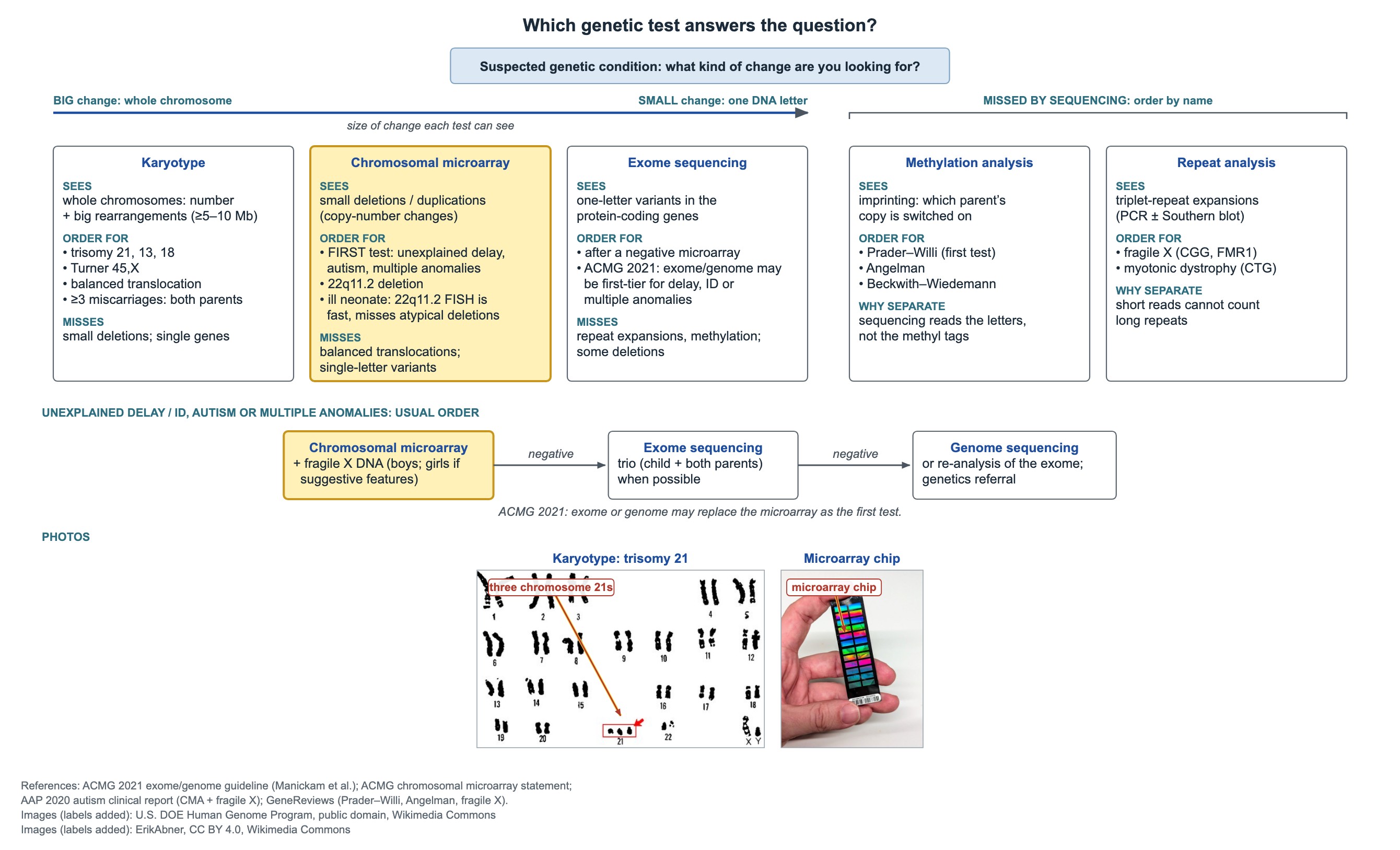
Recurrent pregnancy loss is evaluated with parental karyotypes, not microarray, because a balanced translocation is the change being sought.
| Test | Sees | Use for | Misses |
|---|---|---|---|
| Karyotype | Whole-chromosome number and large rearrangements (about 5–10 Mb or more) | Trisomies 21, 18, 13; Turner; balanced translocations; parents after recurrent pregnancy loss | Small deletions; single-gene changes |
| Chromosomal microarray | Small deletions and duplications | First test for multiple anomalies, unexplained developmental delay or intellectual disability, and autism; 22q11.2 deletion | Balanced translocations; single-letter variants |
| FISH | One targeted region | Fast 22q11.2 answer in a critically ill newborn | Atypical (distal) deletions outside the probe |
| Exome sequencing | Variants in protein-coding exons | After a negative microarray; may be first-tier (ACMG 2021) | Repeat expansions, methylation, some deletions |
| Methylation analysis | Which parent's copy is active | Prader–Willi (first test), Angelman, Beckwith–Wiedemann | Point variants such as UBE3A |
| Repeat analysis | Triplet-repeat length | Fragile X (CGG), myotonic dystrophy (CTG) | Other changes |
Common questions
What is the recurrence risk of Down syndrome?
For free trisomy 21 with normal parental karyotypes, about 1% or the maternal-age risk if higher. For a translocation it depends on the carrier parent: about 10–15% if the mother carries a 14;21 translocation, lower if the father does, and essentially 100% with a 21;21 translocation.
What test confirms Prader–Willi syndrome?
DNA methylation analysis of 15q11–q13 is the first test; it detects deletion, uniparental disomy and imprinting defects.
Why does fragile X need a special test?
Microarray and exome sequencing cannot measure CGG repeat length. FMR1 repeat analysis by PCR with Southern blot is needed.
Does every child with Down syndrome need neck X-rays?
No. Routine X-rays are not advised without symptoms. Neck pain, a change in gait or weakness needs urgent neutral cervical spine X-rays and then MRI.
Study this topic
Sources
- AAP: Health supervision for children and adolescents with Down syndrome (Pediatrics 2022)
- Clinical practice guidelines for the care of girls and women with Turner syndrome (Eur J Endocrinol 2024)
- Updated clinical practice recommendations for children with 22q11.2 deletion syndrome (Genet Med 2023)
- ACMG 2021: Exome and genome sequencing for children with congenital anomalies or intellectual disability
- AAP: Health supervision for children with fragile X syndrome (Pediatrics 2011)
Written for Pedemy in our own words from the sources above; checked November 2026. For education only, not medical advice; check doses and local protocols before treating a patient. Pedemy is independent and not affiliated with or endorsed by the organizations cited.