

Main decision tree
Skin finding: neurocutaneous syndromes
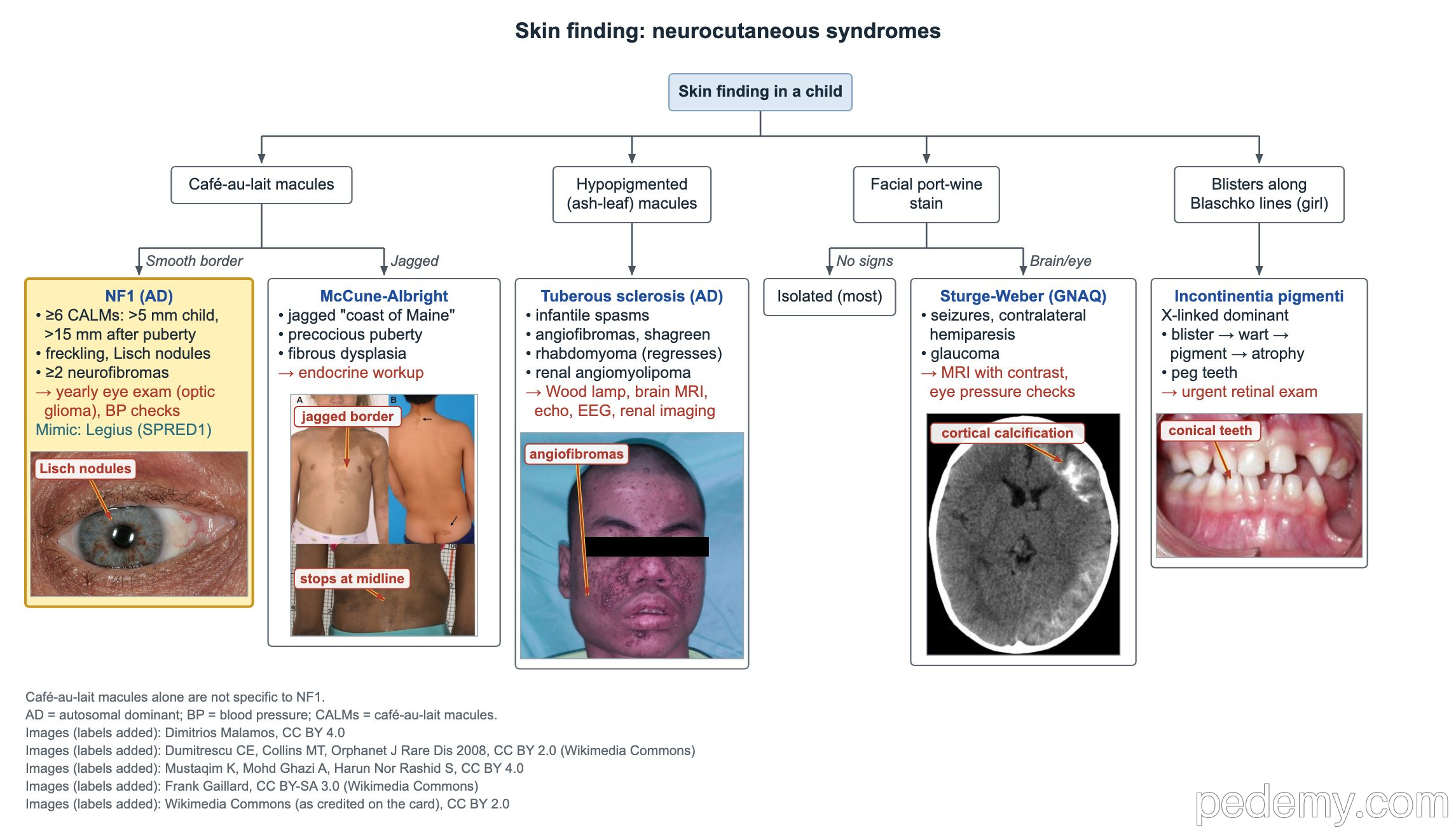
Key points
- NF1 café-au-lait criterion: 6 or more macules, >5 mm before puberty or >15 mm after puberty.
- NF1 needs 2 criteria without a family history, or 1 when a parent has NF1; it is autosomal dominant, gene NF1 on 17q11.2.
- Children with NF1: blood pressure at every visit; eye exams yearly to about age 8, then every 2 years.
- Infantile spasms + ash-leaf macules → tuberous sclerosis complex: Wood lamp exam, brain MRI, echo, EEG and renal imaging.
- Sturge-Weber: facial port-wine stain + ipsilateral leptomeningeal vascular malformation (somatic GNAQ); glaucoma, seizures, contralateral hemiparesis.
- Incontinentia pigmenti is X-linked dominant: blisters → warty lesions → pigment → atrophy; urgent retinal exam.
- WAGR (11p13 deletion, WT1 + PAX6): renal ultrasound every 3 months until age 7 to find Wilms tumor early.
Neurofibromatosis type 1: diagnosis
The tree sorts a child by the skin finding. Smooth-bordered café-au-lait macules point to NF1; café-au-lait macules alone are not specific to NF1.
NF1 is autosomal dominant; about half of cases are de novo. The NF1 gene on 17q11.2 makes neurofibromin, a tumor suppressor that switches off RAS (a RAS-GAP).
- 6 or more café-au-lait macules: >5 mm before puberty, >15 mm after puberty.
- Freckling in the axilla or groin (Crowe sign).
- 2 or more neurofibromas of any type, or 1 plexiform neurofibroma.
- Optic pathway glioma.
- 2 or more Lisch nodules (iris hamartomas) or 2 or more choroidal abnormalities.
- A distinctive bone lesion: sphenoid dysplasia, anterolateral tibial bowing or pseudarthrosis of a long bone.
- A pathogenic NF1 variant at 50% allele fraction in normal tissue.
| Situation | Criteria needed |
|---|---|
| No parent with NF1 | 2 or more of the features listed |
| Parent meets NF1 criteria | 1 or more of the features listed |
| Only café-au-lait macules and freckling | Consider Legius syndrome (SPRED1); genetic testing separates them |
NF1 complications and follow-up
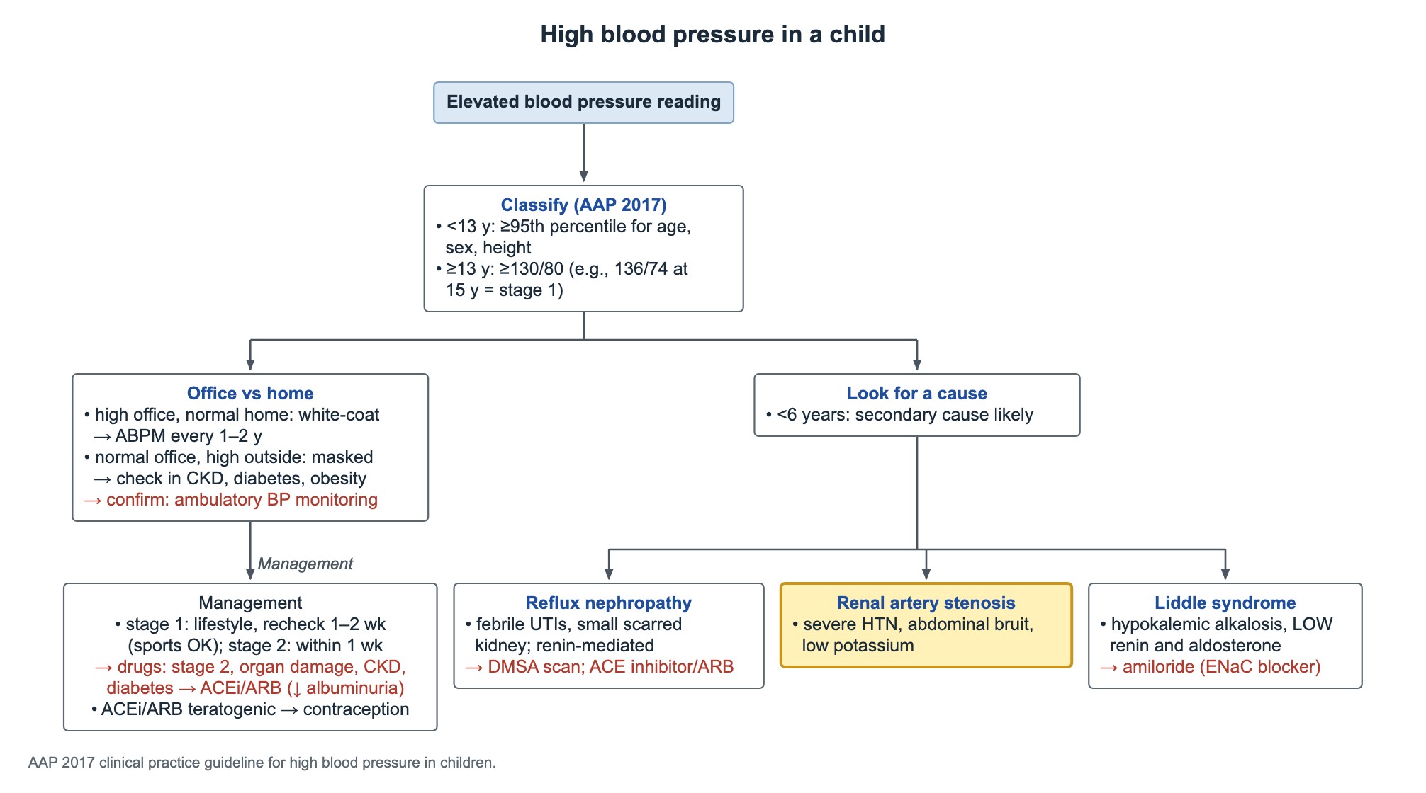
Check blood pressure at every visit. In NF1, a high reading with an abdominal bruit suggests renal artery stenosis; with headaches, sweating or palpitations, think of pheochromocytoma and test plasma free metanephrines. Do not screen for pheochromocytoma when blood pressure is normal.
- Eyes: an ophthalmology exam every year until about age 8, then every 2 years, to find optic pathway glioma.
- A chiasmal optic glioma can cause central precocious puberty: plot growth and puberty at each visit.
- MRI often shows T2-bright spots (focal areas of signal intensity, once called UBOs); routine MRI is not needed without symptoms.
- Learning disability and ADHD are the commonest neurodevelopmental problems; scoliosis is the commonest skeletal complication.
- Plexiform neurofibromas can turn into malignant peripheral nerve sheath tumors: new pain or fast growth needs urgent imaging.
- Women with NF1 have a higher breast cancer risk: yearly breast screening starts at age 30.
Tuberous sclerosis complex
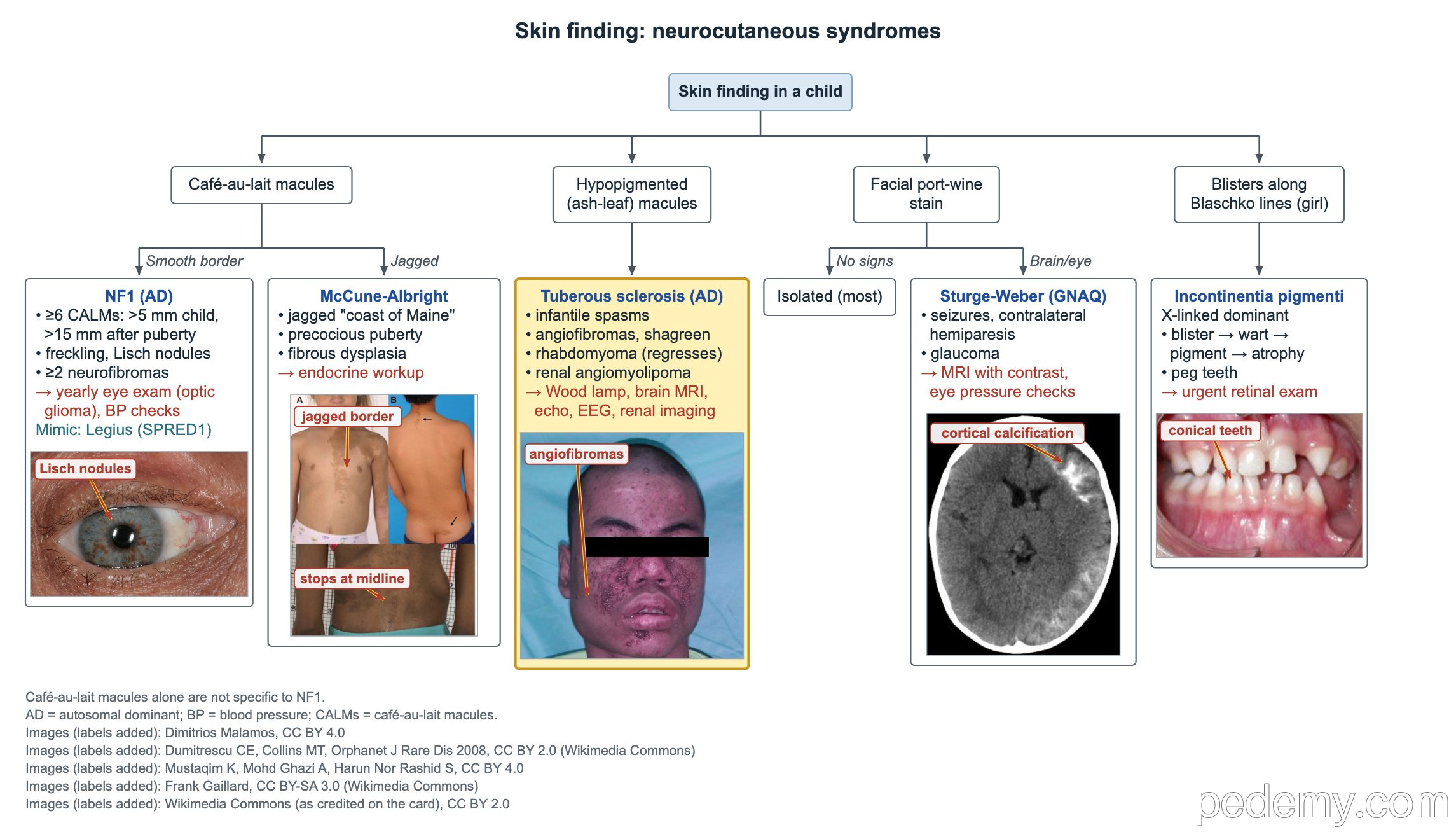
Infantile spasms with hypopigmented (ash-leaf) macules should prompt evaluation for TSC. A Wood lamp helps find faint macules in light skin.
TSC is autosomal dominant, with many de novo cases. TSC1 encodes hamartin and TSC2 encodes tuberin; both restrain mTOR signaling.
- Skin: ash-leaf macules, facial angiofibromas, a shagreen patch (connective-tissue plaque, often on the lower back), ungual fibromas.
- Brain: cortical tubers, subependymal nodules, and subependymal giant cell astrocytoma (SEGA) near the foramen of Monro, which can cause hydrocephalus.
- Heart: cardiac rhabdomyomas, often found before birth; they usually regress in the first years but can cause outflow obstruction and arrhythmias.
- Kidney: angiomyolipomas are the typical renal tumor; cysts also occur.
- Work-up: brain MRI, EEG, echo and ECG, renal imaging, eye and skin exams; repeat brain and kidney imaging every 1–3 years.
- Vigabatrin is first-line for infantile spasms in TSC; mTOR inhibitors treat SEGA and angiomyolipomas.
Sturge-Weber syndrome and port-wine stains
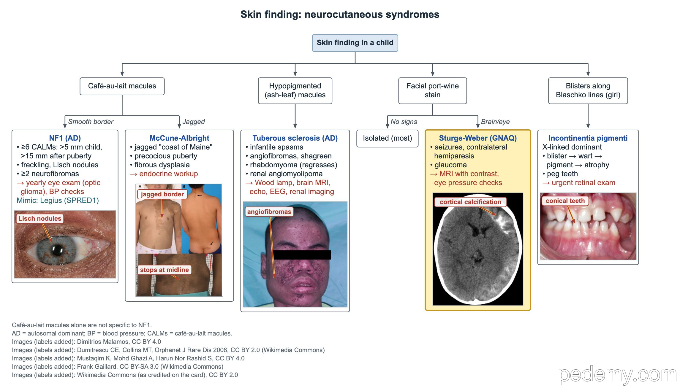
Sturge-Weber syndrome is a facial port-wine stain with an ipsilateral leptomeningeal vascular malformation, often with eye involvement. It is caused by a somatic (not inherited) GNAQ variant.
Most facial port-wine stains are isolated and not Sturge-Weber. Risk is highest when the stain covers the forehead or upper eyelid.
- Brain: seizures, developmental delay and hemiparesis on the side opposite the stain.
- Imaging: MRI with contrast shows the leptomeningeal malformation; CT can show tram-track gyral calcification.
- Eye: glaucoma is the important complication; check eye pressure regularly.
Incontinentia pigmenti and McCune-Albright syndrome
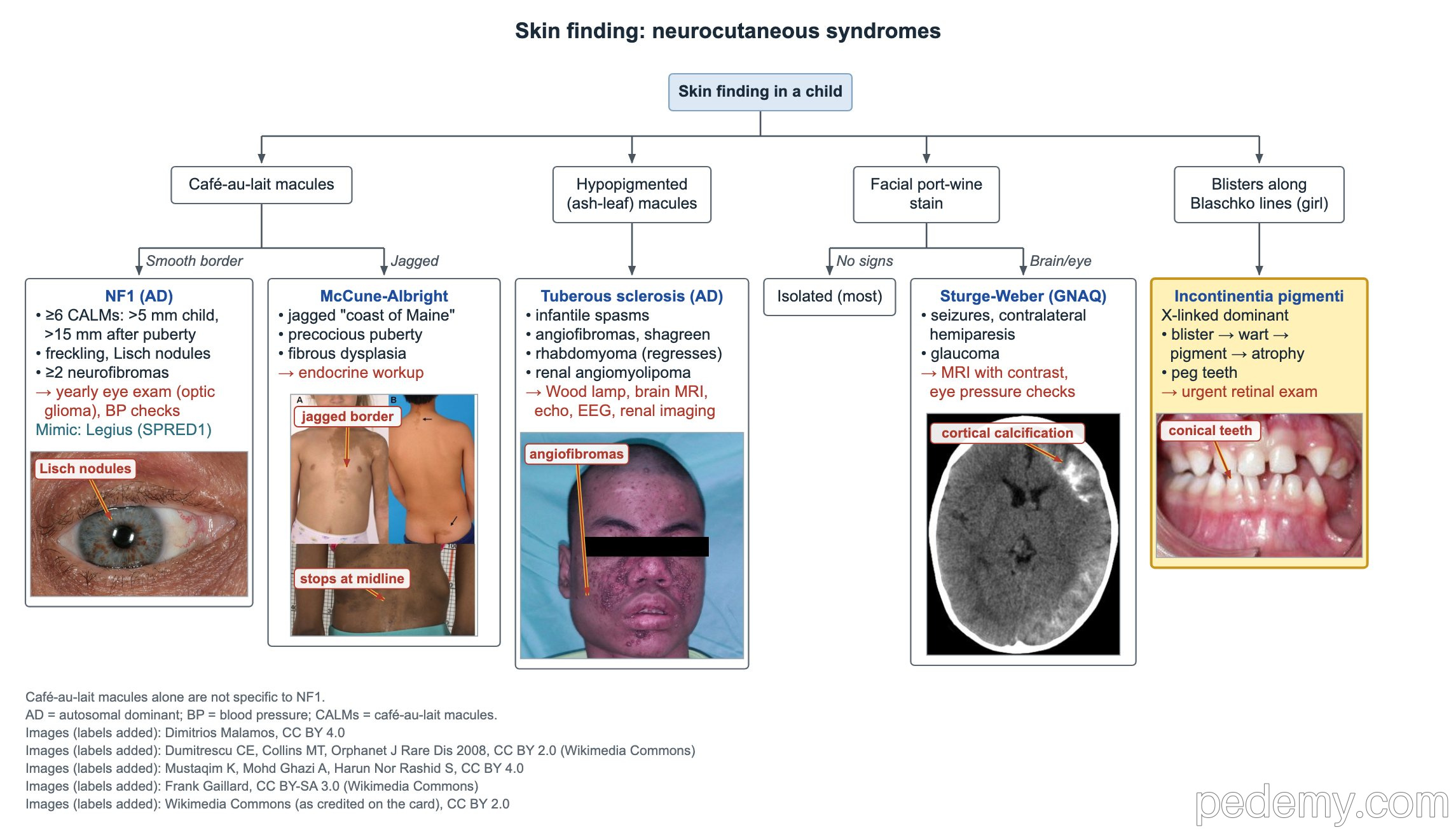
Incontinentia pigmenti affects mainly girls because it is X-linked dominant and usually lethal in males. Skin lesions follow Blaschko lines and pass through stages: blisters, then warty lesions, then swirled pigment, then pale atrophic streaks.
- Teeth: missing teeth and peg- or cone-shaped teeth.
- Eyes: retinal neovascularization can lead to retinal detachment, so an urgent retinal exam is needed after diagnosis.
- Every child needs dental, ophthalmologic and neurologic evaluation.
- McCune-Albright syndrome: café-au-lait macules with jagged "coast of Maine" borders, fibrous dysplasia of bone and precocious puberty; caused by a somatic GNAS variant.
Von Hippel-Lindau and NF2-related schwannomatosis
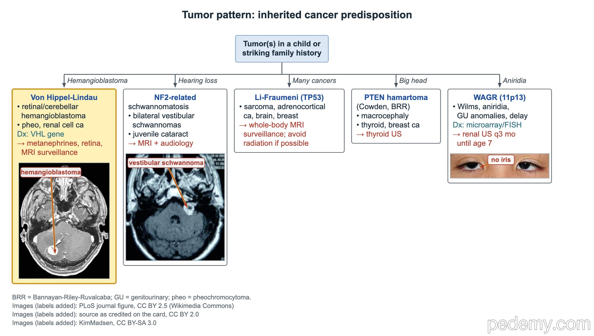
Retinal or cerebellar hemangioblastoma with pheochromocytoma or renal cell carcinoma means von Hippel-Lindau disease. VHL is autosomal dominant, but a new (de novo) variant can occur, so a negative family history does not exclude it.
Confirm with VHL gene testing. Surveillance includes plasma metanephrines, retinal exams and MRI, starting in childhood.
- VHL tumors: retinal and CNS hemangioblastomas, pheochromocytoma, renal cell carcinoma (the important malignant kidney tumor), pancreatic tumors and cysts.
- NF2-related schwannomatosis (formerly NF2) is autosomal dominant: bilateral vestibular schwannomas cause hearing loss, tinnitus and imbalance in young adults.
- In childhood, NF2-related schwannomatosis may first show as a juvenile cataract.
- Follow-up for NF2-related schwannomatosis: MRI and audiology.
Li-Fraumeni, PTEN, WAGR and Carney complex
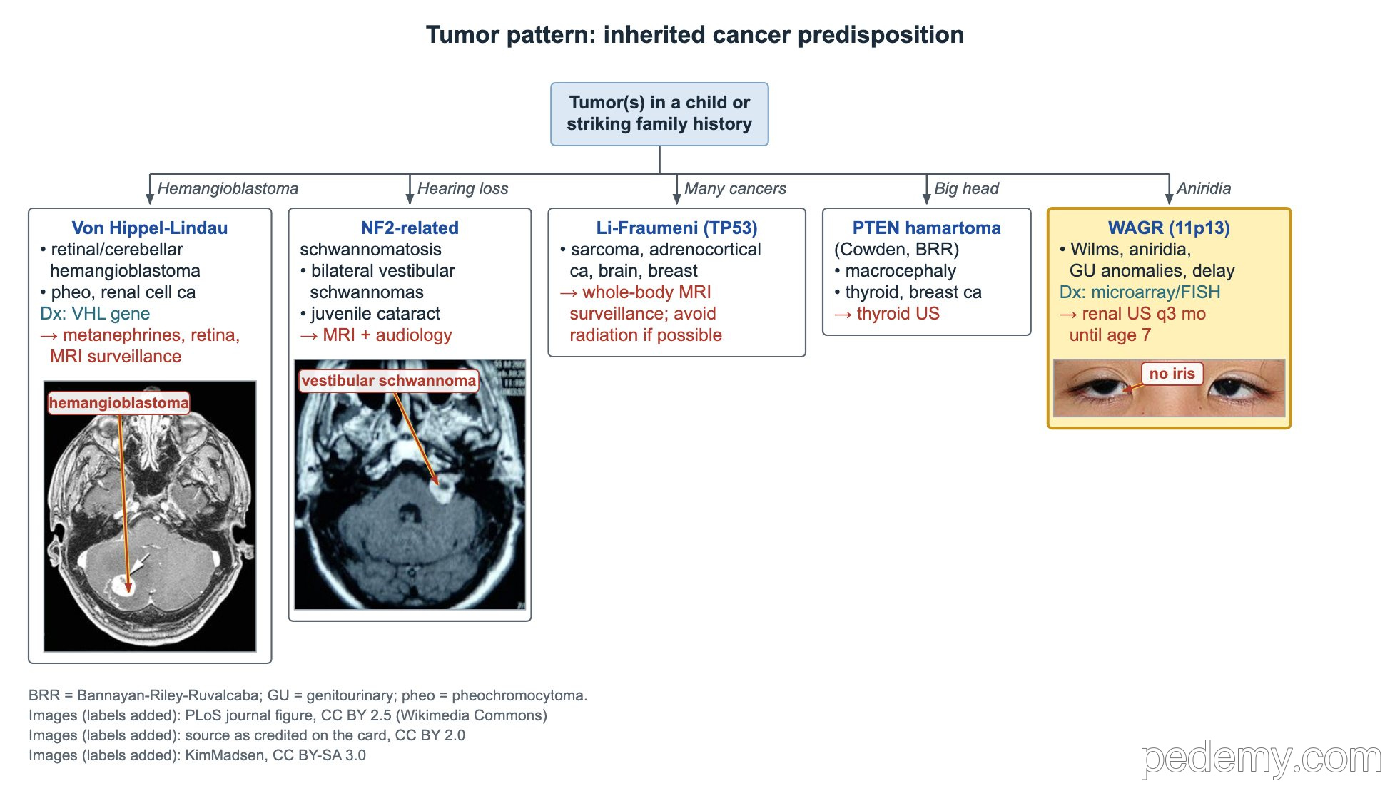
WAGR is a contiguous deletion at 11p13 that removes WT1 (Wilms tumor) and PAX6 (aniridia). A child with aniridia needs microarray or FISH for the deletion, and children with WAGR get a renal ultrasound every 3 months until age 7.
| Syndrome | Clues | Key point |
|---|---|---|
| WAGR (11p13) | Wilms tumor, aniridia, genitourinary anomalies (hypospadias, cryptorchidism), intellectual disability | Renal ultrasound every 3 months until age 7 |
| Li-Fraumeni (TP53) | Sarcoma, adrenocortical carcinoma, brain tumors, early breast cancer | Whole-body MRI surveillance; avoid radiation when possible |
| PTEN hamartoma tumor syndrome | Macrocephaly, hamartomatous polyps, pigmented penile macules (Bannayan–Riley–Ruvalcaba); papillomas, thyroid and breast cancer (Cowden) | Thyroid ultrasound surveillance |
| Carney complex (PRKAR1A) | Spotty pigmentation, cardiac myxomas, ACTH-independent Cushing syndrome | Regular echo for myxomas |
Common questions
How many café-au-lait spots suggest NF1?
Six or more, each larger than 5 mm before puberty or larger than 15 mm after puberty. Spots alone are not specific: Legius syndrome and other conditions can cause them.
How often do children with NF1 need eye exams?
Every year until about age 8, then every 2 years, to look for optic pathway glioma.
Does every port-wine stain mean Sturge-Weber syndrome?
No. Most are isolated. The risk is highest with a stain on the forehead or upper eyelid, which needs eye pressure checks and an assessment for brain involvement.
Study this topic
Sources
- Revised diagnostic criteria for NF1 and Legius syndrome: international consensus (Genet Med 2021)
- AAP clinical report: Health supervision for children with neurofibromatosis type 1 (Pediatrics 2019)
- Updated international TSC diagnostic criteria and surveillance recommendations (Pediatr Neurol 2021)
- Updated diagnostic criteria and nomenclature for NF2 and schwannomatosis (Genet Med 2022)
Written for Pedemy in our own words from the sources above; checked November 2026. For education only, not medical advice; check doses and local protocols before treating a patient. Pedemy is independent and not affiliated with or endorsed by the organizations cited.