

Main decision tree
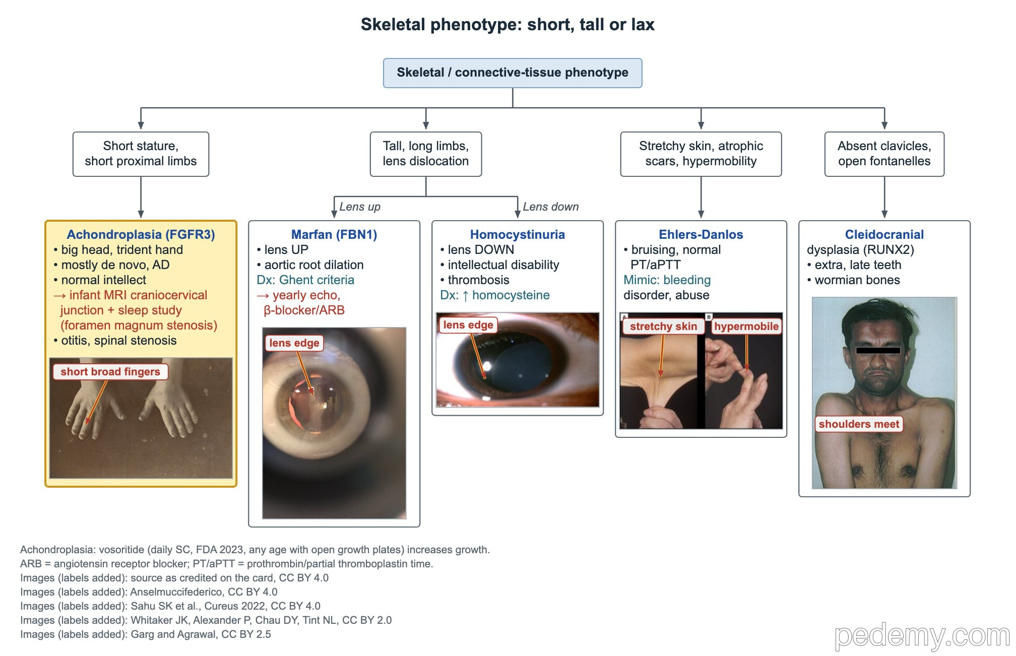
Skeletal phenotype: short, tall or lax
Key points
- Achondroplasia: FGFR3, autosomal dominant, about 80% de novo; rhizomelia, trident hand, big head, normal intellect.
- Infants with achondroplasia need assessment for foramen magnum stenosis: neurologic exam, sleep study and craniocervical MRI.
- Osteogenesis imperfecta type I is the mildest and most common: blue sclerae, low-trauma fractures, hearing loss, autosomal dominant.
- OI type II is lethal in the newborn period from respiratory insufficiency; fractures are often present before birth.
- Marfan without a family history: aortic root z-score ≥2 plus ectopia lentis is enough to diagnose.
- Marfan systemic score ≥7 counts as a major criterion; wrist AND thumb signs together score 3 points.
- Classic Ehlers-Danlos syndrome: COL5A1/COL5A2; easy bruising with a normal PT and aPTT comes from tissue fragility.
Achondroplasia
The tree sorts the main skeletal picture: short proximal limbs, a tall body with lens dislocation, stretchy skin, or absent clavicles. Short proximal limbs (rhizomelia) with a big head point to achondroplasia.
Achondroplasia is caused by a gain-of-function FGFR3 variant. It is autosomal dominant, but most cases are de novo, born to parents of average height.
- Look: macrocephaly with frontal bossing, midface hypoplasia, short broad fingers with a trident hand.
- Intellect is usually normal; motor milestones are often delayed.
- Infancy: foramen magnum (craniocervical) stenosis can cause central apnea, weakness and sudden death; screen with a neurologic exam, sleep study and craniocervical MRI.
- Hydrocephalus with enlarged ventricles; obstructive sleep apnea.
- Ears: serous otitis media is common; check hearing.
- Adults: spinal stenosis.
- Vosoritide, a daily injection, increases growth in children whose growth plates are still open.
Osteogenesis imperfecta: the classic types
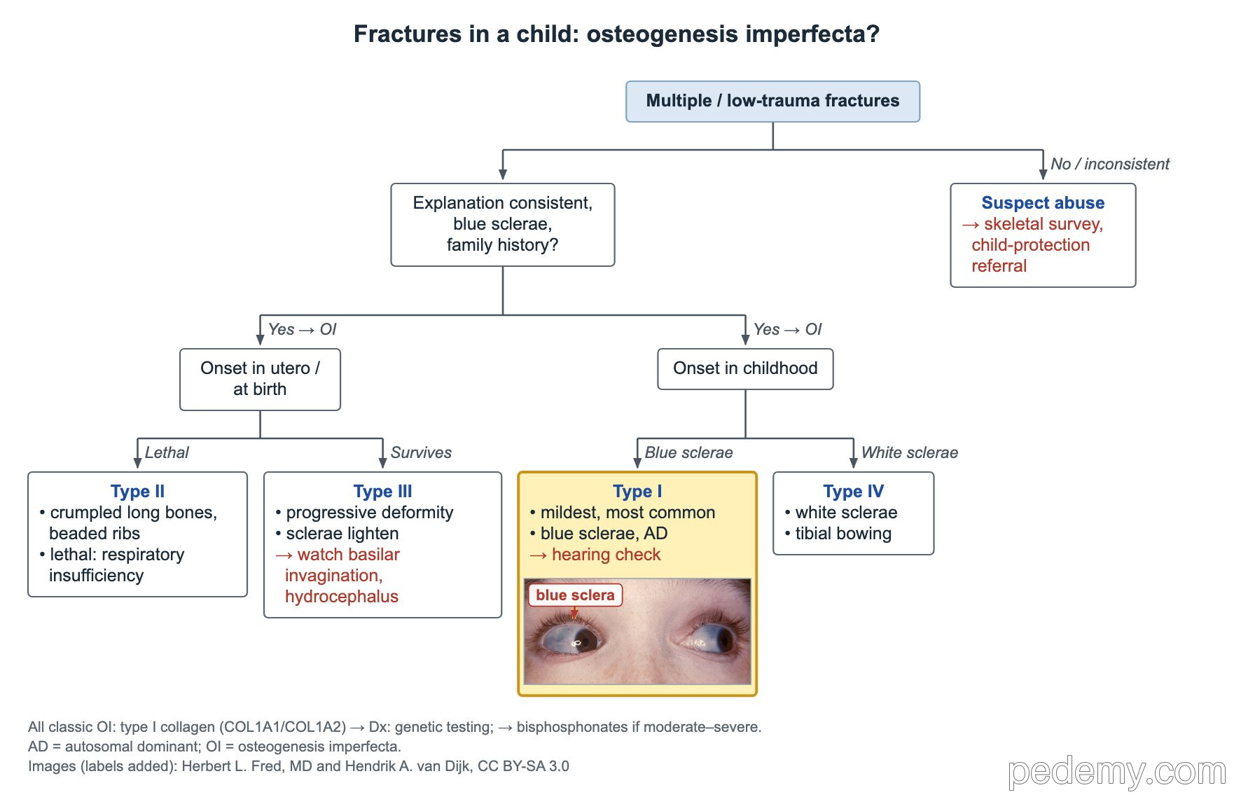
Multiple or low-trauma fractures raise two questions: is the story consistent, and are there blue sclerae or a family history? If not, suspect abuse: get a skeletal survey and make a child-protection referral.
The central problem in all types is bone fragility, often with short stature or deformity. Classic OI involves type I collagen (COL1A1 or COL1A2). Genetic testing confirms the diagnosis; bisphosphonates are used for moderate to severe disease.
- Dentinogenesis imperfecta: discolored teeth that chip easily, a common OI clue.
- Type I: check hearing, because hearing loss often starts in the teens or adulthood.
| Type | Pattern | Clue |
|---|---|---|
| I | Mildest and most common; childhood fractures; autosomal dominant | Blue sclerae, hearing loss; less type I collagen is made |
| II | Fractures before or at birth; lethal | Crumpled long bones, beaded ribs; death from respiratory insufficiency |
| III | Severe, progressively deforming | Sclerae lighten with age; basilar invagination, hydrocephalus |
| IV | Moderate, variable severity | Normal or white sclerae; tibial bowing |
Severe OI in the newborn
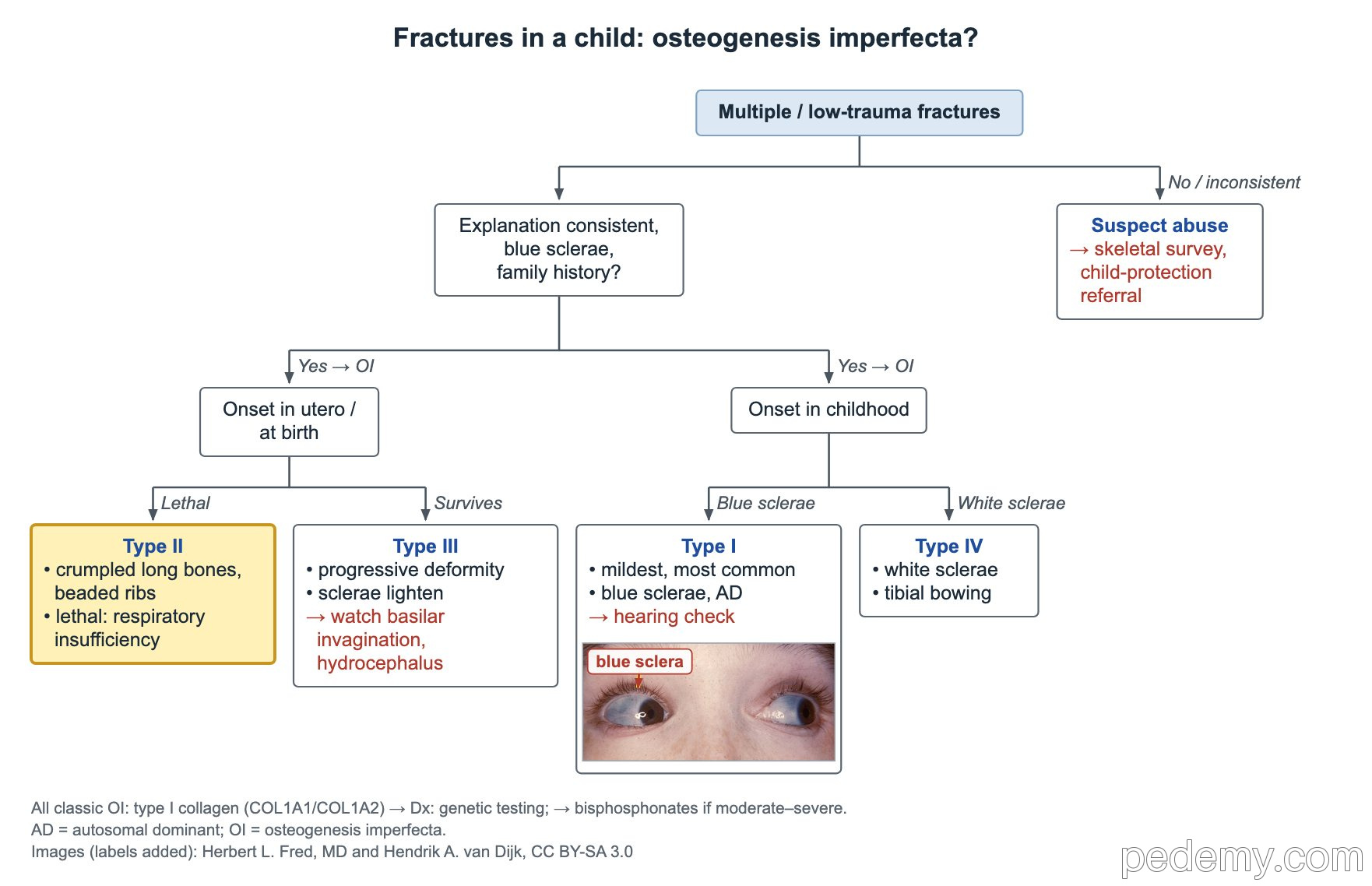
When fractures begin in utero or at birth, the tree splits into the lethal and the surviving severe forms.
- Type II: fractures already present before birth, crumpled long bones and beaded ribs; most infants die from respiratory insufficiency.
- Type III: survives the newborn period, with progressive deformity and short stature.
- Type III: blue sclerae lighten with age; watch for basilar invagination and hydrocephalus.
Marfan syndrome: revised Ghent criteria
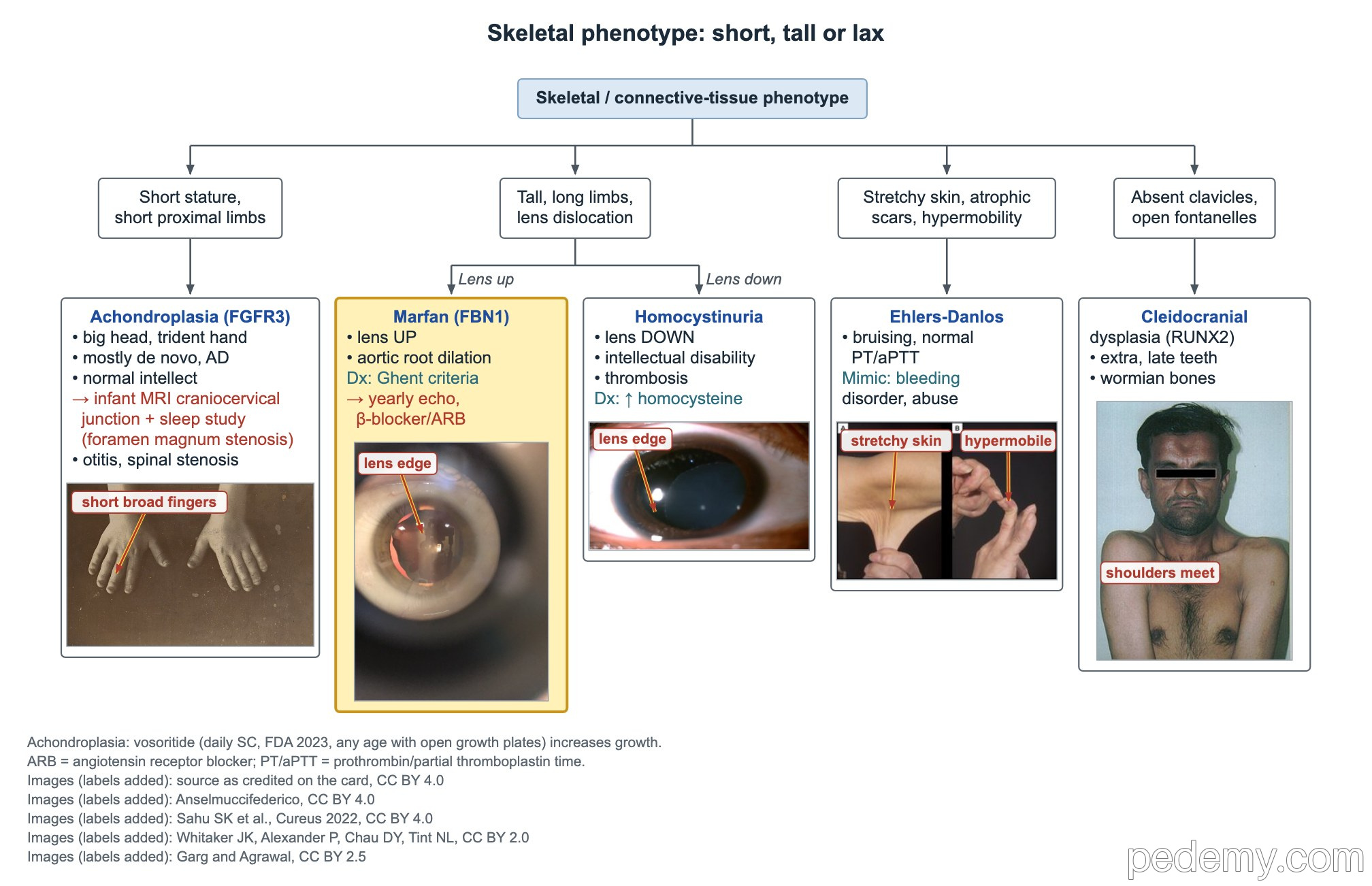
Marfan syndrome is autosomal dominant and caused by FBN1 variants (fibrillin-1). The lens dislocates upward in Marfan and downward in homocystinuria.
A tall teen with long fingers, upward lens dislocation and a parent with aortic dissection needs an echo of the aortic root first.
- Systemic score: wrist AND thumb signs = 3 points (only one = 1).
- 2 points each: pectus carinatum, hindfoot valgus, pneumothorax, dural ectasia, protrusio acetabuli.
- Pectus excavatum or chest asymmetry = 1 point.
- Wrist sign: thumb and fifth finger overlap when wrapped around the opposite wrist. Thumb sign: the thumb tip extends past the edge of the clenched fist.
- Reduced elbow extension scores in Marfan; elbow hypermobility does not count and suggests Ehlers-Danlos syndrome.
| Family history | Any one of these confirms Marfan |
|---|---|
| No family history | Aortic root z ≥2 + ectopia lentis; aortic root z ≥2 + pathogenic FBN1 variant; aortic root z ≥2 + systemic score ≥7; ectopia lentis + FBN1 variant known to cause aortic disease |
| Parent with Marfan | Ectopia lentis; or systemic score ≥7; or aortic root z ≥2 (age 20 or older) or z ≥3 (under 20) |
Marfan aorta, sports and Loeys-Dietz syndrome
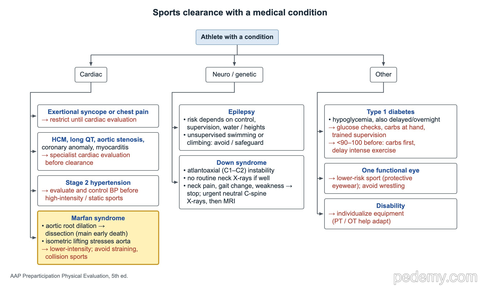
Aortic root dilation leading to dissection is the main cause of early death in Marfan syndrome. Isometric lifting and straining stress the aorta, so athletes are steered to lower-intensity sports and away from collision sports.
Follow the aortic root with an echo at least yearly. A beta-blocker or an angiotensin receptor blocker slows root growth.
- Loeys-Dietz syndrome: aortic root aneurysm with hypertelorism and a bifid uvula or cleft palate; genes TGFBR1 and TGFBR2 (and others in the TGF-beta pathway).
- Loeys-Dietz arteries are tortuous and can dissect at smaller diameters than in Marfan; image the arteries from head to pelvis.
Ehlers-Danlos syndromes
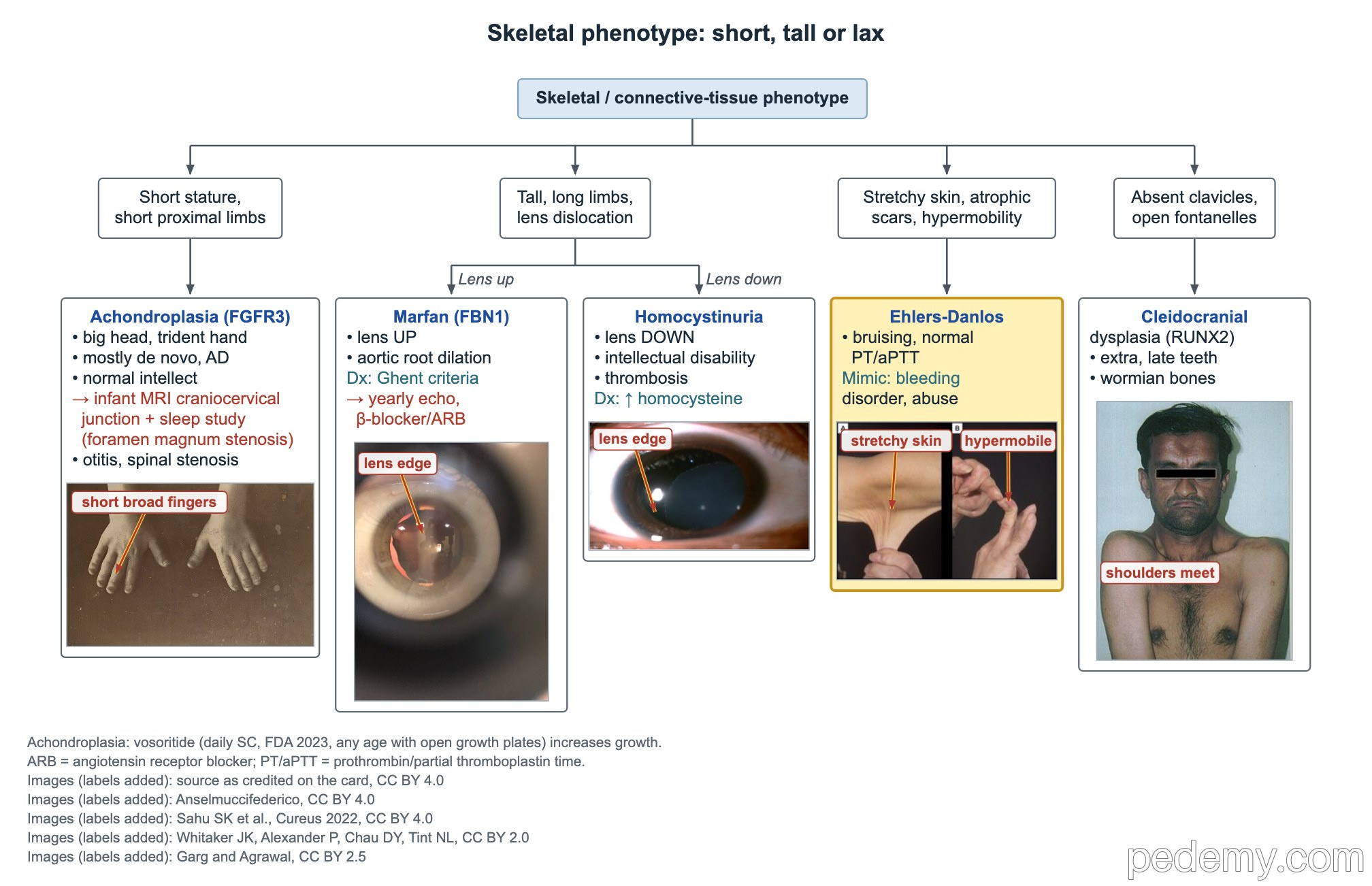
Stretchy, soft (doughy) skin, wide atrophic scars, hypermobile joints and easy bruising suggest Ehlers-Danlos syndrome. The 2017 classification lists 13 types; all except hypermobile EDS are confirmed by genetic testing.
- Classic EDS: COL5A1 or COL5A2.
- Easy bruising with a normal PT and aPTT comes from capillary and tissue fragility, not a coagulation-factor defect.
- Rule out a bleeding disorder and consider abuse before blaming bruises on EDS.
- Mitral valve prolapse (a midsystolic click) can occur.
Cleidocranial dysplasia, Caffey disease and FOP
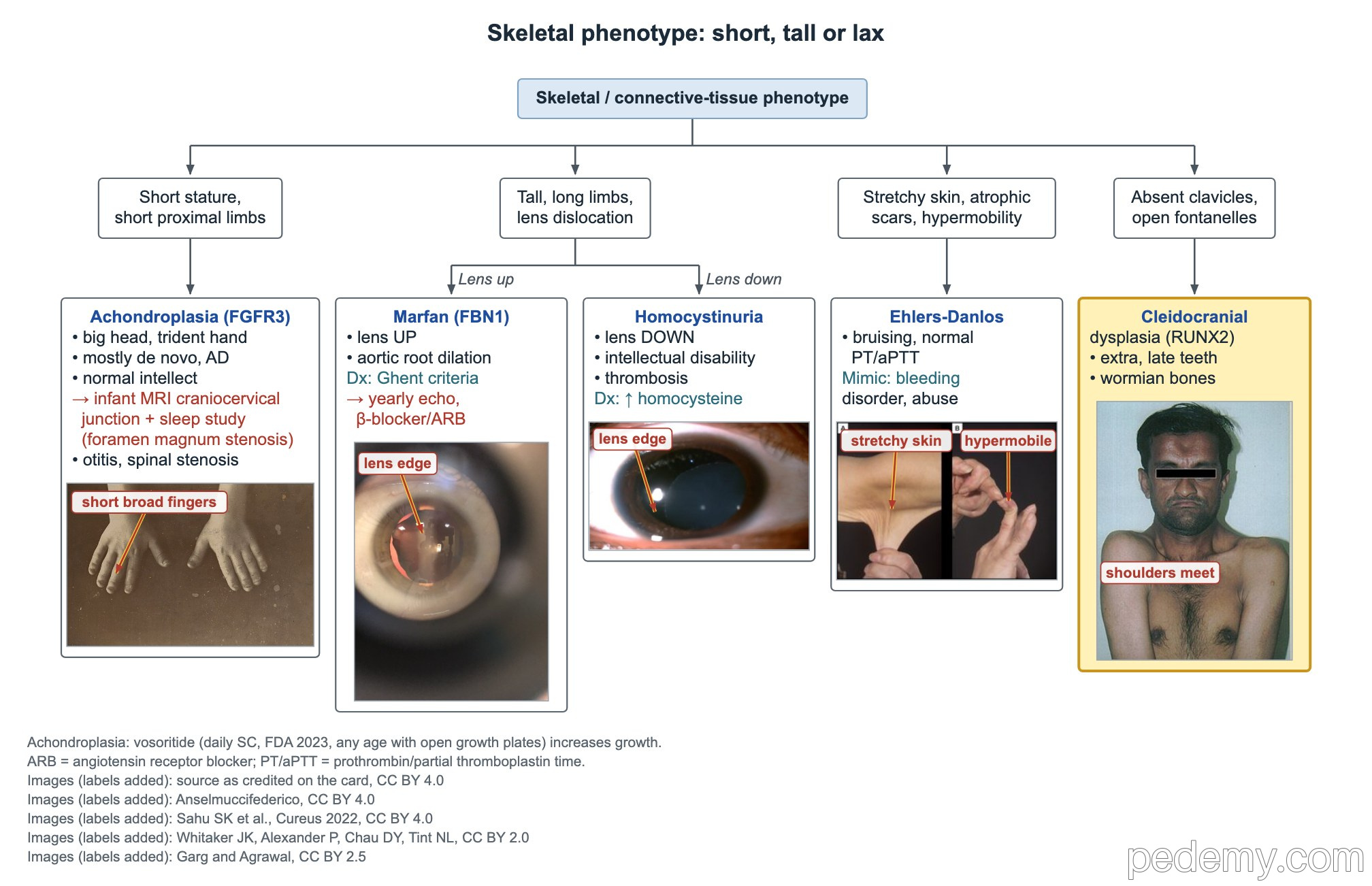
Wormian bones are small extra bones within the skull sutures.
| Disorder | Clues | Key point |
|---|---|---|
| Cleidocranial dysplasia (RUNX2, autosomal dominant) | Absent or small clavicles (shoulders can meet), open fontanelles and delayed skull ossification, wormian bones | Delayed eruption and extra (supernumerary) teeth |
| Caffey disease (infantile cortical hyperostosis) | Infant under 6 months with fever, irritability and hard, tender bone swelling, usually the mandible | Self-limited; usually gone by about age 2 |
| Fibrodysplasia ossificans progressiva (ACVR1) | Short, malformed great toes from birth; flares of painful lumps in the neck and back that turn to bone | Avoid biopsy, surgery and intramuscular injections, which trigger new bone |
Common questions
What are the revised Ghent criteria for Marfan syndrome?
Without a family history, aortic root dilation (z ≥2) plus ectopia lentis is enough. Aortic root dilation can also combine with an FBN1 variant or a systemic score of 7 or more.
What is the most common type of osteogenesis imperfecta?
Type I: the mildest form, with blue sclerae, fractures after minor trauma and hearing loss. It is autosomal dominant and caused by reduced type I collagen.
Is achondroplasia inherited?
It is autosomal dominant, but about 80% of children have a new FGFR3 variant and parents of average height.
Study this topic
Sources
- AAP clinical report: Health supervision for people with achondroplasia (Pediatrics 2020)
- AAP clinical report: Health supervision for children and adolescents with Marfan syndrome (Pediatrics 2023)
- The revised Ghent nosology for the Marfan syndrome (J Med Genet 2010)
- The 2017 international classification of the Ehlers-Danlos syndromes (Am J Med Genet C)
- 2022 ACC/AHA guideline for the diagnosis and management of aortic disease (Circulation)
Written for Pedemy in our own words from the sources above; checked November 2026. For education only, not medical advice; check doses and local protocols before treating a patient. Pedemy is independent and not affiliated with or endorsed by the organizations cited.