

Main decision tree
Abnormal head shape in an infant
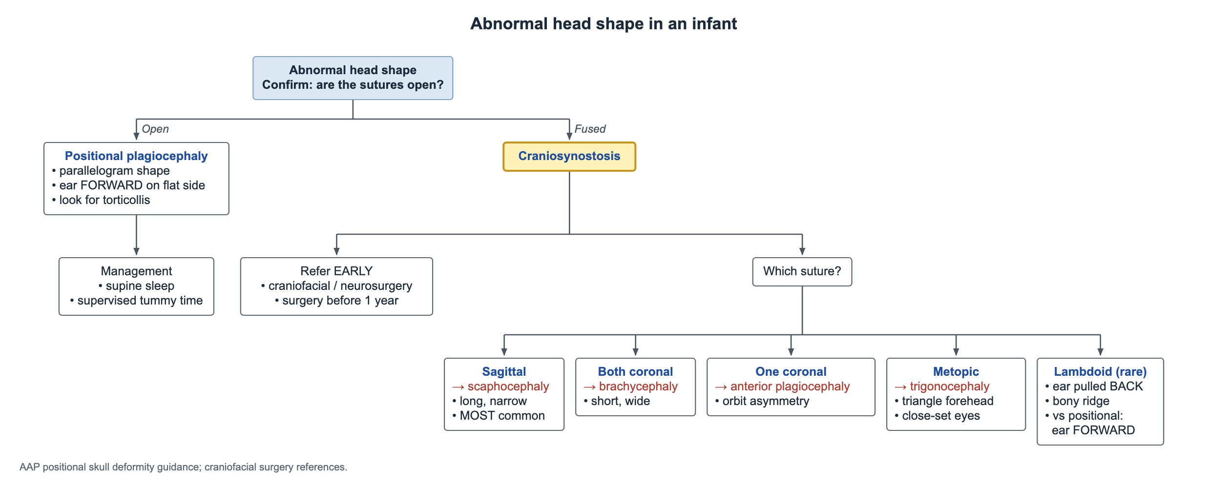
Key points
- Term head circumference averages 35 cm (range about 33–37 cm) and grows fastest in the first 2 months, about 0.5 cm a week.
- Posterior fontanelle closes by about 2 months; anterior fontanelle by 18–24 months.
- Positional plagiocephaly: open sutures, parallelogram head, ear pushed FORWARD on the flat side; look for torticollis.
- Lambdoid synostosis: ear pulled BACK on the flat side with a bony ridge.
- Sagittal synostosis (scaphocephaly) is the most common craniosynostosis; refer early, surgery ideally before 1 year.
- Macrocephaly is a head circumference above +2 SD; microcephaly is below −2 SD for age and sex.
- Large head, normal exam and development: measure the parents' heads first.
Normal head growth, fontanelles and microcephaly
- Average term head circumference is 35 cm, with a usual range of 33–37 cm.
- Growth is fastest from 0 to 2 months, about 0.5 cm a week (2 cm a month), then slows over the first year.
- Posterior fontanelle: usually closed by about 2 months.
- Anterior fontanelle: usually closed by 18–24 months.
- Microcephaly: head circumference below −2 SD for age and sex. First plot the trajectory and measure both parents' heads.
- Congenital CMV and toxoplasmosis can cause microcephaly with cerebral calcifications.
Abnormal head shape: open or fused sutures?
Craniosynostosis is the early, abnormal fusion of one or more skull sutures. The skull cannot grow at right angles to a fused suture, so it grows more along the other sutures and takes a typical shape.
Positional (deformational) plagiocephaly is far more common. The sutures are open; the head flattens from lying in one position.
Positional plagiocephaly
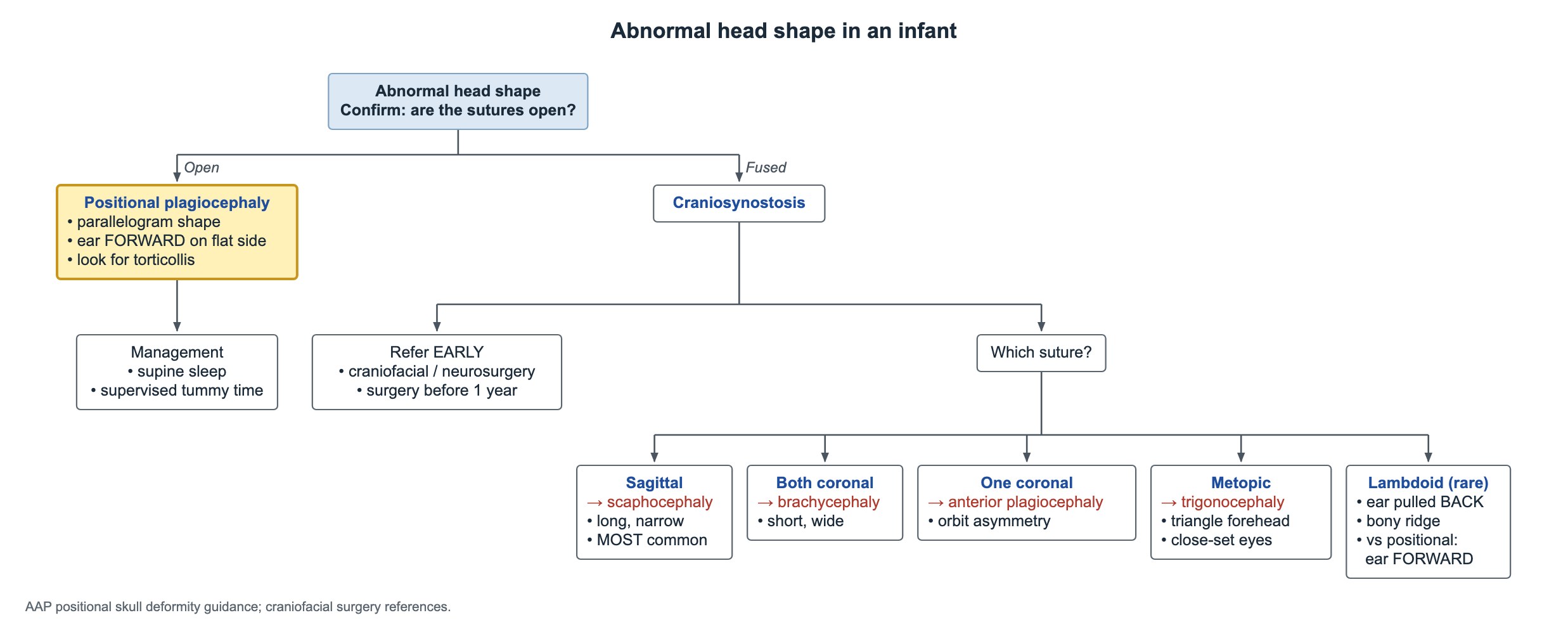
- Seen from above, the head is a parallelogram: flat at the back on one side, with the forehead pushed forward on the same side.
- The ear on the flat side sits forward.
- Look for torticollis: the head tilts toward one shoulder with the chin turned away. Treat it with stretching and physical therapy.
- Keep supine sleep for every sleep; prevent and treat with supervised tummy time while awake and changing head position.
- A helmet is an option for moderate to severe flattening that persists despite repositioning; it is never surgery.
Craniosynostosis by suture
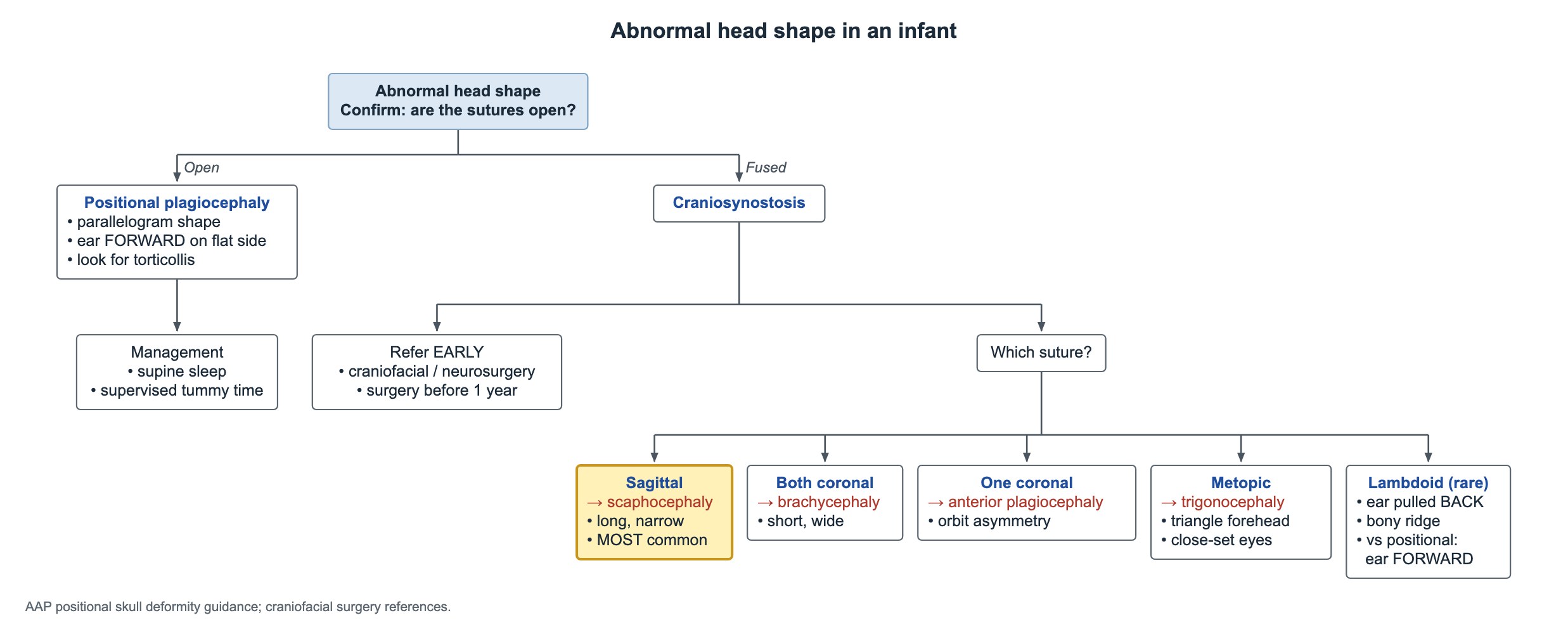
Suspected craniosynostosis needs an early referral to craniofacial surgery or neurosurgery. Surgery works best in the first year of life, before 1 year of age.
| Fused suture | Head shape | Clues |
|---|---|---|
| Sagittal | Scaphocephaly (dolichocephaly) | Long, narrow head; the most common type |
| Both coronal | Brachycephaly | Short, wide head; think of a syndrome |
| One coronal | Anterior plagiocephaly | Flat forehead on one side; orbit asymmetry |
| Metopic | Trigonocephaly | Triangular forehead with a midline ridge; close-set eyes |
| Lambdoid (rare) | Posterior plagiocephaly | Ear pulled back on the flat side; bony ridge behind the ear |
Large head (macrocephaly)
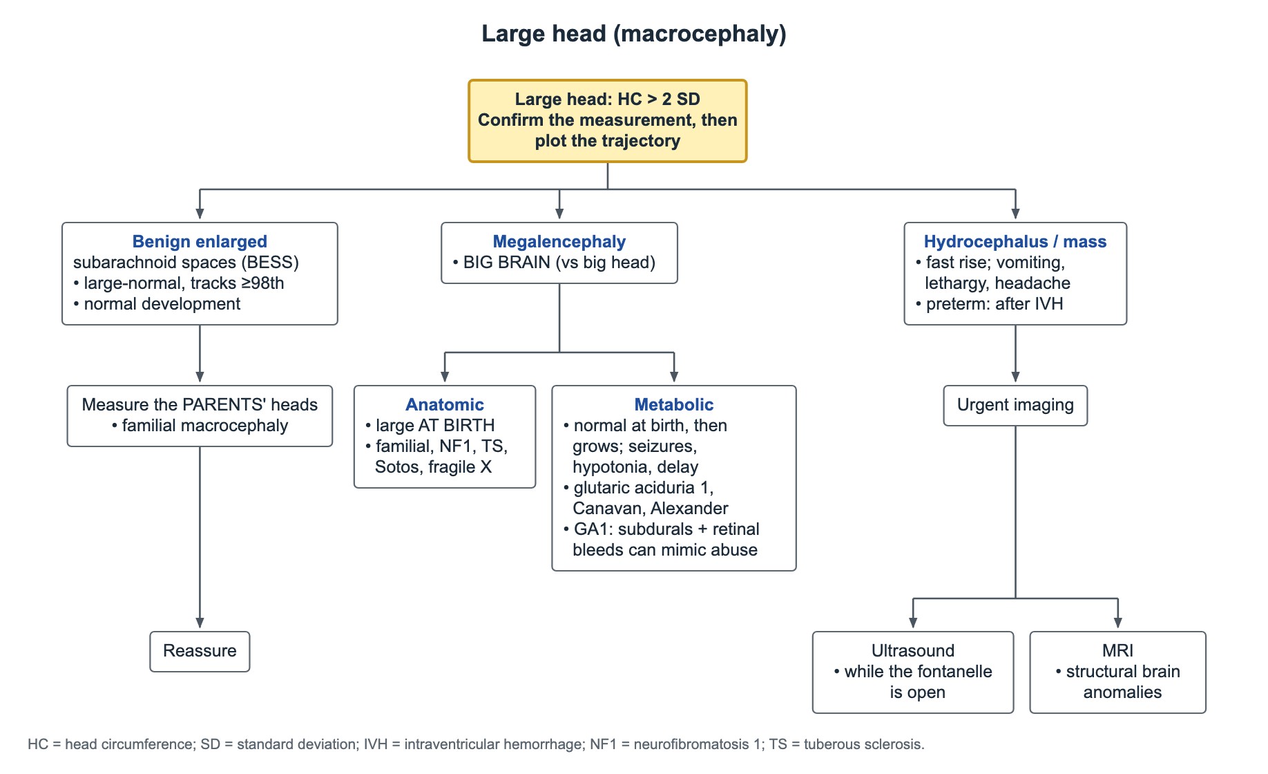
Macrocephaly is a head circumference more than 2 SD above the mean. Confirm the measurement, then plot the trajectory: a head that tracks a high line is very different from one that crosses lines.
- Normal exam and normal development: measure the parents' heads. Familial macrocephaly is common and benign.
- Benign enlargement of the subarachnoid spaces (BESS) is a common benign cause: a large head that tracks at or above the 98th percentile with normal development. Reassure.
- Megalencephaly means a big brain; macrocephaly only means a big head.
Megalencephaly: anatomic or metabolic
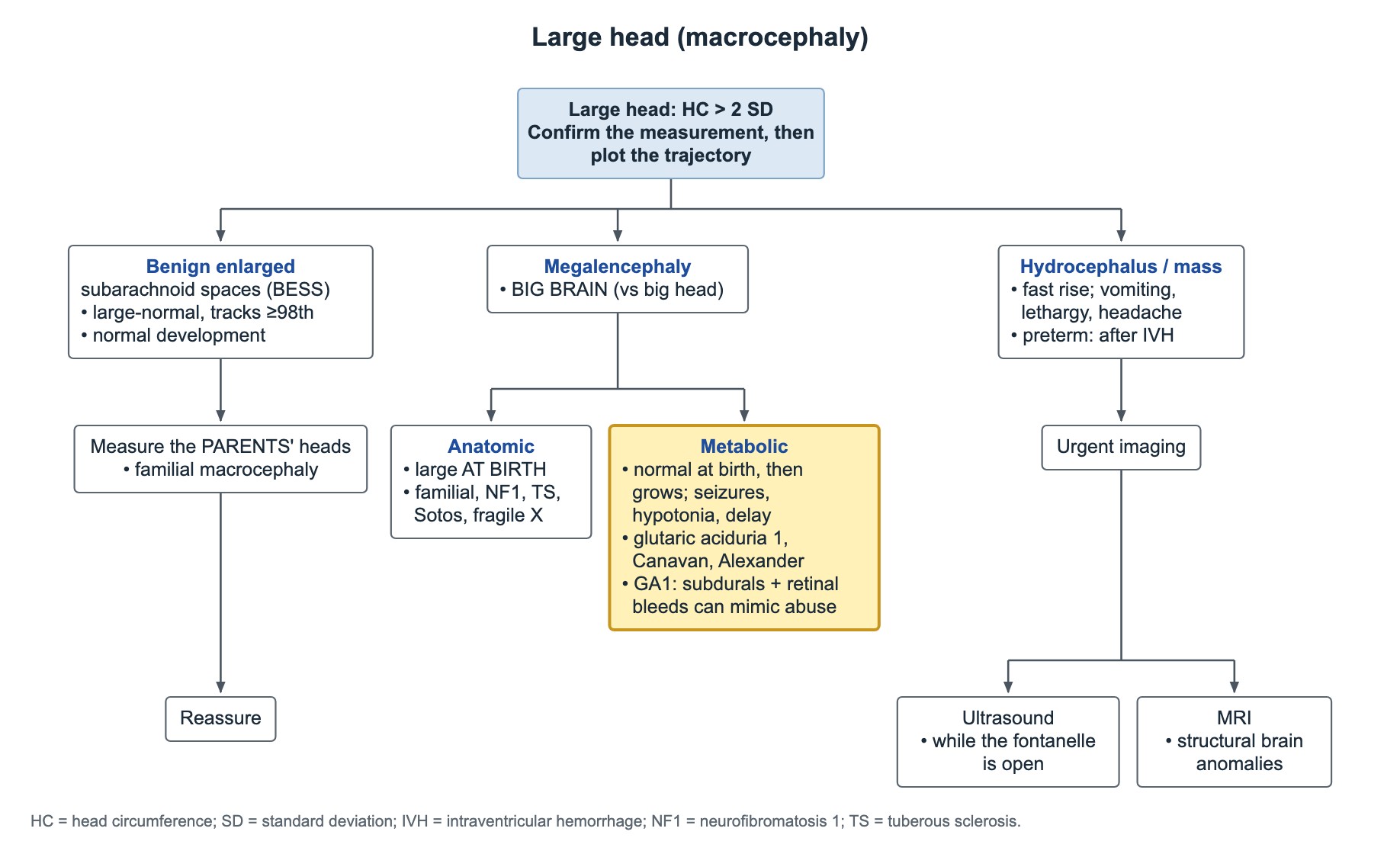
- Canavan disease: macrocephaly, early hypotonia, high urine N-acetylaspartic acid.
- Alexander disease: macrocephaly, seizures and a frontal-predominant leukodystrophy from a GFAP variant.
- Glutaric aciduria type 1: subdural and retinal bleeds can mimic abusive head trauma.
| Type | Pattern | Examples |
|---|---|---|
| Anatomic | Large head already at birth | Familial, neurofibromatosis 1, tuberous sclerosis, Sotos syndrome, fragile X |
| Metabolic | Normal head at birth, then grows too fast, with seizures, hypotonia and delay | Glutaric aciduria type 1, Canavan disease, Alexander disease |
Hydrocephalus and imaging
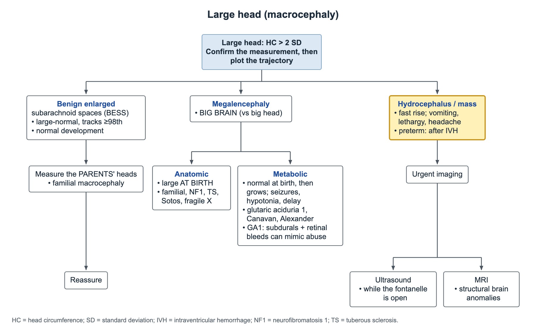
- A fast-rising head with vomiting, lethargy, headache or visual signs suggests raised intracranial pressure: image urgently.
- A flat fontanelle does not exclude hydrocephalus or a mass.
- In preterm infants, hydrocephalus may follow intraventricular hemorrhage.
- Head ultrasound works while the fontanelle is open.
- MRI shows structural brain abnormalities best; CT shows calcifications best.
Common questions
How do you tell positional plagiocephaly from lambdoid craniosynostosis?
In positional plagiocephaly the ear on the flat side is pushed forward and the head is a parallelogram. In lambdoid synostosis the ear is pulled back and there is a bony ridge.
When does the anterior fontanelle close?
Usually by 18–24 months. The posterior fontanelle closes by about 2 months.
What is the most common type of craniosynostosis?
Sagittal synostosis, which gives a long, narrow head called scaphocephaly.
Study this topic
Sources
- AAP 2020: Identifying the misshapen head: craniosynostosis and related disorders (Pediatrics)
- AAP 2011: Prevention and management of positional skull deformities in infants (Pediatrics)
- CDC: Growth charts
Written for Pedemy in our own words from the sources above; checked November 2026. For education only, not medical advice; check doses and local protocols before treating a patient. Pedemy is independent and not affiliated with or endorsed by the organizations cited.