

Main decision tree
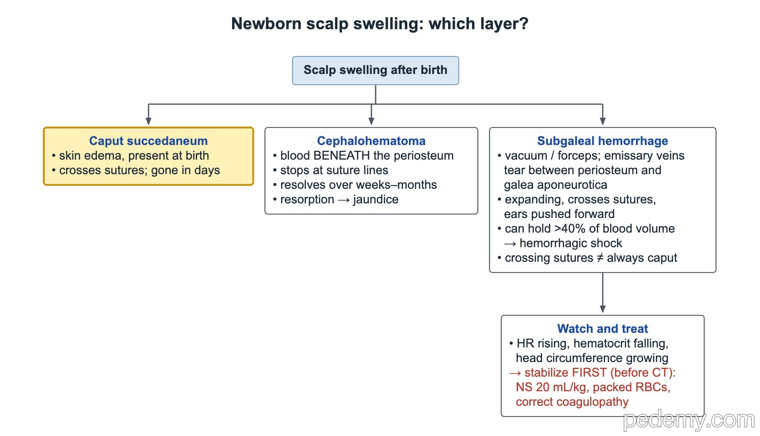
Newborn scalp swelling: which layer?
Key points
- Caput succedaneum: soft scalp edema present at birth, crosses sutures, gone within days.
- Cephalohematoma: blood under the periosteum, stops at suture lines, resolves over weeks to months, can add to jaundice.
- Subgaleal hemorrhage: blood between the galea aponeurotica and the periosteum; the space can hold about 40–50% of a newborn's blood volume.
- Subgaleal bleed with shock: resuscitate first (volume 10 mL/kg, early packed red cells, correct clotting), image later.
- Erb palsy (C5–C6) is about 90% of brachial plexus injuries: waiter's tip posture with grasp preserved.
- Klumpke palsy (C8–T1): claw hand, absent grasp, and Horner syndrome (ptosis, miosis) from T1 injury.
- Brachial plexus palsy not fully recovered by 1 month → refer to a multidisciplinary brachial plexus team.
Scalp swelling: which layer?
Three swellings follow delivery, and each sits in a different layer of the scalp. Caput succedaneum is the most superficial and the most benign: edema of the skin over the presenting part, present at birth, soft and pitting, crossing suture lines and fading within a few days.
- Crossing a suture does not prove the swelling is a harmless caput: a subgaleal hemorrhage also crosses sutures.
- A swelling that keeps growing after birth is blood, not edema, until proven otherwise.
| Swelling | Layer | Crosses sutures? | Course |
|---|---|---|---|
| Caput succedaneum | Skin and subcutaneous edema | Yes | Present at birth; resolves in days |
| Cephalohematoma | Under the periosteum | No (stops at suture lines) | Appears over hours; resolves over weeks to months |
| Subgaleal hemorrhage | Between galea aponeurotica and periosteum | Yes | Expands over hours; can cause hemorrhagic shock |
Cephalohematoma
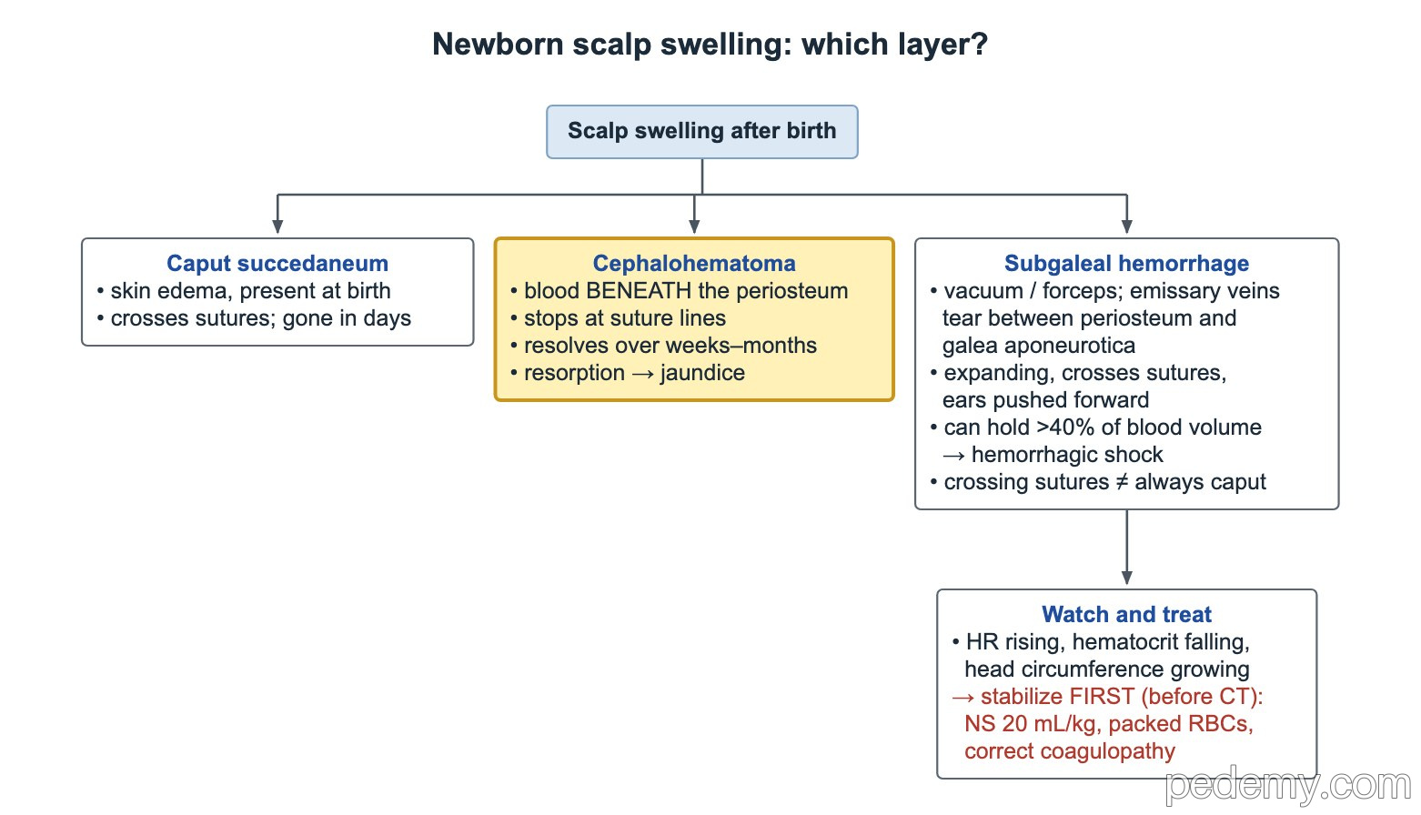
Shearing forces tear vessels under the periosteum. Because the periosteum is fixed at the sutures, the blood stays over one bone, most often the parietal bone, and makes a firm, well-demarcated swelling.
- Usually not visible at birth; it appears over the first hours to days.
- Uncomplicated cephalohematoma needs no treatment and resolves over weeks to months; do not aspirate it.
- Breakdown of the collected blood adds to the bilirubin load: watch for jaundice.
- A small number have an underlying linear skull fracture; this rarely changes management in a well baby.
Subgaleal hemorrhage: recognize it early
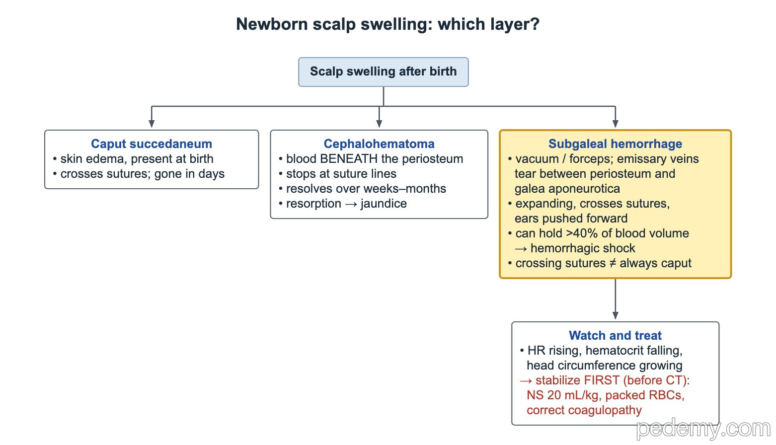
Vacuum or forceps traction tears the emissary veins that link the scalp veins to the dural sinuses. Blood collects in the loose space between the galea aponeurotica and the periosteum, which has no suture boundaries and runs from the orbits to the nape of the neck.
- Diffuse, fluctuant, expanding swelling that crosses sutures; the ears are pushed forward and the swelling can spread to the eyelids and neck.
- The space can hold about 40–50% of a newborn's blood volume, so the immediate threat is hemorrhagic shock.
- Signs of blood loss: pallor, tachycardia, poor perfusion, falling hematocrit.
- Any baby born by vacuum needs regular scalp and vital-sign checks in the first hours.
Subgaleal hemorrhage: stabilize first
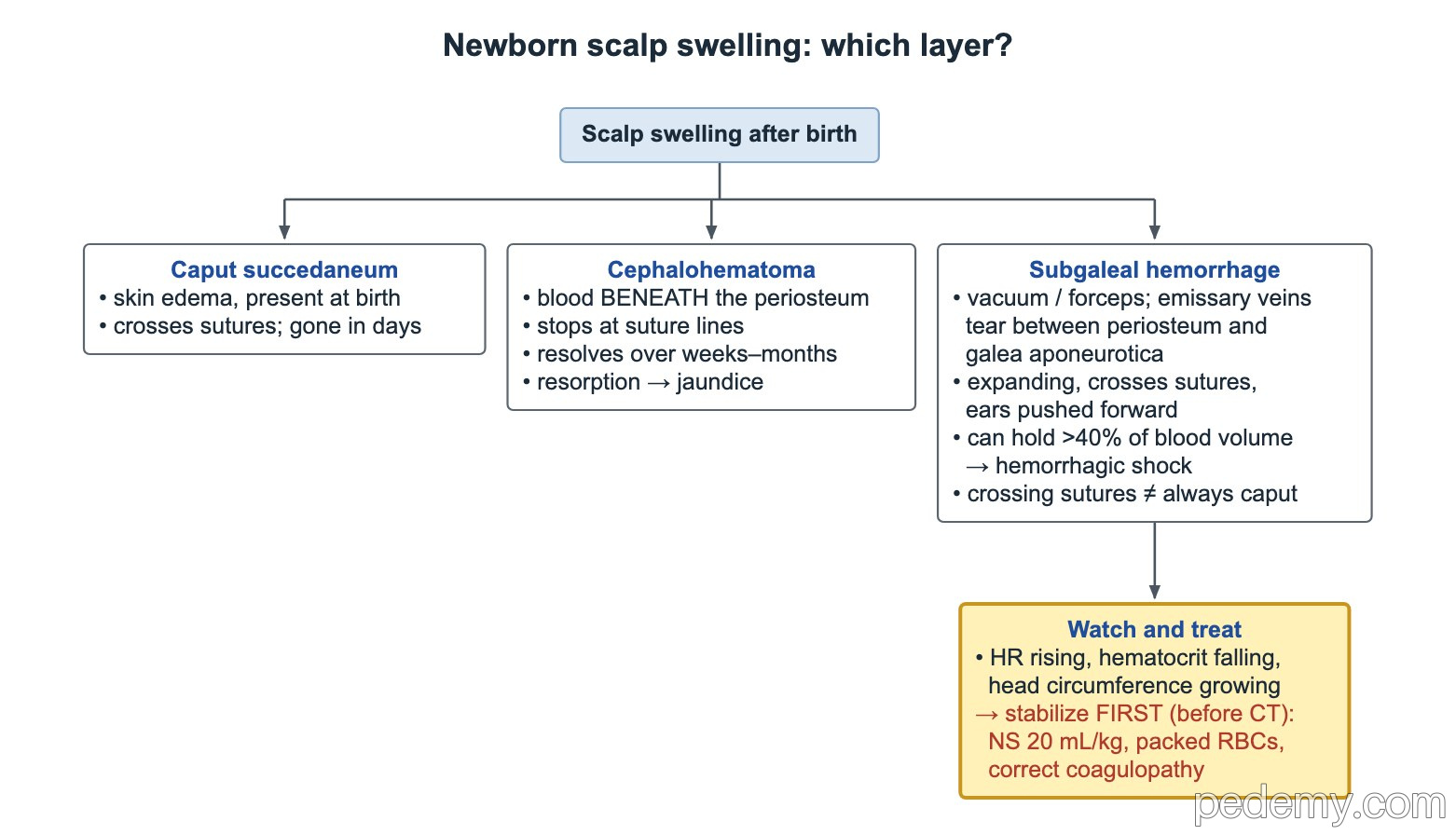
An unstable baby is resuscitated before any imaging. Imaging (head ultrasound or CT) can wait until the circulation and clotting are under control.
- Track three bedside trends: heart rate (rising), hematocrit (falling) and head circumference (growing).
- Give a volume bolus of 10 mL/kg (normal saline or blood) over 5–10 minutes, as in neonatal resuscitation, and repeat as needed.
- Shock that persists after crystalloid needs packed red blood cell transfusion; give blood early.
- Check and correct coagulopathy (platelets, PT/PTT, fibrinogen) with plasma, cryoprecipitate or platelets as needed.
- Watch for jaundice as the blood is reabsorbed.
Brachial plexus and phrenic nerve injuries
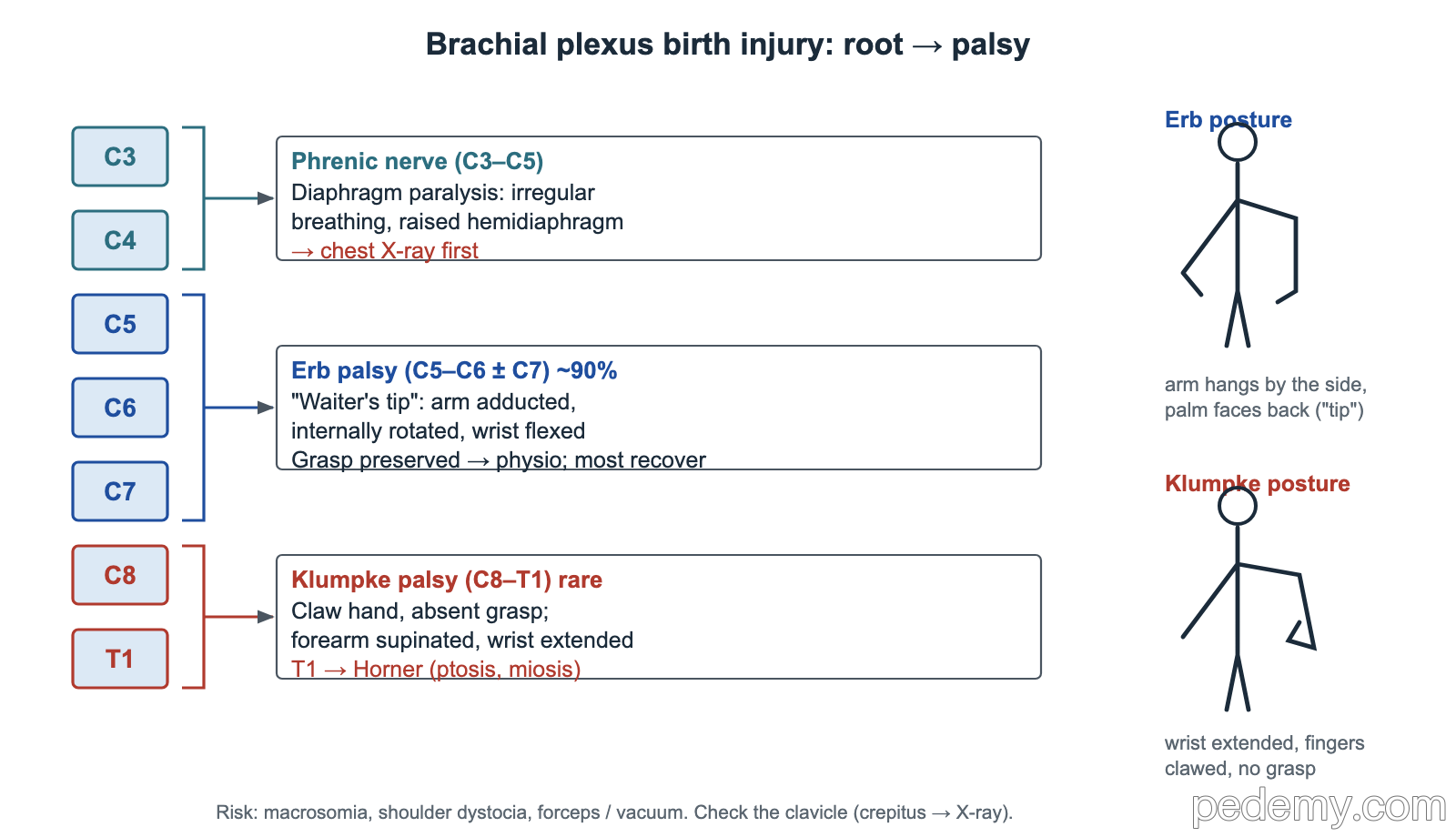
Stretching of the neck during a difficult delivery injures the nerve roots. Risk factors are macrosomia, shoulder dystocia and instrumented delivery. The level of the injured roots decides the arm posture.
- Feel the clavicle: crepitus or a step suggests a clavicle fracture, which can mimic or accompany a plexus palsy.
- A baby with Erb palsy and breathing trouble needs a chest X-ray first: look for a raised hemidiaphragm, a pneumothorax or a clavicle fracture.
- Most Erb palsies recover within about 3 months with gentle range-of-motion exercises and physiotherapy.
| Injury | Roots | Findings |
|---|---|---|
| Erb palsy (about 90%) | C5–C6 (± C7) | Arm adducted and internally rotated, elbow extended, wrist flexed (waiter's tip); Moro absent on that side; grasp preserved |
| Klumpke palsy (rare) | C8–T1 | Claw hand, absent grasp, forearm supinated and wrist extended; T1 injury → Horner syndrome (ptosis, miosis) |
| Phrenic nerve injury | C3–C5 | Labored, irregular breathing and cyanosis from a paralyzed, raised hemidiaphragm; often with Erb palsy |
When to refer
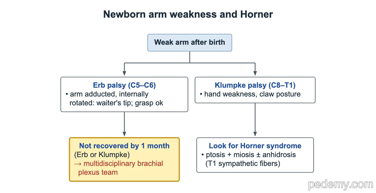
- Any brachial plexus palsy (Erb or Klumpke) that has not fully recovered by 1 month → refer to a multidisciplinary brachial plexus team.
- Klumpke palsy and Horner syndrome mean a lower-root, more severe injury with a poorer outlook; refer early.
- Babies with persistent weakness may need nerve surgery in the first months of life, so late referral loses options.
Common questions
How do you tell caput from cephalohematoma?
Caput is soft edema present at birth that crosses sutures and goes in days. Cephalohematoma is a firm collection of blood under the periosteum that stops at the suture lines and lasts weeks to months.
Why is a subgaleal hemorrhage dangerous?
The subgaleal space has no suture boundaries and can hold about 40–50% of a newborn's blood volume, so the baby can go into hemorrhagic shock within hours.
Does Erb palsy get better?
Most babies recover within about 3 months. A palsy that is not fully recovered by 1 month should be seen by a multidisciplinary brachial plexus team.
Study this topic
Sources
- AHA/AAP 2025: Part 5, Neonatal Resuscitation (Circulation)
- ACOG Task Force 2014: Neonatal brachial plexus palsy, executive summary (Obstet Gynecol)
- AAP 2022: Management of hyperbilirubinemia in the newborn infant 35 or more weeks of gestation (Pediatrics)
Written for Pedemy in our own words from the sources above; checked November 2026. For education only, not medical advice; check doses and local protocols before treating a patient. Pedemy is independent and not affiliated with or endorsed by the organizations cited.