

Main decision tree
Juvenile dermatomyositis: recognize, confirm, treat
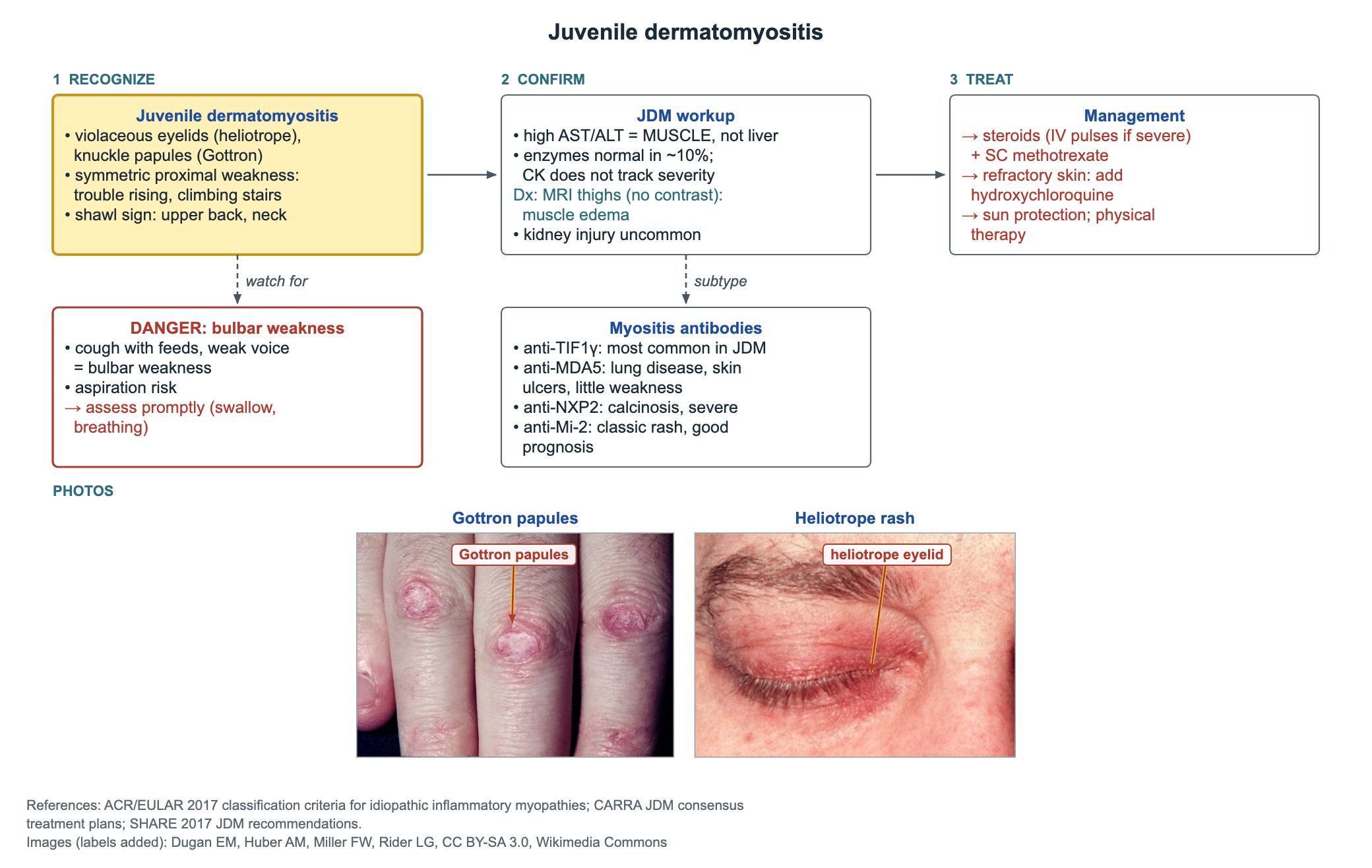
Key points
- Classic picture: heliotrope eyelids, Gottron papules over the knuckles and symmetric proximal weakness (Gowers sign).
- Raised AST and ALT in JDM come from inflamed muscle, not the liver.
- Muscle enzymes are normal in about 10% of children, and CK does not track severity.
- MRI of the thighs (T2/STIR, no contrast needed) shows muscle edema and can guide a biopsy.
- First-line treatment: corticosteroids (IV pulses if severe) plus subcutaneous methotrexate once weekly.
- Anti-MDA5: lung disease and skin ulcers with little weakness; anti-NXP2: calcinosis and severe disease.
- Coughing with feeds or a new nasal or weak voice means bulbar weakness and aspiration risk.
Recognize juvenile dermatomyositis
Think of JDM in a child with a rash and new trouble rising from the floor, climbing stairs or lifting the arms. The weakness is proximal and symmetric. The rash often comes first, sometimes months before the weakness.
- Heliotrope rash: violaceous color of the upper eyelids, often with eyelid swelling.
- Gottron papules: pink to violet papules over the MCP and PIP joints; also over elbows and knees.
- Shawl sign: red patches over the upper back, shoulders and back of the neck; a V-sign over the upper chest.
- Nailfold changes: dilated, tortuous capillaries with redness around the nails.
- Gowers sign: the child climbs up the legs with the hands to stand from the floor.
Workup: muscle enzymes and MRI
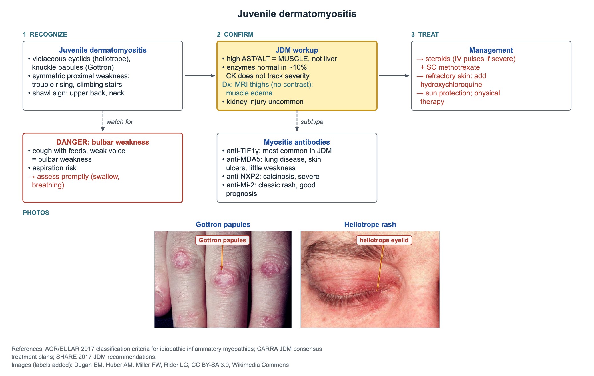
Check all the muscle enzymes, not only CK: sometimes only one of them is high. Normal enzymes do not rule out JDM, and the CK level does not show how severe the disease is.
- Enzymes: CK, aldolase, LDH, AST and ALT.
- High AST and ALT with normal bilirubin and GGT point to muscle, not liver injury.
- MRI of the thighs, T2/STIR without contrast: patchy muscle edema; useful when enzymes are normal and to choose a biopsy site.
- Kidney injury is uncommon in JDM, unlike rhabdomyolysis.
- Myositis-specific antibodies help predict the course (next section).
Myositis antibodies
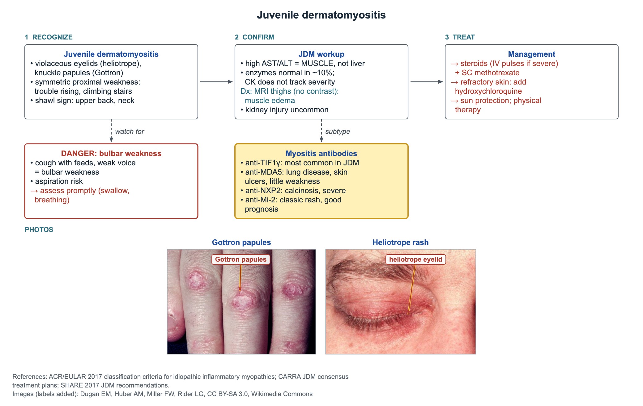
| Antibody | What it predicts |
|---|---|
| Anti-TIF1γ | Most common in JDM; marked skin disease, often a chronic course |
| Anti-NXP2 | Calcinosis and more severe muscle disease |
| Anti-MDA5 | Interstitial lung disease and skin ulcers, often with little weakness; screen the lungs |
| Anti-Mi-2 | Classic rash with a good response to treatment and a good prognosis |
Treatment
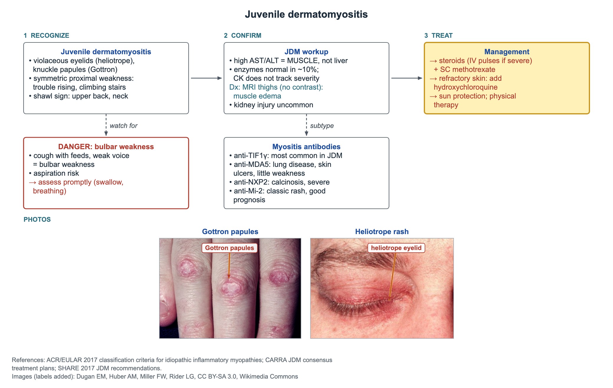
- Corticosteroids from the start: IV methylprednisolone pulses for severe disease, then oral steroids with a slow taper.
- Add subcutaneous methotrexate once weekly at diagnosis; it controls disease and spares steroids.
- Skin disease that persists: add hydroxychloroquine.
- Severe or refractory disease: IVIG or other agents chosen by rheumatology.
- Sun protection every day, because UV light worsens the rash.
- Physical therapy to rebuild strength once inflammation is controlled.
Danger sign: bulbar and breathing weakness
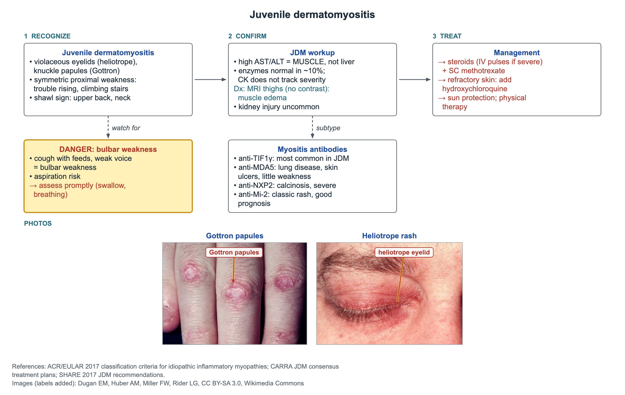
Weak throat and palate muscles cause coughing or choking with feeds, a nasal or weak voice and pooling of secretions. This puts the child at risk of aspiration.
Assess swallowing and breathing promptly. These children need urgent, intensified treatment, and some need a feeding tube or breathing support for a time.
Long-term complications
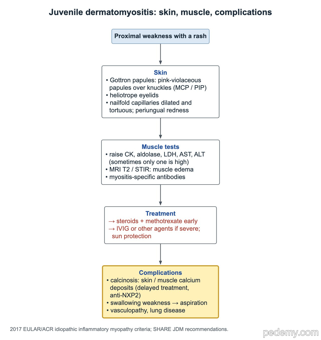
- Calcinosis: calcium deposits in skin, fat and muscle; linked to delayed or under-treated disease and to anti-NXP2.
- Swallowing weakness with aspiration.
- Vasculopathy: skin ulcers and, rarely, bowel wall ulceration or perforation.
- Interstitial lung disease, especially with anti-MDA5.
- Steroid side effects: poor growth, bone loss, weight gain, high blood pressure.
Common questions
Can a child have dermatomyositis with a normal CK?
Yes. About 10% of children with JDM have normal muscle enzymes. Check aldolase, LDH, AST and ALT too, and use thigh MRI to look for muscle edema.
Why are liver enzymes high in juvenile dermatomyositis?
AST and ALT are also found in muscle. In JDM they leak from inflamed muscle, so the liver is usually normal.
What is the first-line treatment for juvenile dermatomyositis?
Corticosteroids plus once-weekly subcutaneous methotrexate, started early. Hydroxychloroquine is added for persistent skin disease.
Study this topic
Sources
- SHARE consensus recommendations for the management of juvenile dermatomyositis (Ann Rheum Dis 2017)
- 2017 EULAR/ACR classification criteria for adult and juvenile idiopathic inflammatory myopathies (Ann Rheum Dis)
Written for Pedemy in our own words from the sources above; checked November 2026. For education only, not medical advice; check doses and local protocols before treating a patient. Pedemy is independent and not affiliated with or endorsed by the organizations cited.