

Main decision tree
Lupus and connective tissue disease
Key points
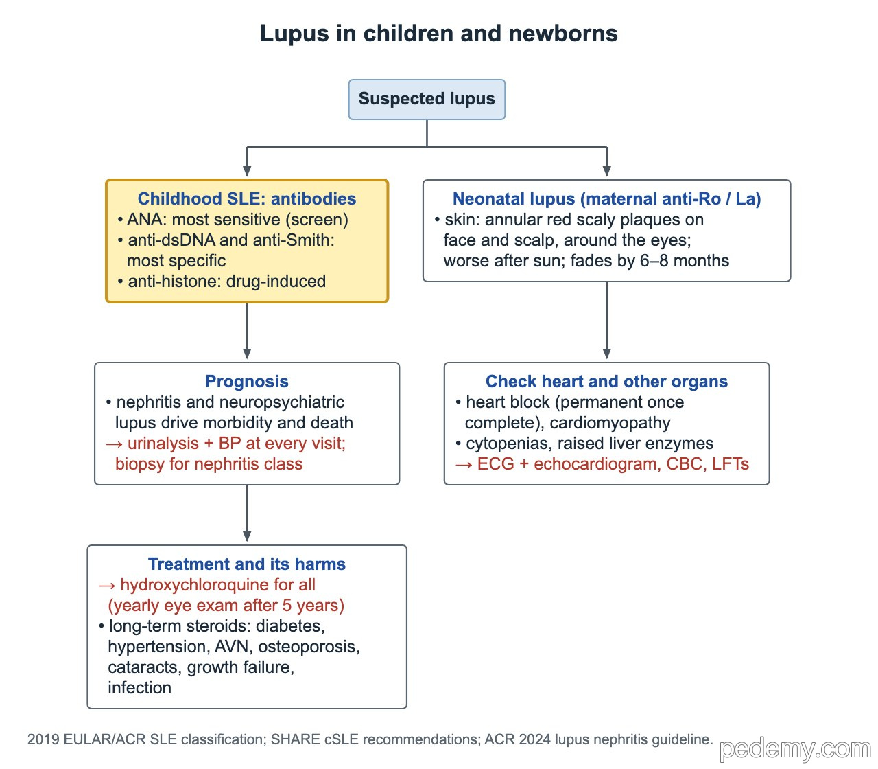
- ANA is the most sensitive lupus test; anti-dsDNA and anti-Smith are the most specific.
- An isolated positive ANA does not establish lupus or JIA.
- 2019 EULAR/ACR classification: ANA ≥1:80 at least once, then ≥10 weighted points with ≥1 clinical criterion.
- Kidney (lupus nephritis) and neuropsychiatric disease drive prognosis: check BP and urinalysis at every visit.
- Neonatal lupus: maternal anti-Ro ± anti-La; the rash fades, but complete heart block is permanent.
- Drug-induced lupus: months into minocycline, hydralazine, procainamide or isoniazid; anti-histone positive; stop the drug.
- Raynaud: warming first, then a calcium channel blocker (amlodipine or nifedipine); ulcers suggest secondary Raynaud.
Recognize systemic lupus erythematosus
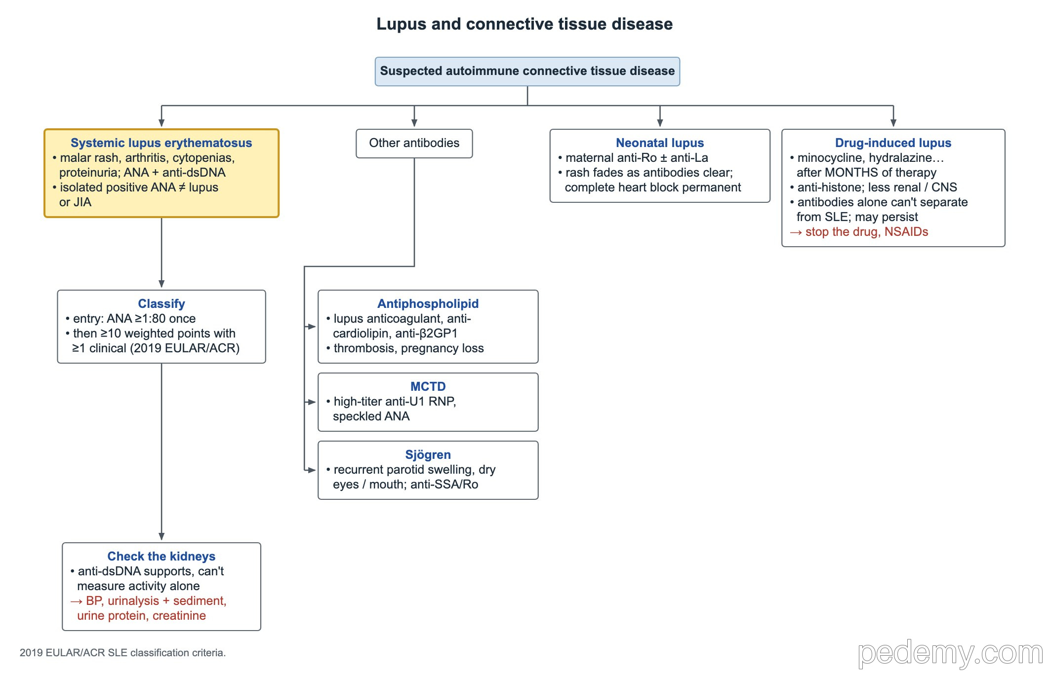
Suspect SLE in a teen, usually a girl, with a photosensitive malar rash, arthritis, cytopenias and proteinuria, plus a positive ANA and anti-dsDNA. Fever, fatigue, oral ulcers, hair loss and serositis are common.
Lupus arthritis is painful but usually nonerosive, unlike JIA.
Antibodies: what each one tells you
| Antibody | Meaning |
|---|---|
| ANA | Most sensitive: a good screen, but often positive in healthy children |
| Anti-dsDNA | Specific for SLE; linked to nephritis; does not measure kidney activity by itself |
| Anti-Smith | Most specific for SLE |
| Anti-histone | Drug-induced lupus |
| Anti-Ro (SSA) ± anti-La (SSB) | Sjögren disease; in the mother, neonatal lupus and heart block |
| Anti-U1 RNP (high titer) | Mixed connective tissue disease, with a speckled ANA |
| Antiphospholipid antibodies | Lupus anticoagulant, anticardiolipin, anti-β2-glycoprotein I: thrombosis and pregnancy loss |
2019 EULAR/ACR classification criteria
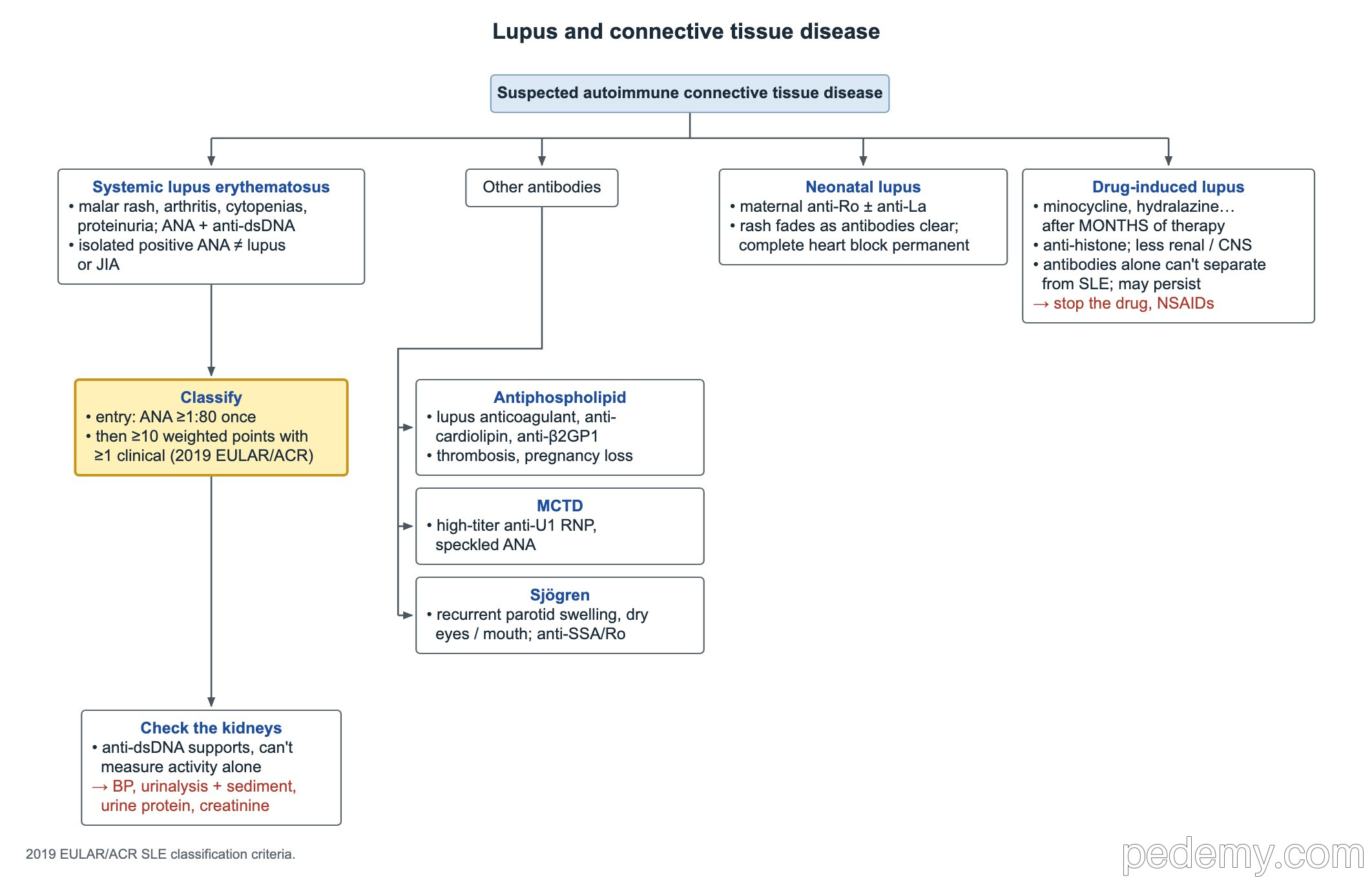
- Entry criterion: ANA titer ≥1:80 at least once.
- Then add weighted points (2 to 10) from 7 clinical domains (constitutional, hematologic, neuropsychiatric, mucocutaneous, serosal, musculoskeletal, renal) and 3 immunologic domains (antiphospholipid antibodies, low complement, SLE-specific antibodies).
- ≥10 points, with at least 1 clinical criterion, classifies SLE.
- These are classification criteria for research; diagnosis remains a clinical judgment.
Check the kidneys: lupus nephritis and treatment
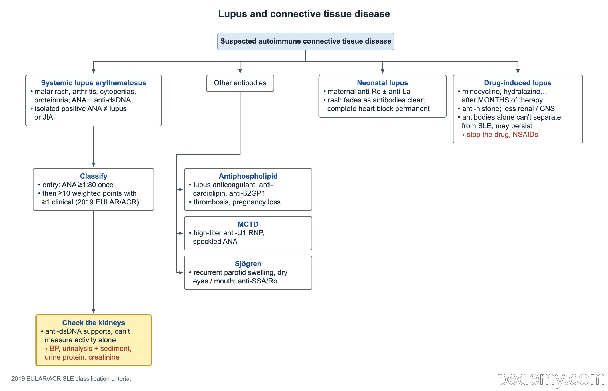
Kidney disease and neuropsychiatric lupus cause most of the long-term harm in childhood SLE. Anti-dsDNA and low complement support active disease, but they cannot show kidney activity alone.
- Every visit: blood pressure and urinalysis with sediment; also urine protein quantification and creatinine.
- Persistent proteinuria or active sediment: kidney biopsy to define the nephritis class.
- Hydroxychloroquine for every patient with SLE; yearly eye exams after 5 years of use (earlier if risk factors).
- Lupus nephritis (ACR 2024): IV steroid pulses, then an oral taper, plus two more immunosuppressive drugs, continued for years.
- Long-term steroid harms: diabetes (hyperglycemia), hypertension, avascular necrosis, osteoporosis, cataracts, growth failure and infection.
Neonatal lupus
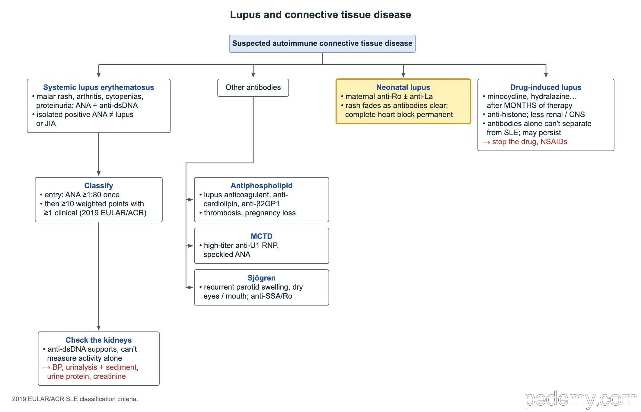
Maternal anti-Ro (SSA), often with anti-La (SSB), cross the placenta. Many mothers have no diagnosis yet.
- Skin: annular, red, scaly plaques on the face and scalp, often around the eyes; worse after sun; fades by 6–8 months as maternal antibodies clear.
- Heart: congenital heart block, which is permanent once complete; cardiomyopathy can also occur.
- Other: anemia, low platelets, neutropenia and raised liver enzymes.
- Evaluate an affected infant with ECG and echocardiogram, CBC and liver tests.
Drug-induced lupus
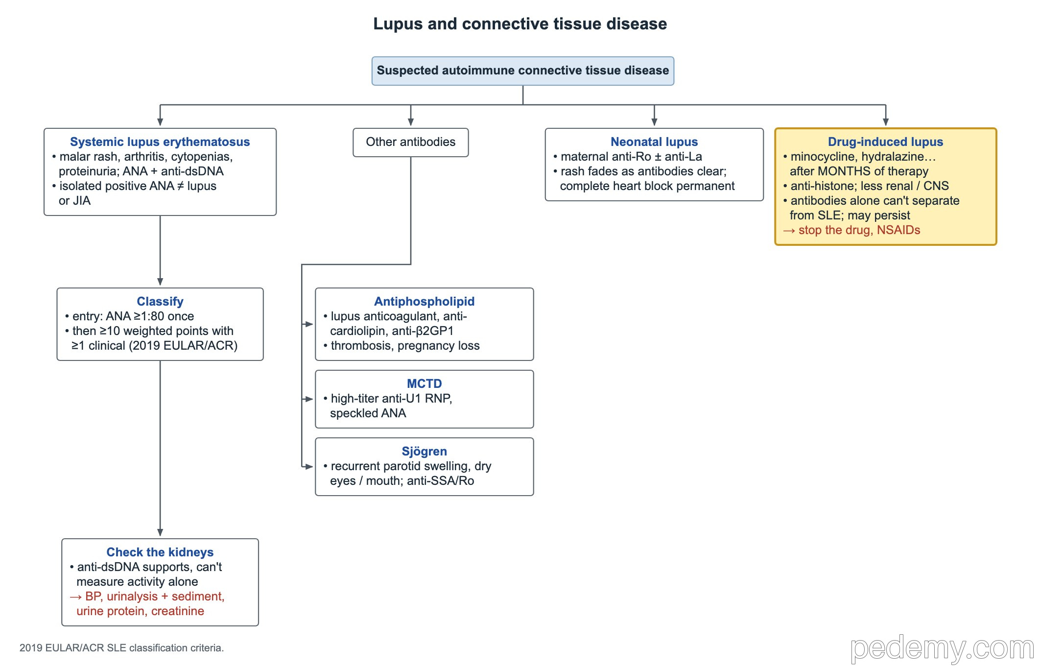
- Drugs: minocycline (common in teens treated for acne), hydralazine, procainamide, isoniazid.
- Onset after months of therapy, not right after starting the drug.
- Arthralgia, fever and serositis; kidney and CNS disease are uncommon.
- Anti-histone antibodies are typical, but antibodies alone cannot separate drug-induced lupus from SLE.
- Management: stop the drug; NSAIDs, sometimes a brief course of steroids.
- Symptoms settle in weeks, but the ANA can stay positive for years without active disease.
Other connective tissue diseases and Raynaud
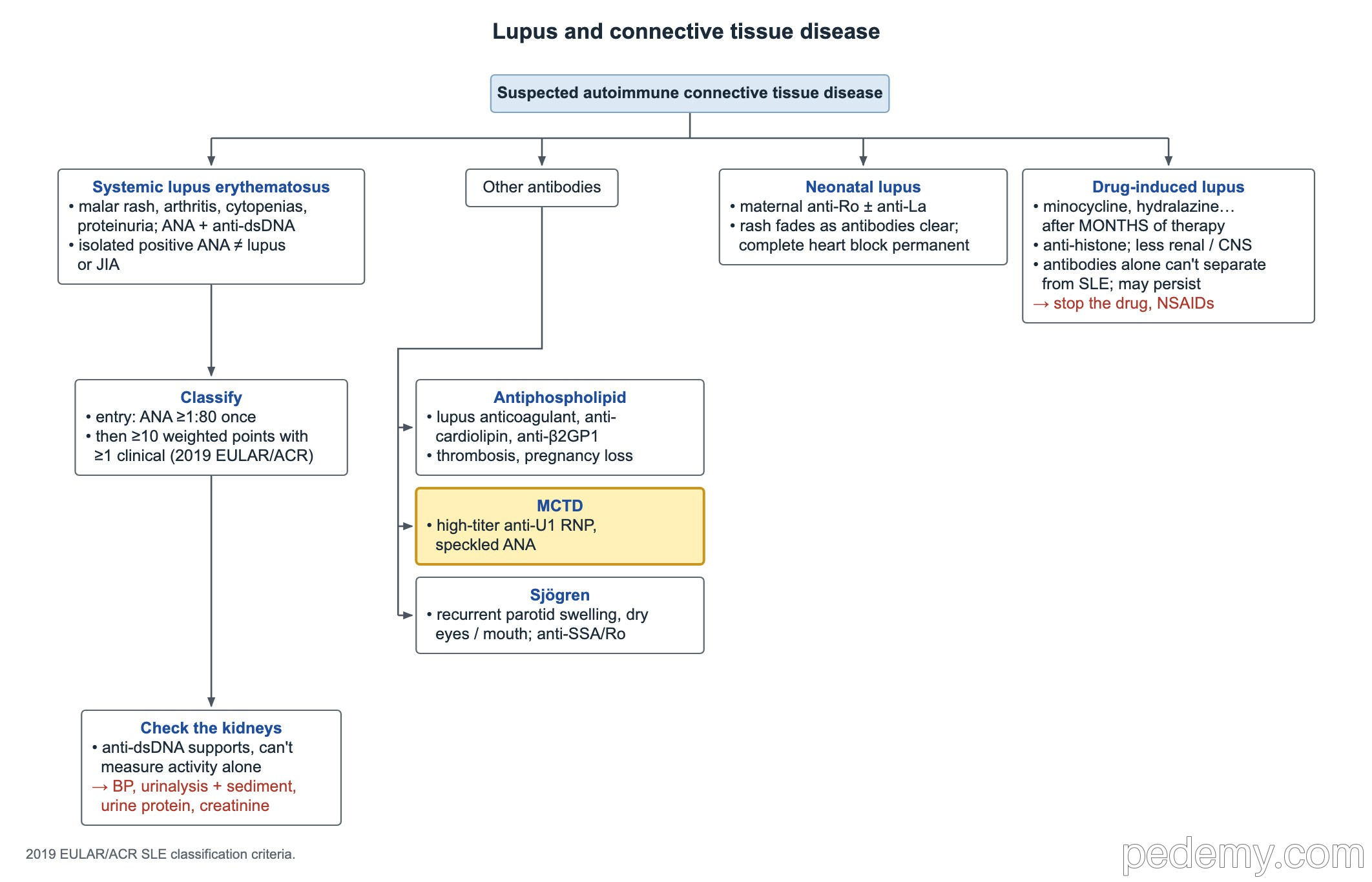
- Mixed connective tissue disease: features of lupus, scleroderma and myositis with high-titer anti-U1 RNP.
- Childhood Sjögren disease: recurrent parotid swelling, dry eyes or mouth, anti-Ro (SSA).
- Antiphospholipid syndrome: thrombosis or pregnancy morbidity with persistent antiphospholipid antibodies, with or without lupus.
- Raynaud phenomenon: cold- or stress-triggered color change, white (pallor), then blue (cyanosis), then red (reperfusion).
- Primary Raynaud (brief, normal exam, no ulcers): warm the hands, feet and core, and stay hydrated; stimulant ADHD medicines are a common trigger in teens.
- Not controlled by warming: calcium channel blocker (amlodipine or nifedipine); next, a PDE5 inhibitor (sildenafil) or topical nitroglycerin.
- Digital ulcers, abnormal nailfold capillaries or systemic symptoms suggest secondary Raynaud: check autoimmune serology and refer to rheumatology.
Common questions
What is the most specific antibody for lupus?
Anti-dsDNA and anti-Smith. ANA is the most sensitive but is not specific.
Does a positive ANA mean my child has lupus?
No. Many healthy children have a low-titer positive ANA. Lupus needs typical clinical features and usually more specific antibodies.
Is heart block from neonatal lupus permanent?
Complete heart block is usually permanent, and many infants need a pacemaker. The skin rash, by contrast, fades as maternal antibodies clear by 6–8 months.
Study this topic
Sources
- 2019 EULAR/ACR classification criteria for systemic lupus erythematosus (Arthritis Rheumatol)
- 2024 ACR guideline for the screening, treatment and management of lupus nephritis (Arthritis Rheumatol 2025)
Written for Pedemy in our own words from the sources above; checked November 2026. For education only, not medical advice; check doses and local protocols before treating a patient. Pedemy is independent and not affiliated with or endorsed by the organizations cited.