

Main decision tree
Inborn errors of immunity 2: T cells and phagocytes
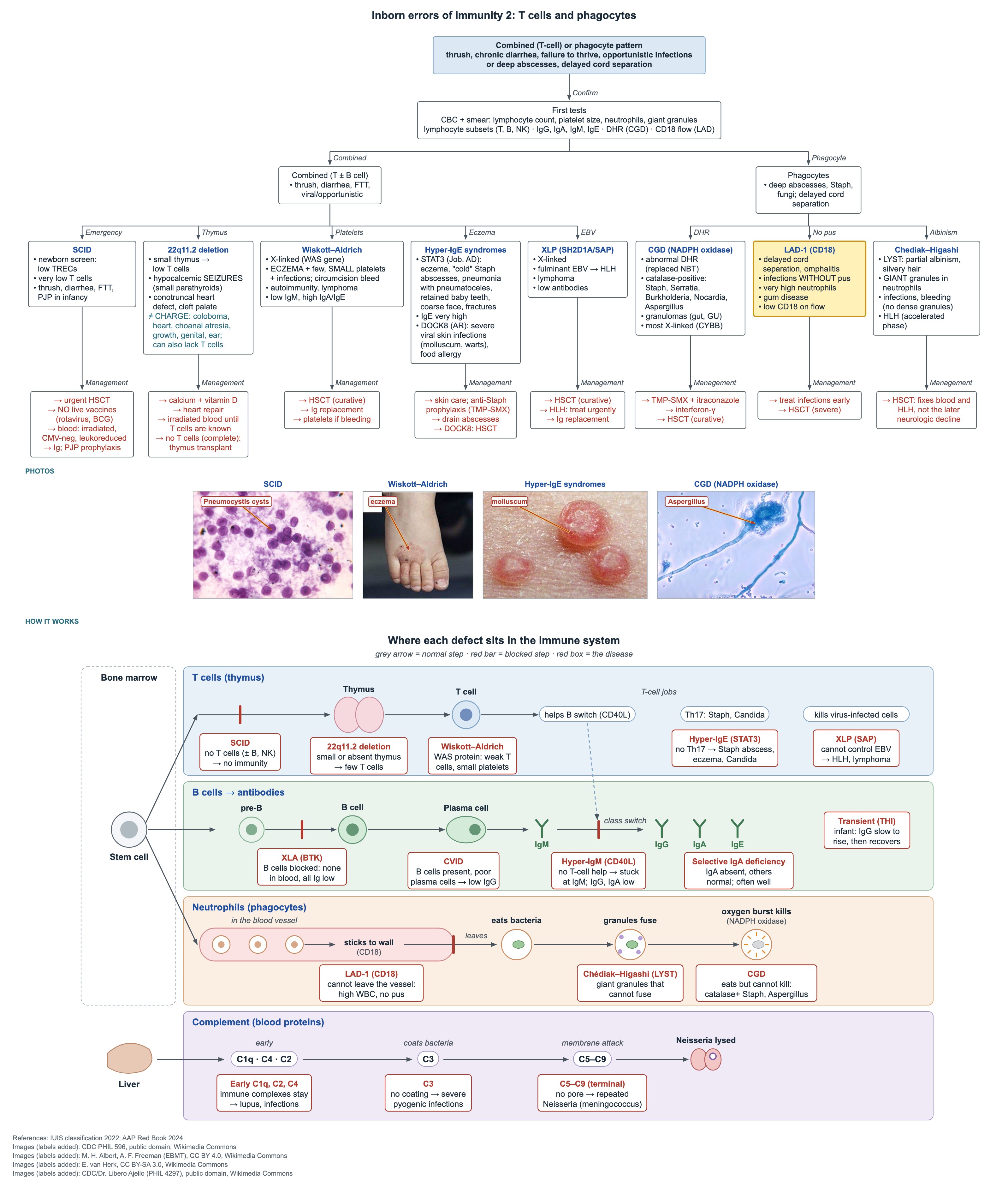
Key points
- LAD-1 (CD18): delayed cord separation, infections with little pus and marked neutrophilia even without infection.
- CGD: catalase-positive organisms (Staphylococcus, Serratia, Burkholderia, Nocardia, Aspergillus); screen with DHR flow cytometry.
- CGD is most often X-linked (CYBB); lifelong prophylaxis is TMP-SMX plus itraconazole.
- Chédiak–Higashi (LYST): partial albinism, silvery hair, giant neutrophil granules, bleeding from absent platelet dense granules.
- Chédiak–Higashi: HLH is the main threat; stem cell transplant cures the blood and immune problems but not the later neurologic decline.
- Low CH50 with normal AH50 = early classical pathway (C1q, C2, C4); both low = C3, terminal components or consumption.
- Terminal complement (C5–C9) deficiency: recurrent meningococcal disease; give MenACWY and MenB with boosters.
Leukocyte adhesion deficiency type 1 (LAD-1)
Neutrophils cannot stick to the vessel wall because CD18 (a beta-2 integrin) is missing or low. They stay in the blood, so the count is high but little pus forms at the site of infection.
- Delayed separation of the umbilical cord, often with omphalitis.
- Severe skin, mucosal and soft-tissue bacterial infections with little pus and poor wound healing.
- Marked neutrophilia that persists even between infections.
- Severe gum disease (gingivitis, periodontitis) in older children.
- Diagnosis: low CD18 on neutrophils by flow cytometry.
- Management: treat infections early; stem cell transplant for severe disease.
Chronic granulomatous disease (CGD)
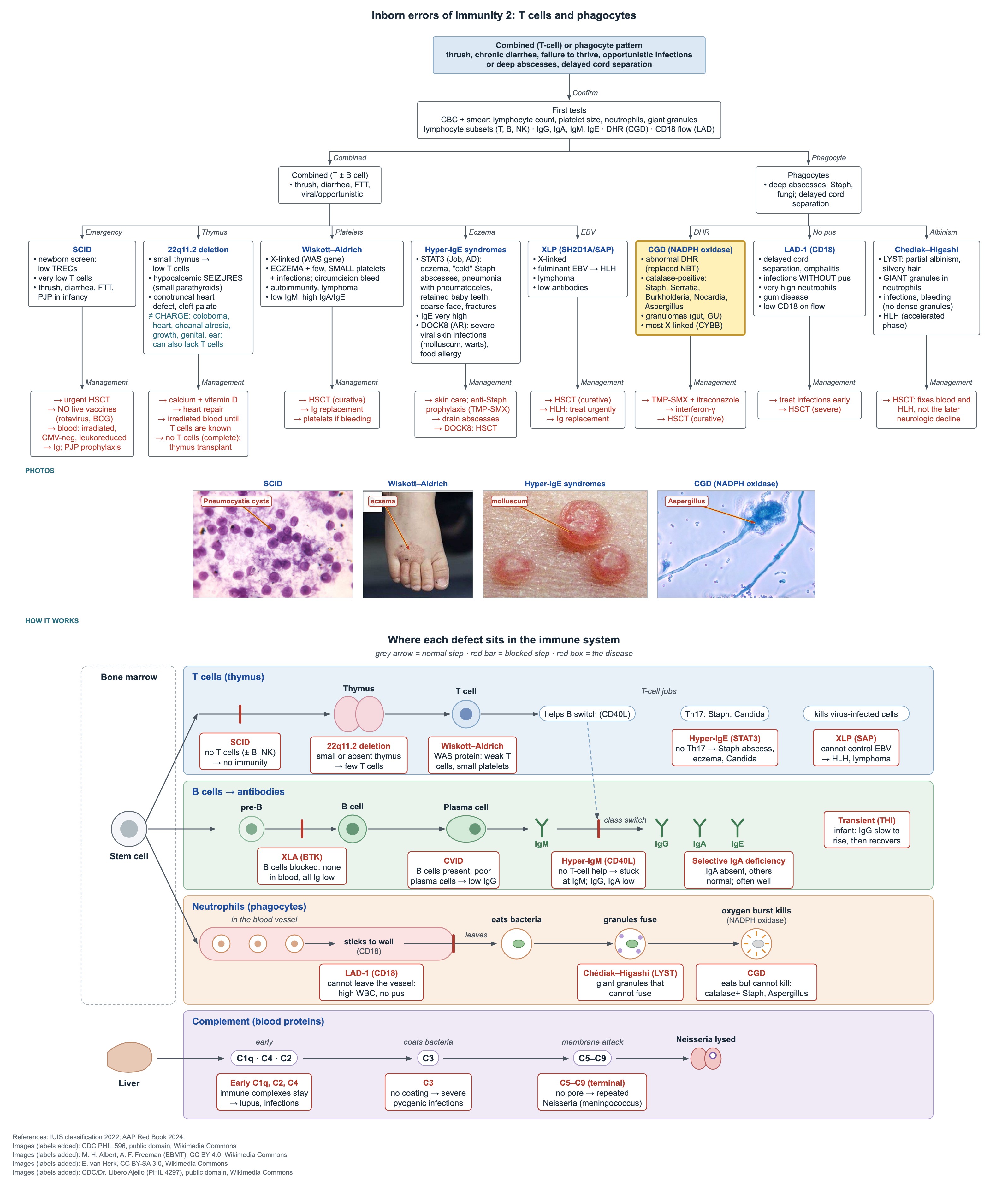
Phagocytes engulf bacteria but cannot kill them, because NADPH oxidase does not make the oxidative burst. Organisms that make catalase break down the little peroxide they produce themselves, so these germs cause deep, recurrent infections.
- Typical organisms: Staphylococcus aureus, Serratia, Burkholderia cepacia, Nocardia and Aspergillus.
- Typical sites: lung, liver and skin abscesses, lymphadenitis, osteomyelitis.
- Granulomas can block the gut or urinary tract; colitis can look like Crohn disease.
- Inheritance: most often X-linked (CYBB, gp91phox); the rest are autosomal recessive.
- Screening test: dihydrorhodamine (DHR) oxidative-burst test by flow cytometry, which has replaced the NBT test.
- A 6-week-old with worsening pneumonia whose aspirate grows Aspergillus has CGD until proven otherwise: order DHR.
CGD: prophylaxis and cure
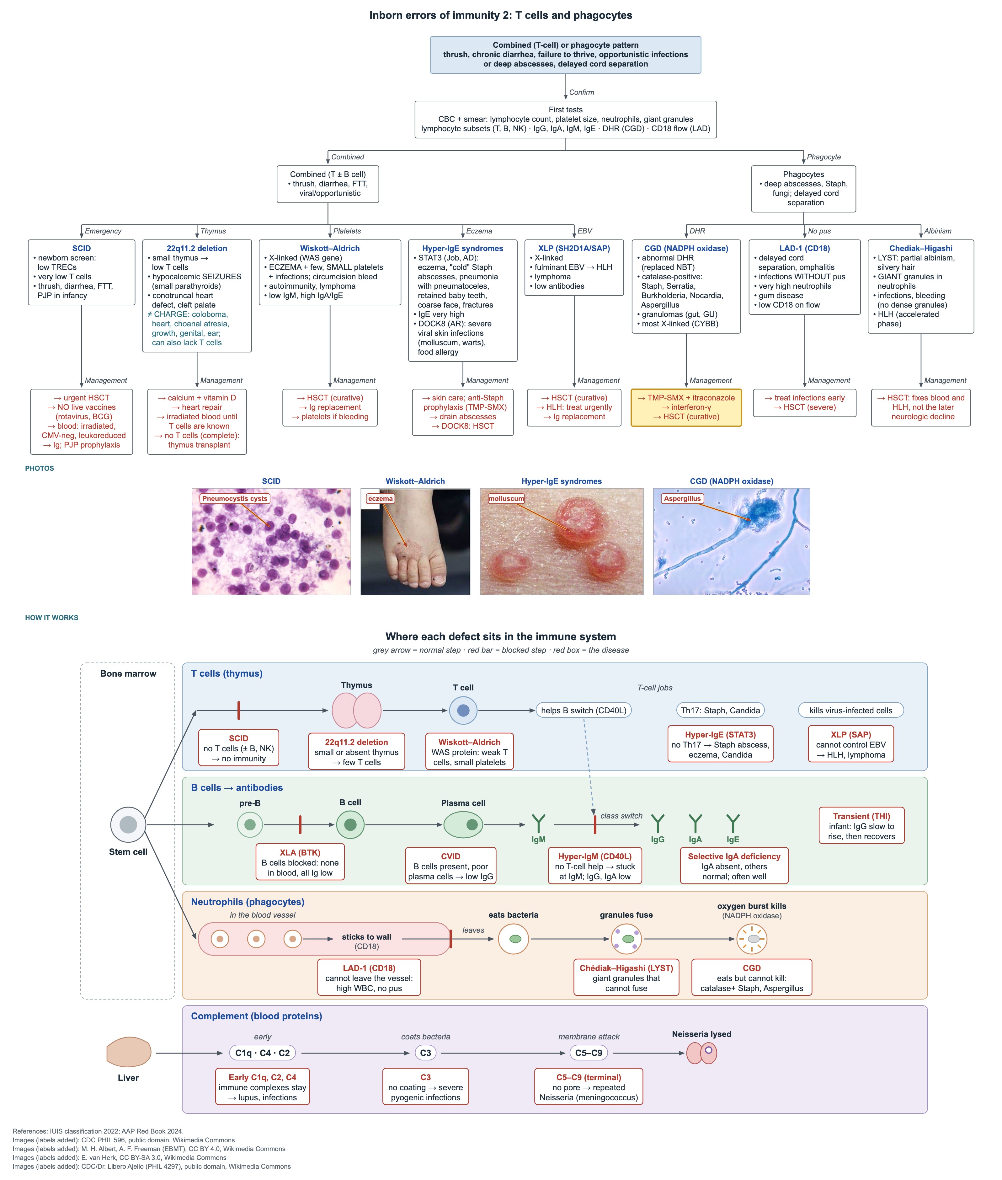
- Lifelong antibacterial prophylaxis with trimethoprim-sulfamethoxazole.
- Lifelong antifungal prophylaxis with itraconazole.
- Interferon-gamma injections are often added to reduce serious infections.
- Treat any fever promptly and look hard for an abscess or fungal infection.
- Hematopoietic stem cell transplant is curative.
Chédiak–Higashi syndrome
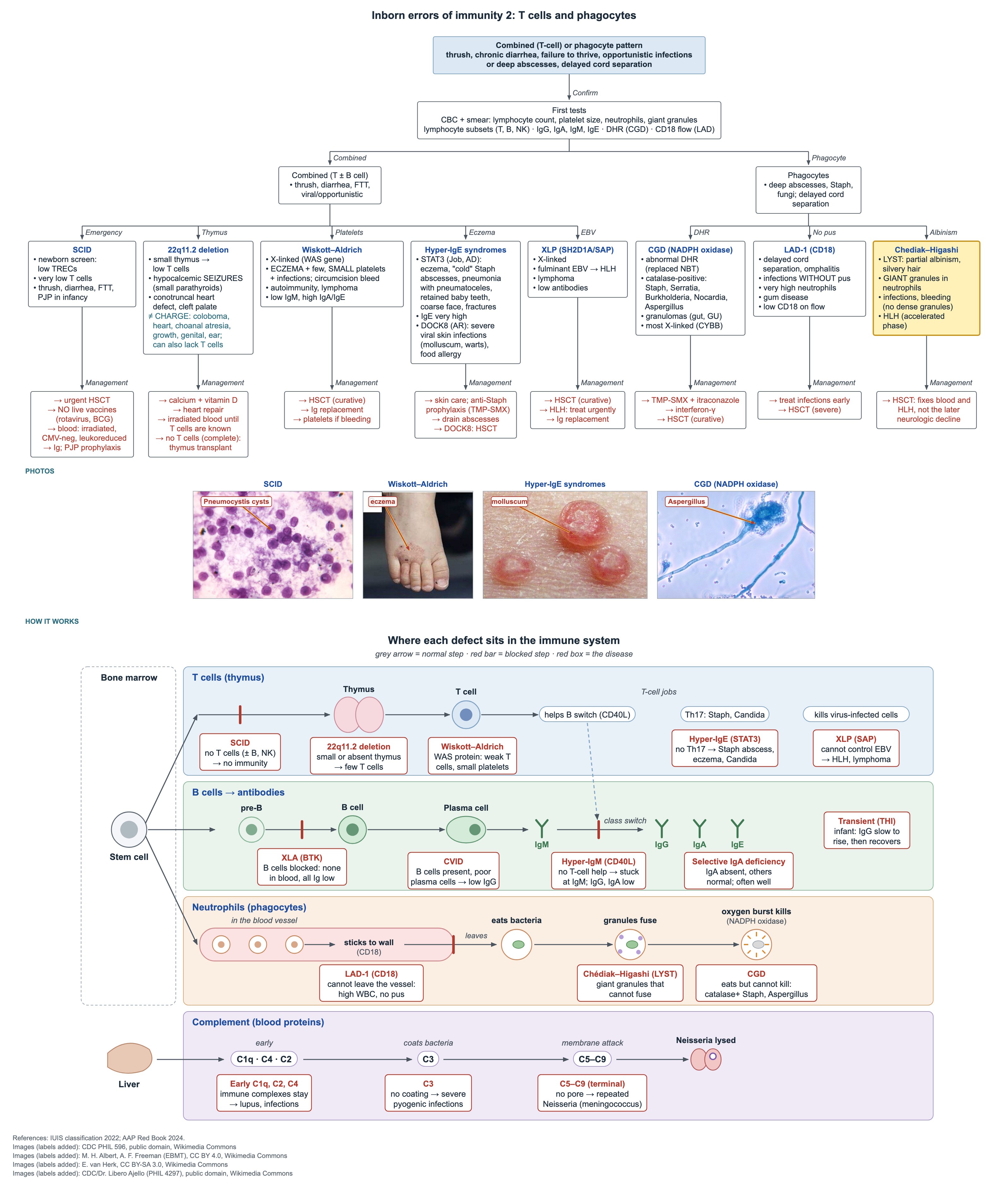
An autosomal recessive defect in LYST (lysosomal trafficking regulator) stops granules from fusing normally. Neutrophils carry giant granules they cannot use, and melanin and platelet granules are abnormal too.
- Partial oculocutaneous albinism and silvery hair.
- Recurrent pyogenic infections, mainly skin and respiratory.
- Peripheral smear clue: giant granules in neutrophils and other granulocytes.
- Easy bruising with a normal platelet count: platelets lack dense (delta) granules.
- Accelerated phase: fever, splenomegaly, cytopenias and a very high ferritin mean hemophagocytic lymphohistiocytosis (HLH).
- Later life: progressive neurologic problems such as neuropathy, ataxia and cognitive decline.
Chédiak–Higashi: treatment
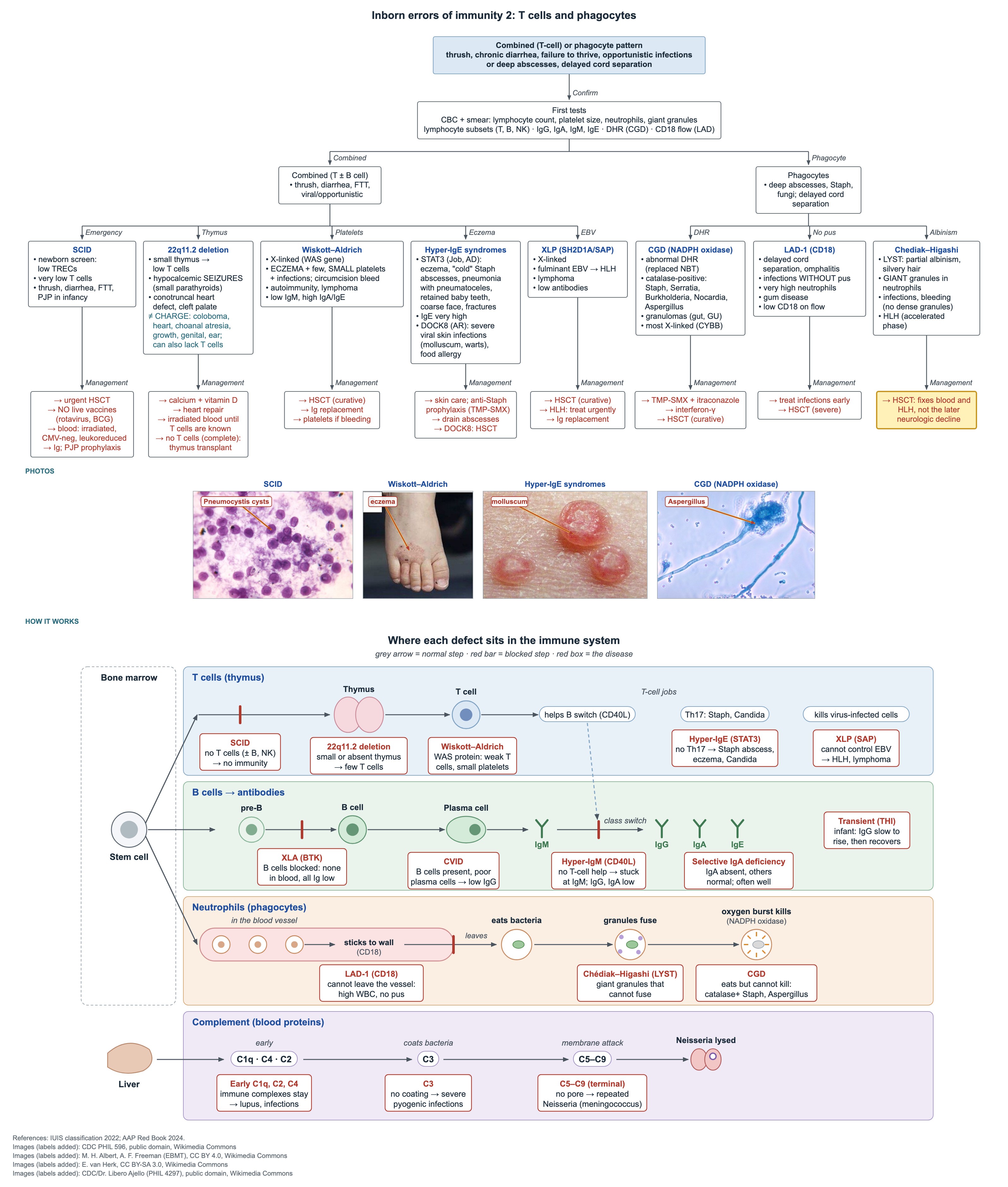
Hematopoietic stem cell transplant is the only cure. Done before HLH starts, it corrects the infections, the bleeding tendency and the HLH risk.
It does not stop the later neurodegeneration, because nerve cells do not come from the donor marrow. Neurologic follow-up continues after a successful transplant.
HLH is treated urgently with HLH-directed therapy as a bridge to transplant.
Complement tests and early classical pathway defects
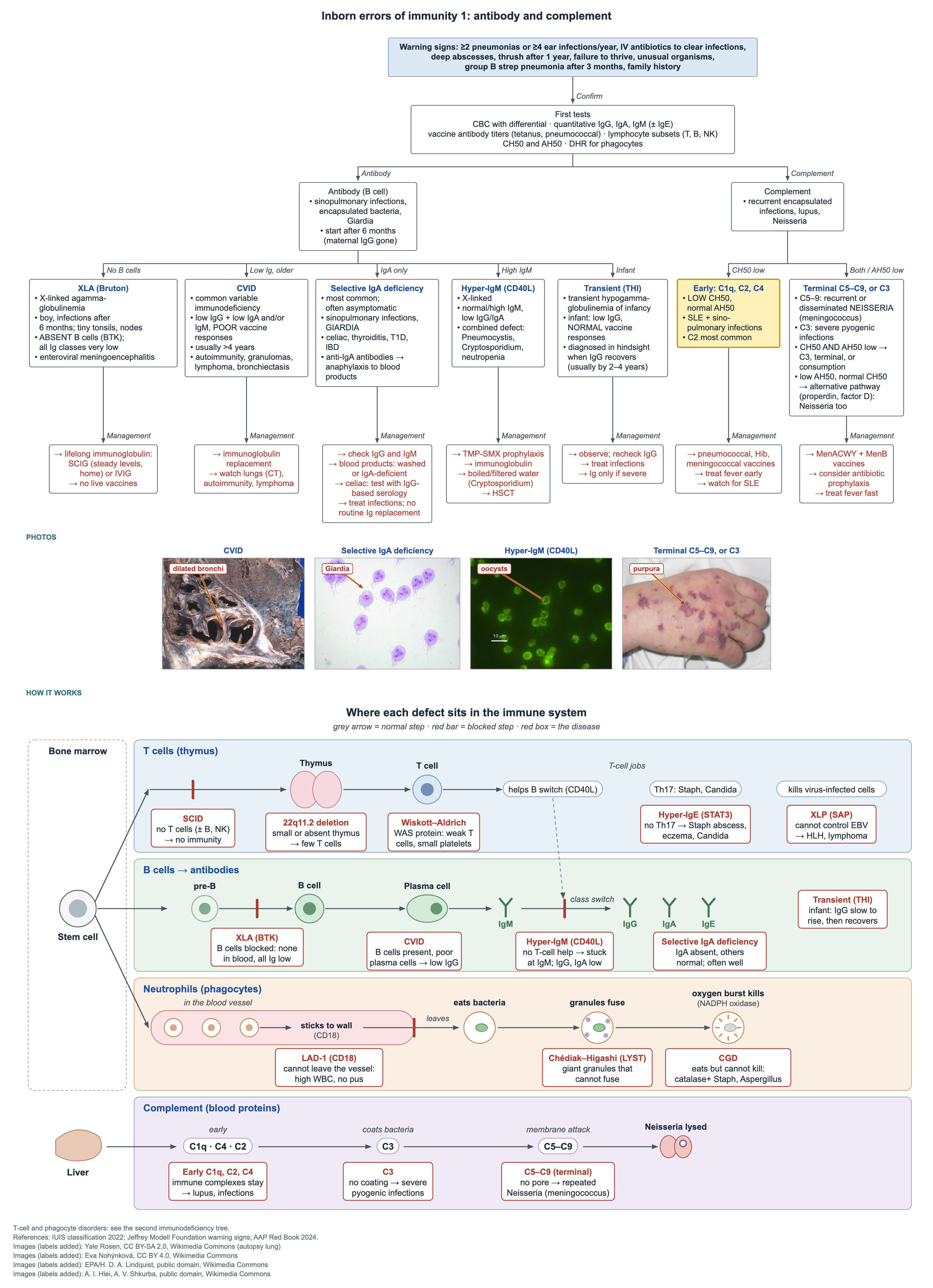
CH50 tests the classical pathway and AH50 the alternative pathway. Read the 2 together to place the defect.
- Early classical defects: early-onset lupus plus recurrent sinopulmonary infections with encapsulated bacteria.
- C1q deficiency carries the highest lupus risk; C2 deficiency is the most common.
- Management: pneumococcal, Hib and meningococcal vaccines, early treatment of fever and monitoring for lupus.
| CH50 | AH50 | Where the defect is |
|---|---|---|
| Low | Normal | Early classical pathway: C1, C2 or C4 |
| Normal | Low | Alternative pathway: factor B, factor D or properdin |
| Low | Low | Shared component (C3 or C5–C9) or complement consumption |
C3 and terminal complement defects
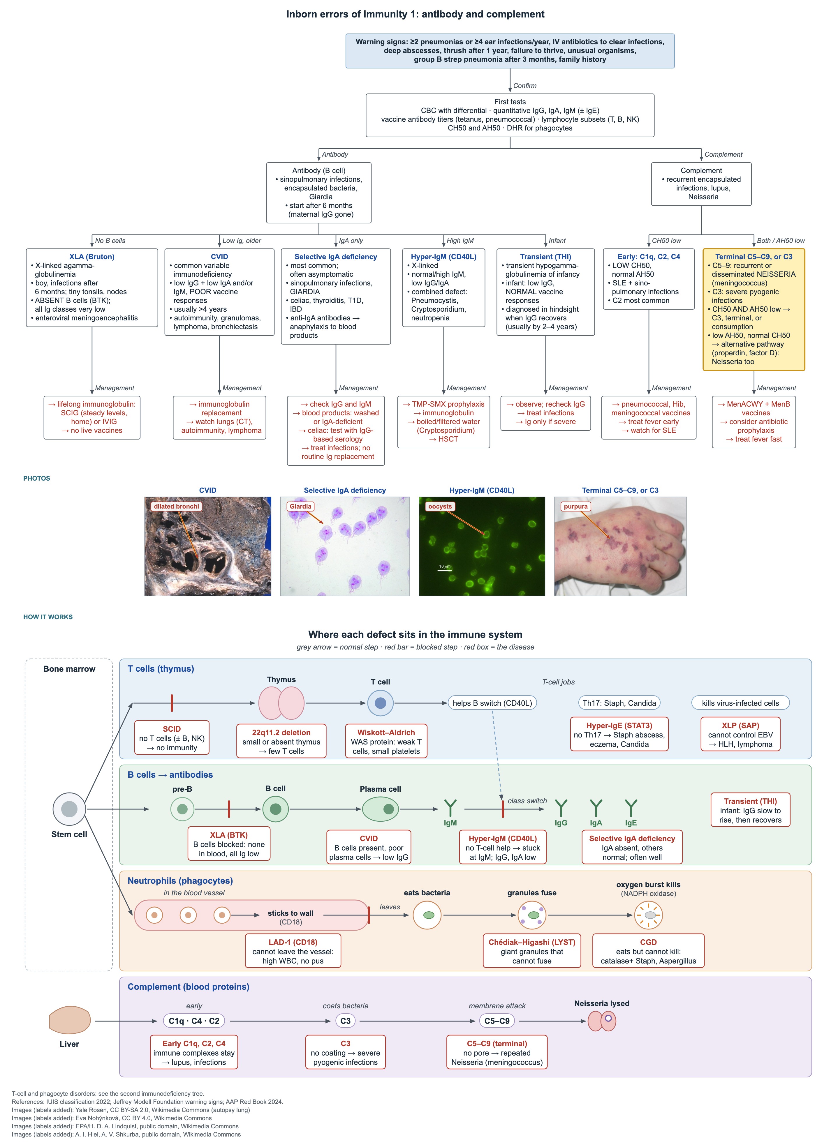
- C3 deficiency: no opsonization, so severe recurrent pyogenic infections.
- Terminal (C5–C9) deficiency: no membrane attack complex, so recurrent or disseminated Neisseria infections, mainly meningococcal.
- Alternative pathway (properdin, factor D) defects also cause Neisseria infections.
- Vaccines: MenACWY and MenB series, with boosters for as long as the risk lasts (complement deficiency is a high-risk condition for meningococcal vaccines).
- Consider antibiotic prophylaxis and teach the family to seek care fast for fever.
Common questions
What is the best test for chronic granulomatous disease?
The dihydrorhodamine (DHR) test by flow cytometry. It measures the neutrophil oxidative burst and has replaced the older NBT test.
Which complement deficiency causes recurrent meningococcal infections?
Deficiency of the terminal components C5–C9, and also properdin or factor D. These children need MenACWY and MenB vaccines with boosters.
Does a stem cell transplant cure Chédiak–Higashi syndrome?
It cures the immune, bleeding and HLH problems, but it does not prevent the neurologic decline that appears later.
Study this topic
Sources
- AAAAI/ACAAI/CIS 2025: Inborn errors of immunity practice parameter (Ann Allergy Asthma Immunol)
- Practice parameter for inborn errors of immunity: what is new in the 2025 update (J Allergy Clin Immunol)
- GeneReviews: Chédiak–Higashi syndrome (NCBI)
Written for Pedemy in our own words from the sources above; checked November 2026. For education only, not medical advice; check doses and local protocols before treating a patient. Pedemy is independent and not affiliated with or endorsed by the organizations cited.