

Main decision tree
Newborn pustules and bumps: benign or needs action?
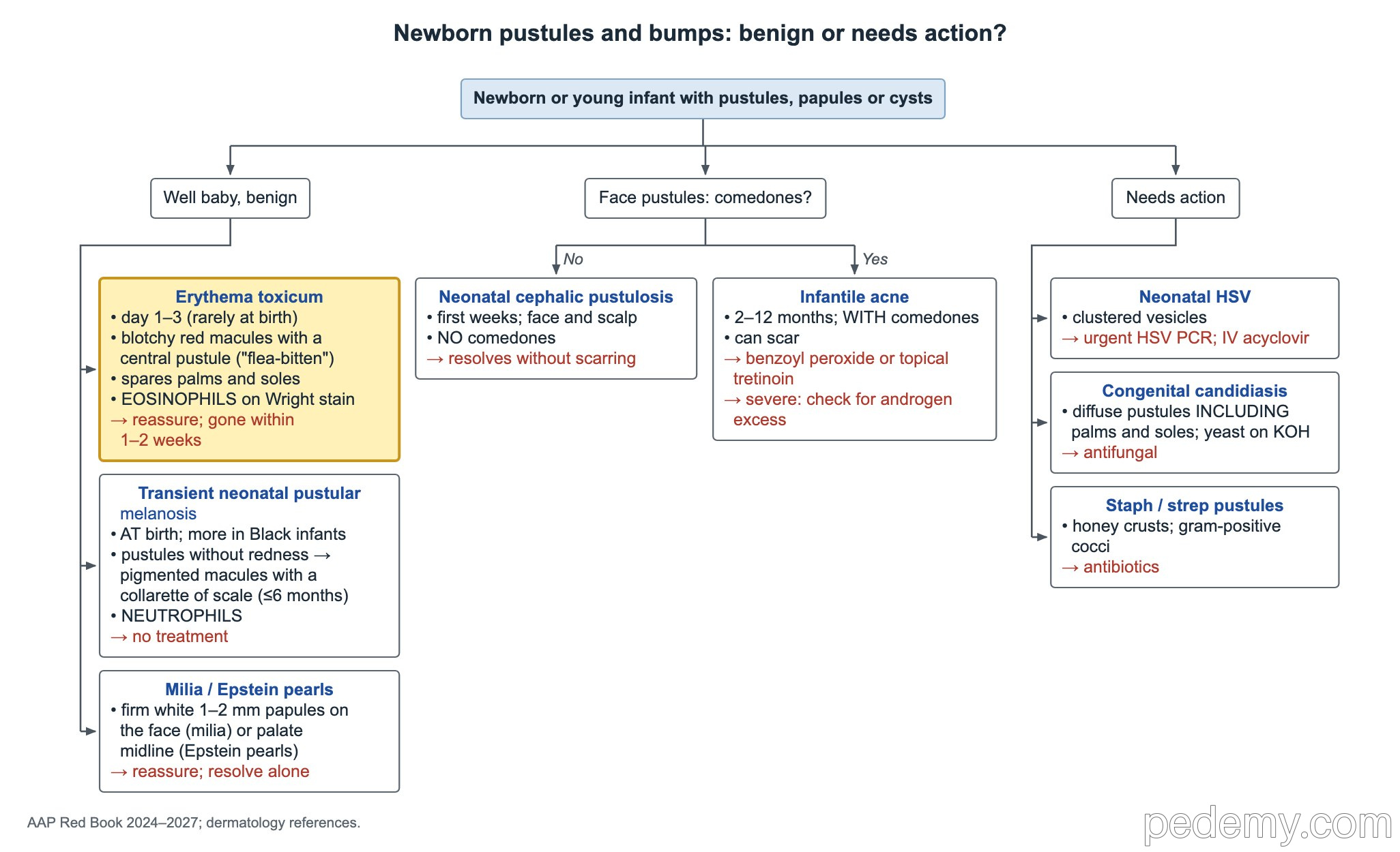
Key points
- Erythema toxicum: day 1–3, blotchy red macules with central pustules, spares palms and soles, eosinophils on smear, gone within 1–2 weeks.
- Transient neonatal pustular melanosis: present at birth, more common in Black infants, pustules without redness → pigmented macules with a collarette of scale; neutrophils.
- Neonatal cephalic pustulosis has no comedones and resolves without scars; infantile acne (2–12 months) has comedones and can scar.
- Grouped vesicles on a red base in a newborn = neonatal HSV until proven otherwise → urgent HSV PCR and IV acyclovir.
- Pustules on the palms and soles at birth with yeast on KOH = congenital cutaneous candidiasis.
- Irritant diaper rash spares the creases; candida involves the folds with satellite papules.
- Scalp aplasia cutis that is >4 cm, has a hair collar or a vascular stain → MRI.
Benign newborn pustules
Pustules on the palms and soles do not fit erythema toxicum: think of congenital candidiasis or TNPM.
| Feature | Erythema toxicum | Transient neonatal pustular melanosis (TNPM) |
|---|---|---|
| Onset | First few days (day 1–3); rarely at birth | Present at birth |
| Who | Term infants | More common in Black infants |
| Look | Blotchy red macules with a central pustule ('flea-bitten') | Pustules without redness that rupture, leaving pigmented macules with a collarette of scale |
| Palms and soles | Spared | Can be involved |
| Smear | Eosinophils | Neutrophils, no organisms |
| Course | Gone within 1–2 weeks | Pustules gone in days; macules fade over weeks to months |
| Management | Reassure and educate | Reassure; no treatment |
Milia, Epstein pearls and other bumps
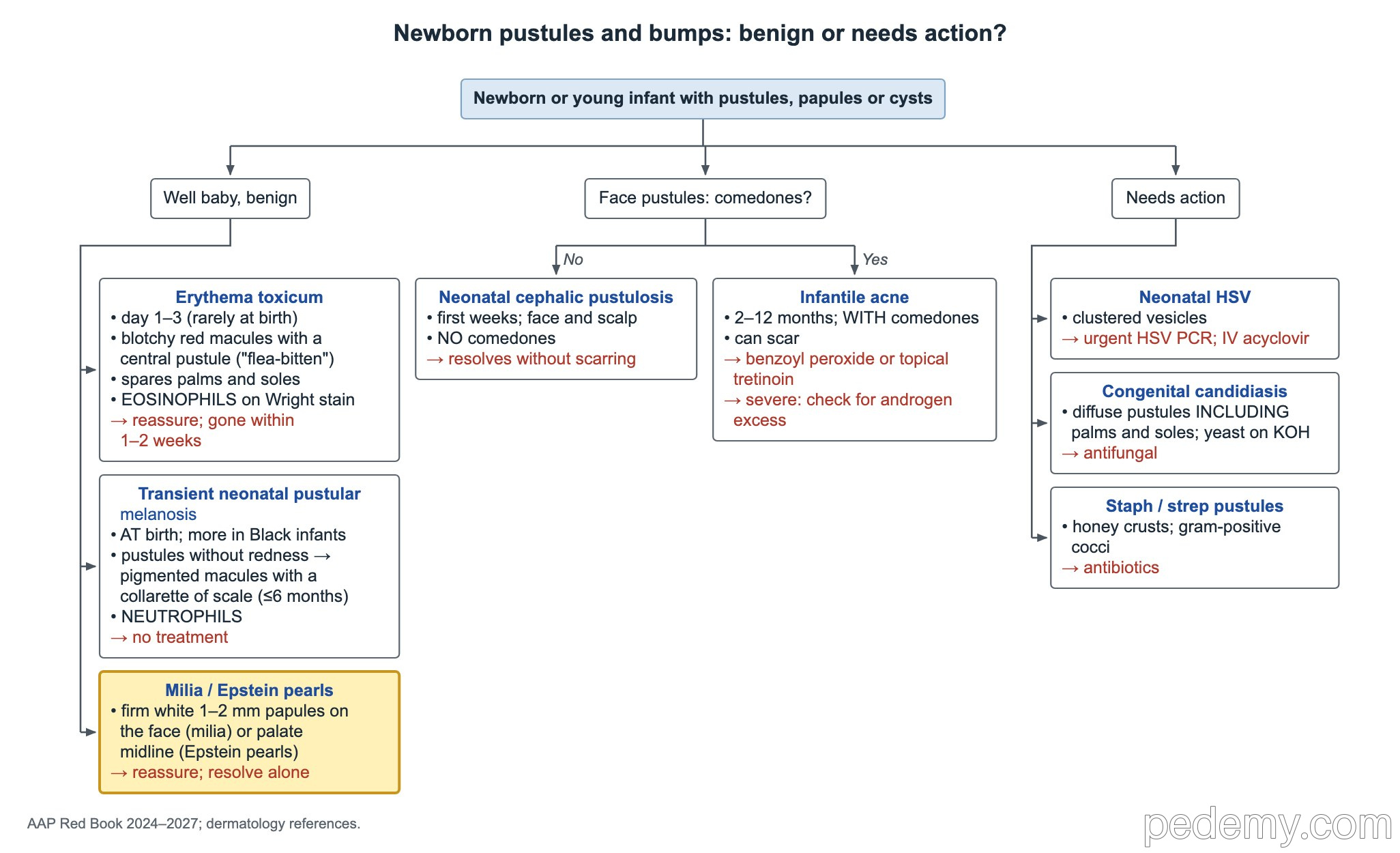
- Milia: firm white 1–2 mm papules on the face; no treatment, they resolve on their own.
- Epstein pearls: tiny white-yellow cysts along the midline of the palate (midpalatal raphe); reassure, no treatment.
- Dermoid cyst: firm, painless lump at the outer eyebrow in an infant. Midline nasal or scalp dermoids need imaging before any surgery because they can connect to the brain.
Face pustules: cephalic pustulosis or infantile acne?
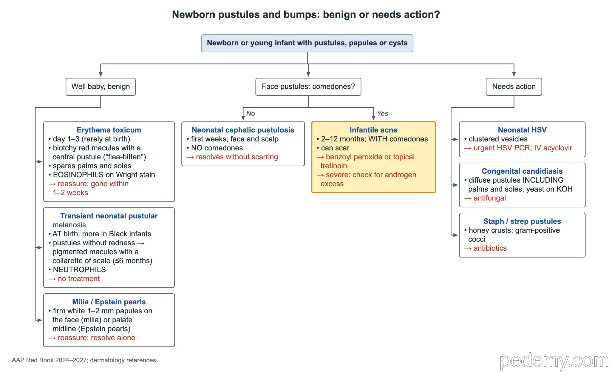
Severe infantile acne, or acne with signs of virilization, needs an evaluation for androgen excess.
| Feature | Neonatal cephalic pustulosis | Infantile acne |
|---|---|---|
| Age | First weeks of life | About 2–12 months |
| Lesions | Pustules on the face and scalp, no comedones | Comedones, papules and pustules on the cheeks |
| Scarring | No, resolves on its own | Can scar |
| Treatment | None needed | Benzoyl peroxide or topical tretinoin |
Newborn rashes that need action
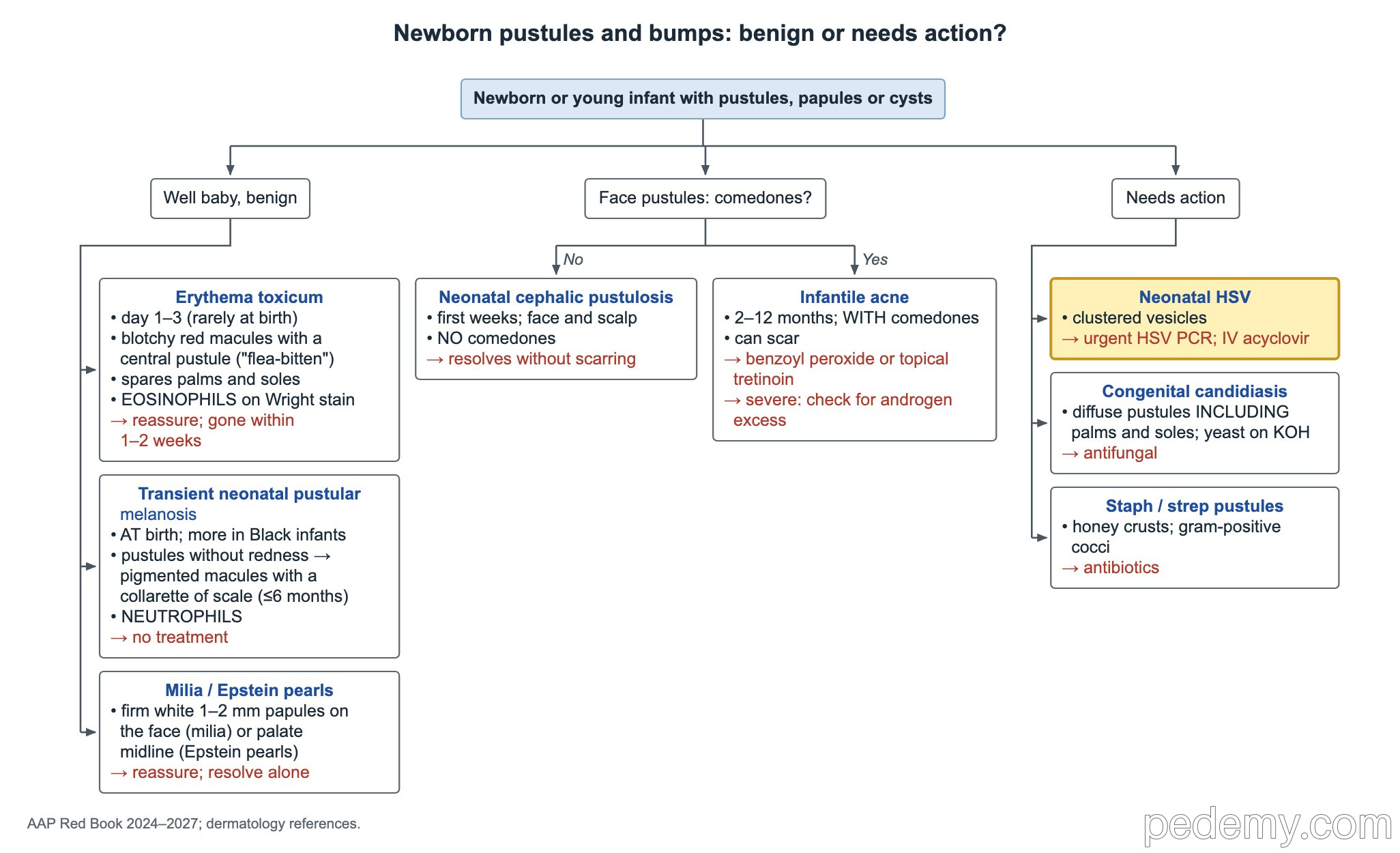
- Neonatal HSV: grouped vesicles on a red base. Send HSV PCR of the lesions and blood and start IV acyclovir right away.
- Congenital cutaneous candidiasis: widespread pustules at birth including the palms and soles; yeast on KOH. Treat with an antifungal.
- Staphylococcal or streptococcal pustules (neonatal impetigo): honey-colored crusted pustules; Gram stain shows gram-positive cocci, mostly S. aureus. Treat with antibiotics.
Diaper-area rashes
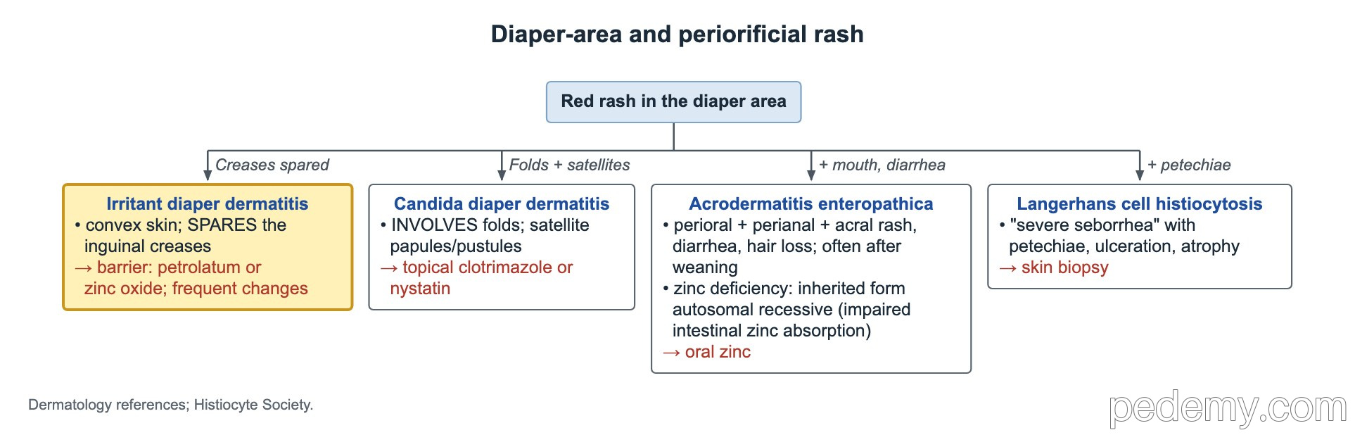
| Rash | Clues | Treatment |
|---|---|---|
| Irritant diaper dermatitis | Red skin on convex surfaces; creases spared | Barrier ointment (zinc oxide, petrolatum); frequent diaper changes |
| Candida diaper dermatitis | Beefy red rash involving the folds, with satellite papules and pustules | Topical clotrimazole (or another azole) or nystatin |
| Acrodermatitis enteropathica | After weaning: crusted rash around the mouth, anus and on the hands and feet, plus diarrhea and hair loss | Oral zinc; lifelong if inherited (autosomal recessive defect in zinc absorption) |
| Langerhans cell histiocytosis | 'Severe seborrhea' with petechiae, ulceration or atrophy | Skin biopsy |
Scalp lesions: cradle cap, nevus sebaceous, aplasia cutis
- Seborrheic dermatitis (cradle cap): greasy yellow scale and redness on the scalp of a young infant; can also affect the face, trunk and diaper area. Emollients soften and loosen the scale; it resolves in months.
- Nevus sebaceous: yellow-orange, hairless, waxy plaque on the scalp, present from birth. Secondary tumors appear mostly after puberty and are usually benign (trichoblastoma); the risk of a malignant tumor such as basal cell carcinoma is low, about 1% or less. Options are observation with periodic skin exams or elective excision later in childhood or adolescence.
- Large nevus sebaceous with eye, bone or brain anomalies: nevus sebaceous (Schimmelpenning) syndrome.
- Aplasia cutis congenita: a well-defined area of absent skin, usually on the scalp vertex. Small lesions need only local wound care and observation.
- Aplasia cutis that is larger than 4 cm, has a hair collar or an overlying vascular stain → MRI to look for skull and brain defects.
- Multiple areas of scalp aplasia cutis: think of trisomy 13. Also linked to Wolf–Hirschhorn syndrome (4p deletion) and Adams–Oliver syndrome.
Common questions
What is erythema toxicum and is it dangerous?
It is a harmless rash of blotchy red spots with small central pustules that appears in the first days of life. It spares the palms and soles and goes away within 1–2 weeks without treatment.
How do you tell neonatal acne from infantile acne?
Neonatal cephalic pustulosis appears in the first weeks with pustules and no comedones and clears without scars. Infantile acne starts at about 2–12 months, has comedones and can scar.
Which newborn rash is an emergency?
Grouped vesicles on a red base. Neonatal herpes must be ruled out with HSV PCR, and IV acyclovir is started before the result comes back.
Study this topic
Sources
- AAP Clinical Report: Management of neonates born to women with active genital herpes lesions (Pediatrics, 2013)
- AAP Red Book: Report of the Committee on Infectious Diseases
Written for Pedemy in our own words from the sources above; checked November 2026. For education only, not medical advice; check doses and local protocols before treating a patient. Pedemy is independent and not affiliated with or endorsed by the organizations cited.