

Main decision tree
Blue-gray spots
Key points
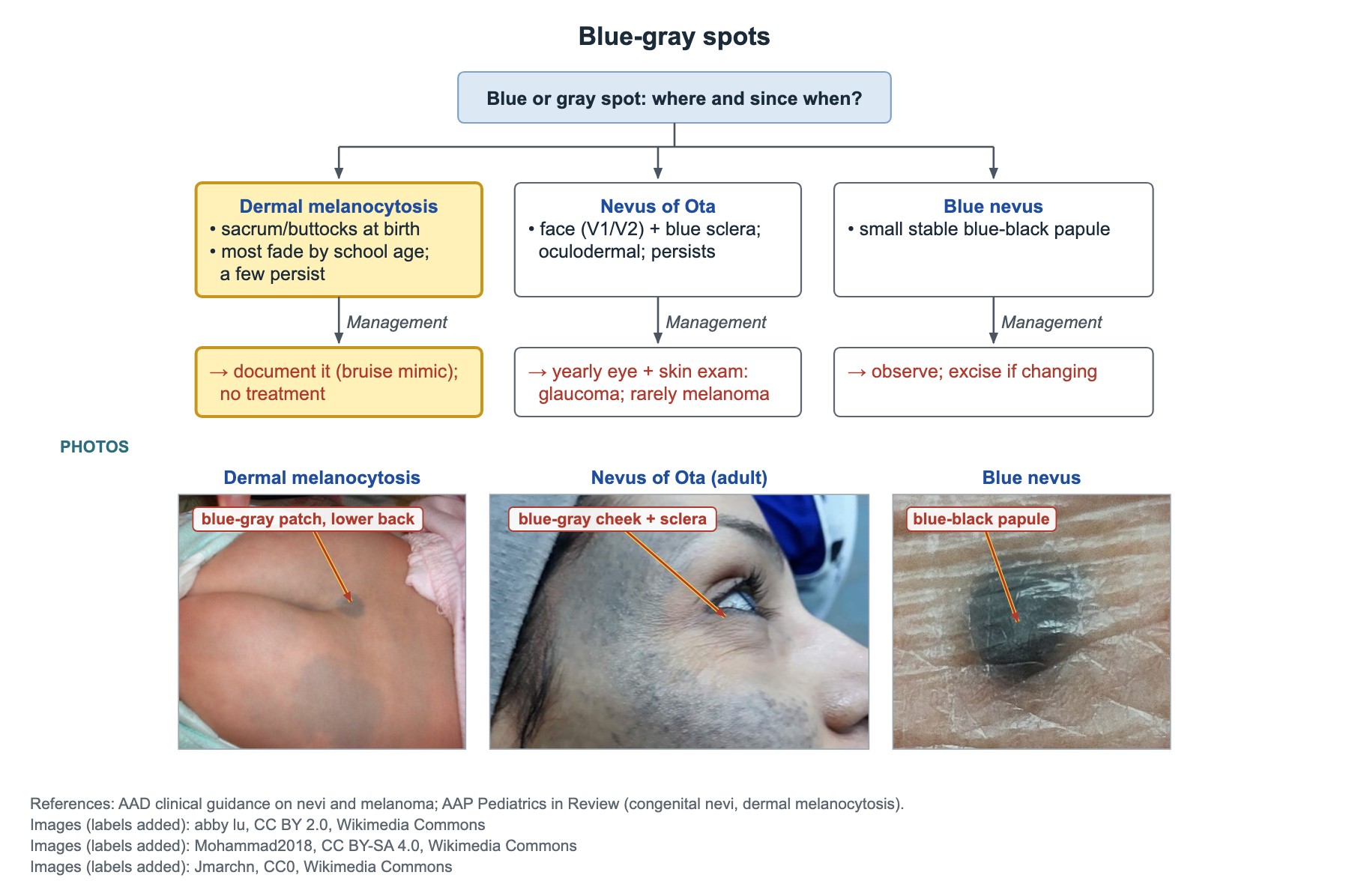
- Congenital dermal melanocytosis: blue-gray patch over the sacrum and buttocks at birth; no treatment; document it because it mimics bruises.
- Nevus of Ota (oculodermal melanocytosis): blue-gray patch in the V1/V2 area plus blue sclera; yearly eye and skin exams for glaucoma and, rarely, melanoma.
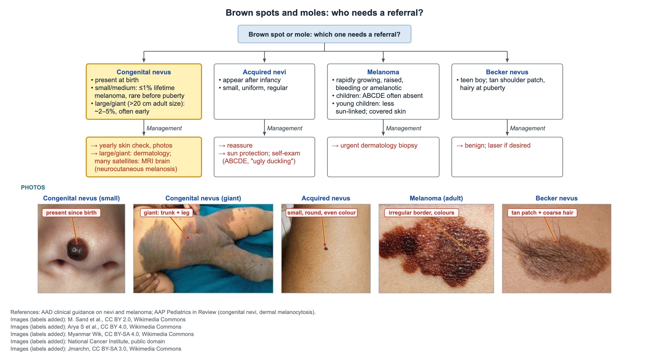
- Congenital melanocytic nevi: small and medium carry about 1% or less lifetime melanoma risk; large or giant (>20 cm projected adult size) about 2–5%, often in childhood.
- Large or giant nevus with many satellite nevi: brain MRI for neurocutaneous melanosis.
- Childhood melanoma often does not follow ABCDE: it can be raised, uniform in color, amelanotic and bleeding; refer urgently for excisional biopsy.
- Vitiligo: chalk-white, sharply bordered patches around the eyes, mouth and hands; autoimmune; check TSH.
- Pilomatricoma is a rock-hard, slowly growing, bluish nodule on the head or neck; it does not regress, so it is excised.
Blue-gray spots
Congenital dermal melanocytosis is a flat blue-gray patch, most often over the sacrum and buttocks, present at birth. It is most common in Black, Asian, Hispanic and Native American infants.
- Course: most fade during early childhood and are usually gone by about age 10; a few persist, especially away from the sacrum.
- Management: no treatment. Record the size and site in the chart and explain it to the parents.
- Why it matters: the blue-gray color can be mistaken for bruising and lead to a child abuse evaluation. Bruises are tender, change color over days and fit an injury pattern; dermal melanocytosis does not.
Nevus of Ota and blue nevus
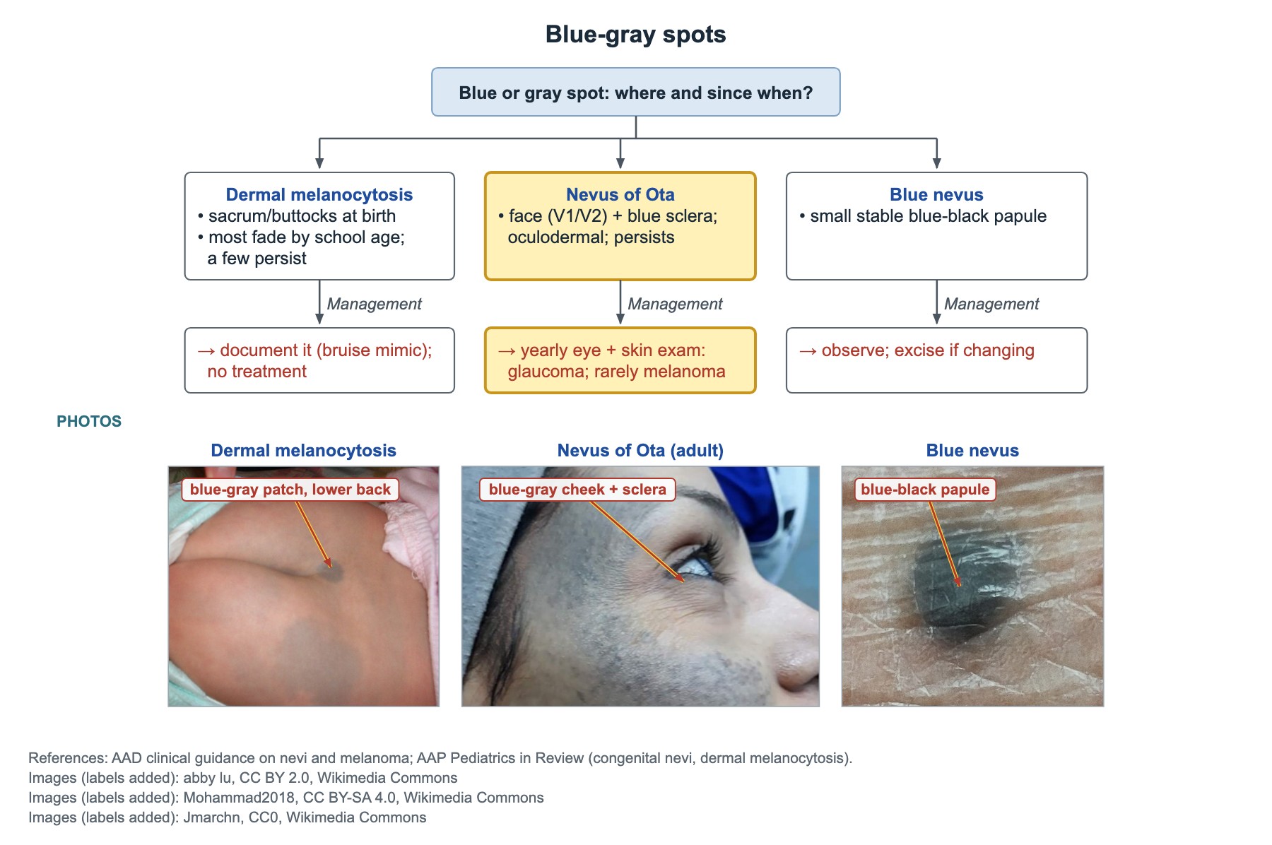
- Nevus of Ota (oculodermal melanocytosis): blue-gray patch on one side of the face in the V1/V2 area, often with blue-gray sclera. It persists for life.
- Nevus of Ota risks: glaucoma and, rarely, ocular or skin melanoma. Arrange a yearly eye exam and regular skin exams.
- Laser treatment can lighten the skin patch if wanted.
- Blue nevus: small, well-circumscribed, stable blue-black papule, often on the hand or face. Observe it; excise only if it changes or looks atypical.
Congenital and acquired nevi
| Lesion | Key facts | Management |
|---|---|---|
| Small or medium congenital nevus | Present at birth; lifetime melanoma risk about 1% or less, rare before puberty | Yearly skin check, photos |
| Large or giant congenital nevus (>20 cm projected adult size) | Melanoma risk about 2–5%, often in early childhood; risk of neurocutaneous melanosis | Dermatology follow-up for life; brain MRI if many satellite nevi |
| Acquired nevi | Appear after infancy; small, round, uniform color | Reassure; sun protection; self-exam (ABCDE, 'ugly duckling') |
| Becker nevus | Teen boy; large tan patch on the shoulder or chest that becomes hairy at puberty | Benign; laser for pigment or hair if desired |
Melanoma and other skin cancers
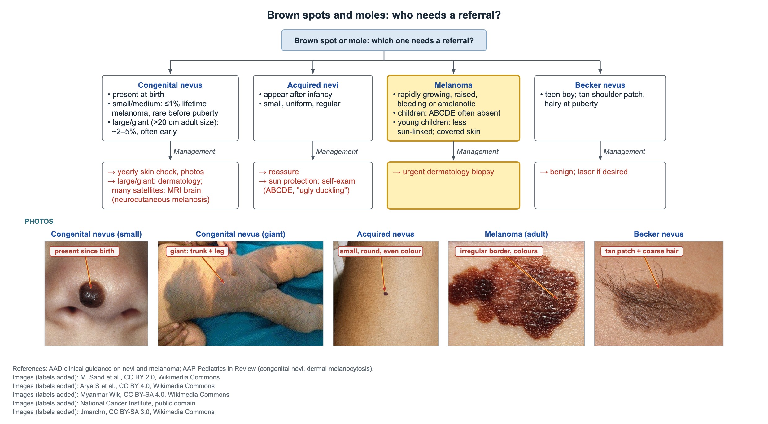
A rapidly enlarging, raised pigmented lesion in an adolescent is melanoma until proven otherwise. In younger children, melanoma often lacks the adult ABCDE features: it may be amelanotic (pink or red), raised, uniform in color and bleeding. It is also less tied to sun exposure than other skin cancers and can arise on covered skin.
- Next step for any lesion concerning for melanoma: prompt dermatology referral for excisional biopsy.
- Solar lentigines: uniform brown macules with irregular borders on the face of a fair teen with freckles; a sign of sun damage.
- Basal cell carcinoma: pearly red papule on sun-exposed skin that bleeds or ulcerates.
- Squamous cell carcinoma: scaly or bleeding papule or plaque on sun-exposed skin.
- Prevention: sun protection with shade, clothing and broad-spectrum sunscreen SPF 30 or more; no tanning beds.
White patches: vitiligo
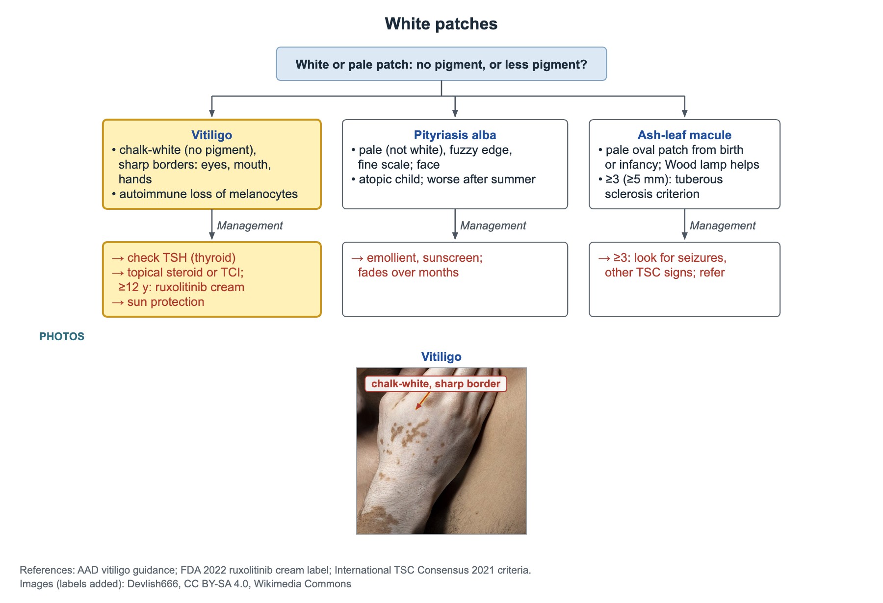
- Vitiligo: chalk-white (no pigment), sharply bordered patches around the eyes and mouth and on the hands. It is autoimmune destruction of melanocytes.
- Check TSH: autoimmune thyroid disease is the most common associated condition.
- Treatment: topical steroid or topical calcineurin inhibitor; ruxolitinib cream from age 12; sun protection of the white skin.
- Pityriasis alba: pale (not white), fuzzy-edged, finely scaly patches on the face of an atopic child; treat with emollient and sunscreen.
- Ash-leaf macule: pale oval patch from birth or infancy; 3 or more (at least 5 mm) is a tuberous sclerosis criterion.
Skin lumps
| Lump | Clues | Management |
|---|---|---|
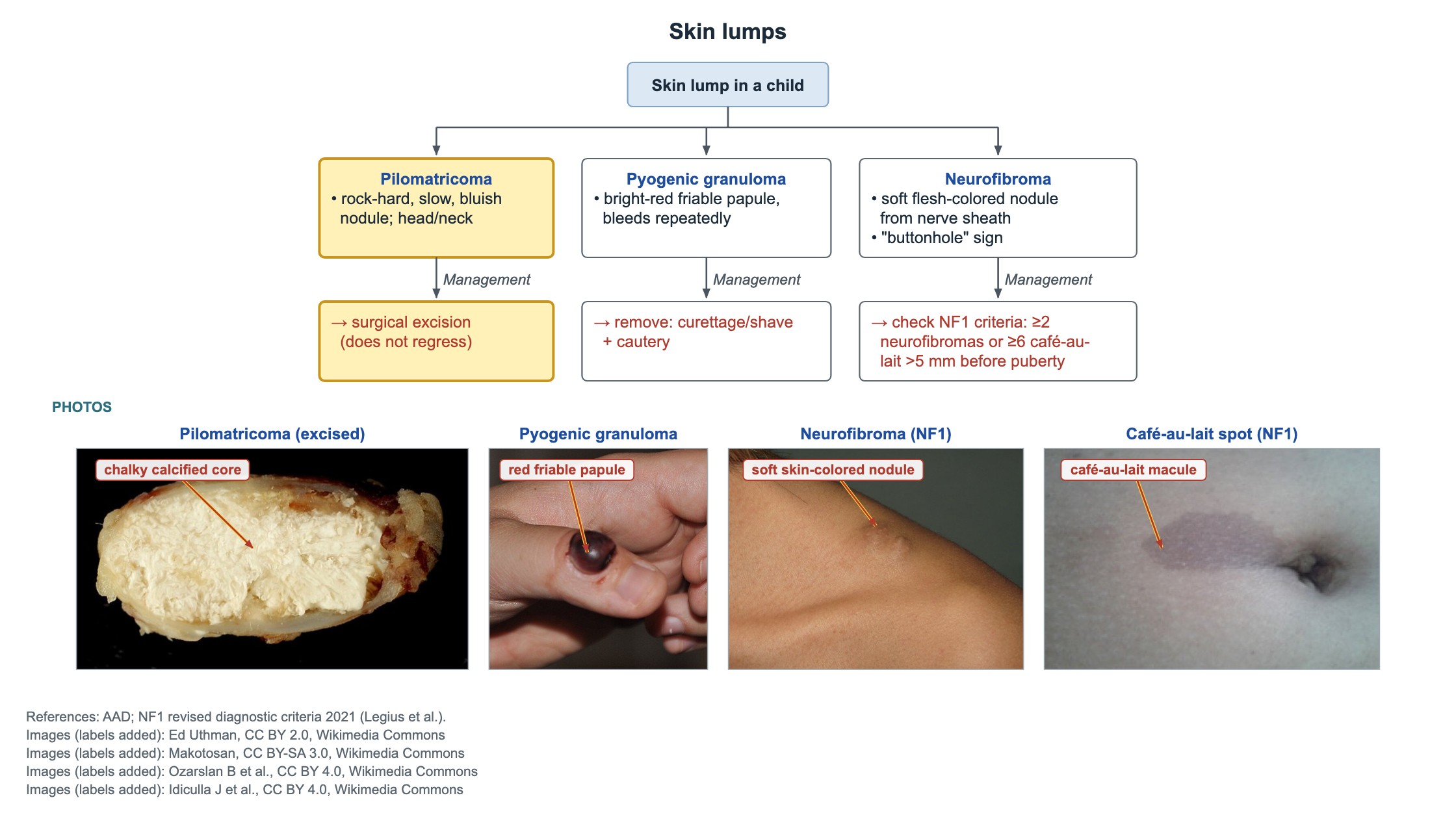
| Pilomatricoma (pilomatrixoma) | Rock-hard, slowly enlarging, bluish nodule on the face or neck | Surgical excision; it does not regress |
| Pyogenic granuloma (lobular capillary hemangioma) | Bright red, friable papule that bleeds easily and repeatedly | Remove by curettage or shave with cautery |
| Neurofibroma | Soft skin-colored papule from a nerve sheath; 'buttonhole' sign (pushes in with a finger) | Check NF1 criteria: 2 or more neurofibromas, or 6 or more café-au-lait macules >5 mm before puberty |
Common questions
Do Mongolian spots go away?
Most congenital dermal melanocytosis patches fade during early childhood and are gone by about age 10. Patches outside the sacral area are more likely to persist.
Does melanoma in children look like melanoma in adults?
Often not. Childhood melanoma is frequently raised, pink or uniform in color, and may bleed, so the ABCDE rules can miss it. A new, growing or bleeding bump needs prompt dermatology review.
What is the melanoma risk of a giant congenital nevus?
About 2–5% over a lifetime, often in early childhood. Small and medium congenital nevi carry a risk of about 1% or less.
Study this topic
Sources
- Risk of melanoma in congenital melanocytic nevi of all sizes: a systematic review (J Eur Acad Dermatol Venereol, 2022)
- AAP Clinical Report: The evaluation of suspected child physical abuse (Pediatrics, 2015)
- Revised diagnostic criteria for neurofibromatosis type 1 and Legius syndrome (Genet Med, 2021)
Written for Pedemy in our own words from the sources above; checked November 2026. For education only, not medical advice; check doses and local protocols before treating a patient. Pedemy is independent and not affiliated with or endorsed by the organizations cited.