

Main decision tree
Hair loss in a child
Key points
- Hair-pull test is positive when more than 10% of grasped hairs come out easily.
- Alopecia areata: smooth round bald patches with exclamation-mark hairs; ask about autoimmune thyroid disease, vitiligo and atopy.
- Trichotillomania: irregular patches of broken hairs of different lengths → CBT with habit reversal.
- Telogen effluvium: diffuse shedding 2–3 months after fever, surgery or weight loss; regrows in 6–12 months.
- "Severe seborrhea" with petechiae, ulceration or atrophy → think Langerhans cell histiocytosis → skin biopsy.
- Acrodermatitis enteropathica: autosomal recessive defect in intestinal zinc absorption; periorificial and acral rash, diarrhea, hair loss.
- Behçet disease: recurrent oral ulcers plus genital ulcers, uveitis or erythema nodosum.
Hair loss: patchy, diffuse or scarring
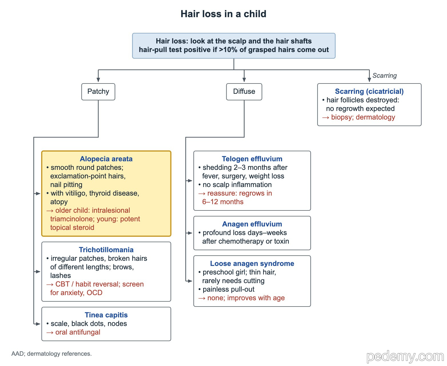
Look at the scalp and the hair shafts, then decide whether the loss is patchy, diffuse or scarring. A hair-pull test is positive when more than 10% of the grasped hairs come out easily.
Alopecia areata is autoimmune. It gives smooth, well-circumscribed round bald patches with exclamation-mark hairs at the edge, and sometimes nail pitting.
- Associations: autoimmune thyroid disease, vitiligo, atopy and Down syndrome. Ask about a family history of thyroid disease.
- Worse prognosis: total scalp (totalis) or body (universalis) loss, ophiasis (band along the back and sides of the scalp), young age at onset, and atopy.
- A few patches in an older child: intralesional triamcinolone. Young child: a potent topical steroid.
- Severe disease in teens 12 years and older: the oral JAK inhibitor ritlecitinib is FDA approved.
- Scarring (cicatricial) alopecia destroys the follicle, so no regrowth is expected → biopsy and dermatology referral.
- Tinea capitis is the other common cause of patchy loss: scale, black dots and enlarged nodes → oral antifungal.
Hair pulling and diffuse shedding
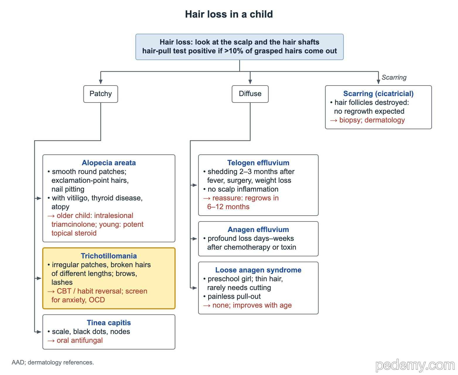
Trichotillomania gives irregular patches with broken hairs of different lengths, sometimes in the eyebrows and eyelashes too. First-line care is cognitive behavioral therapy with habit reversal, plus screening for anxiety, depression and OCD.
| Diffuse loss | Clues | Management |
|---|---|---|
| Telogen effluvium | Diffuse shedding 2–3 months after a febrile illness, surgery or weight loss; no scalp inflammation | Reassure; regrowth in 6–12 months |
| Anagen effluvium | Sudden heavy loss within days to weeks of chemotherapy or a toxin | Expected with cytotoxic drugs; regrows after treatment ends |
| Loose anagen syndrome | Preschool girl with thin hair that rarely needs cutting; hairs pull out painlessly | No treatment; improves with age |
Langerhans cell histiocytosis: the "seborrhea" that is not
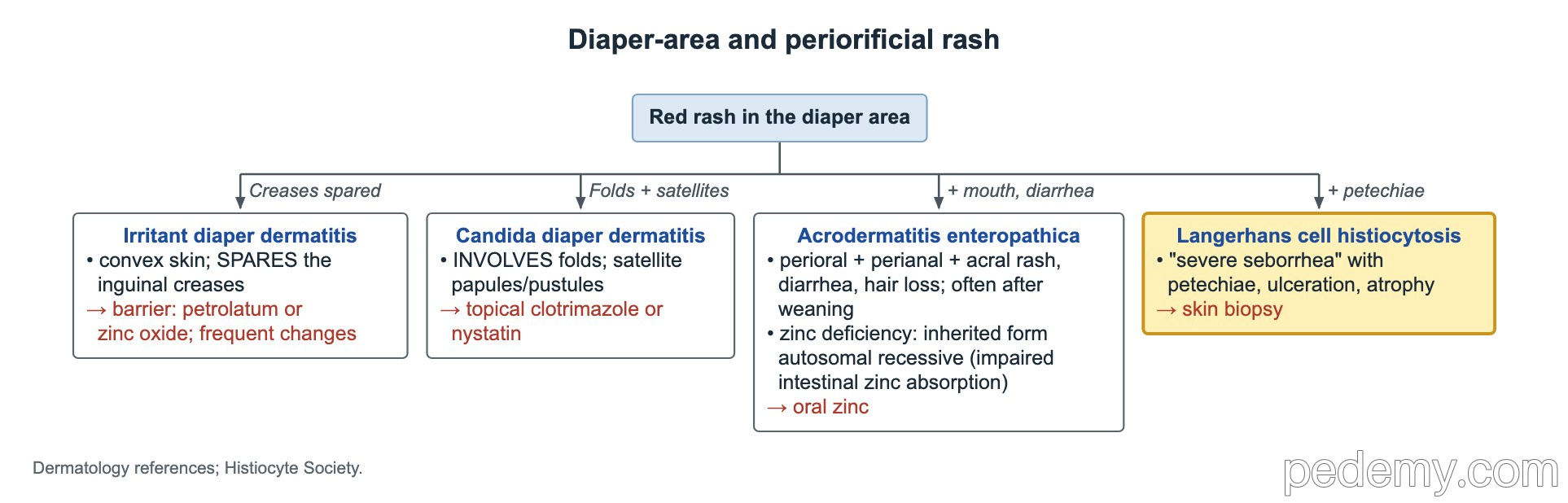
Seborrheic dermatitis in an infant is greasy, scaly and harmless. Very severe "seborrhea" on the scalp, behind the ears or in the diaper folds with petechiae, ulceration or atrophy should make you think of Langerhans cell histiocytosis (LCH).
The diagnostic step is a skin biopsy (CD1a and langerin-positive cells). A confirmed case then needs a full check for disease in bone, liver, spleen, marrow and lung, because skin may be the first sign of multisystem LCH.
Acrodermatitis enteropathica (zinc deficiency)
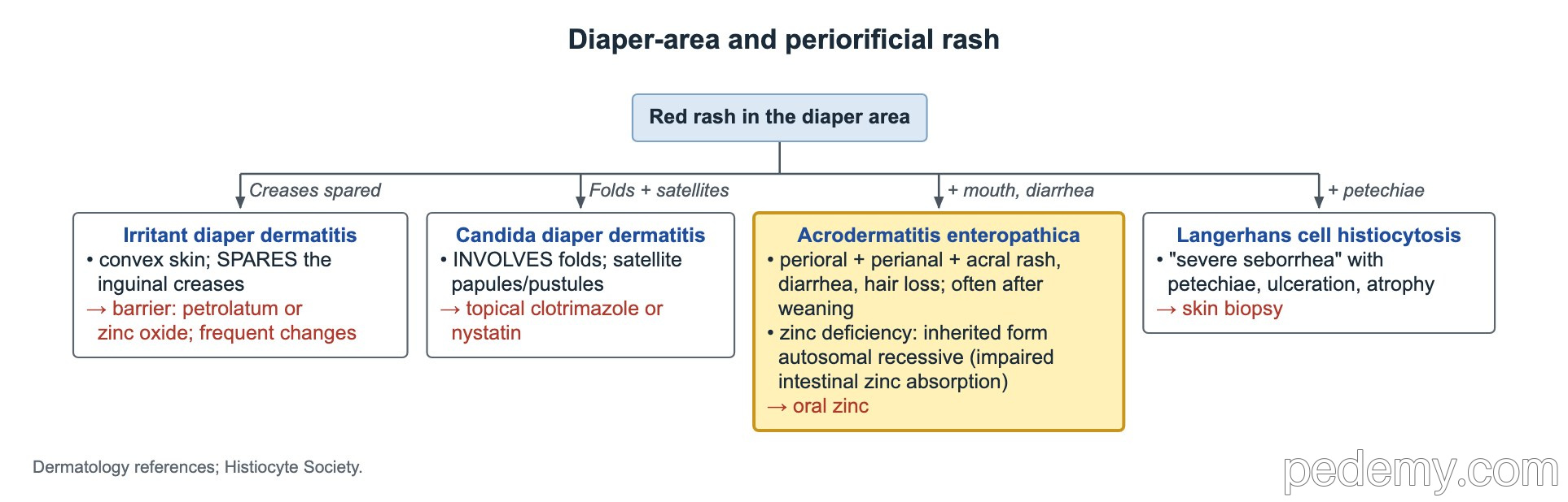
Zinc deficiency gives a sharply marginated, crusted red rash around the mouth and anus and on the hands and feet, with diarrhea, hair loss and irritability. It often starts after weaning from breast milk.
The inherited form is autosomal recessive: the gut cannot absorb zinc normally. Serum zinc (and often alkaline phosphatase) is low. Oral zinc clears the rash within days and is lifelong in the inherited form.
| Diaper-area rash | Clue | Treatment |
|---|---|---|
| Irritant dermatitis | Convex skin; spares the creases | Barrier cream (petrolatum or zinc oxide), frequent changes |
| Candida | Involves the folds; satellite papules and pustules | Topical clotrimazole or nystatin |
| Acrodermatitis enteropathica | Periorificial + acral rash, diarrhea, hair loss | Oral zinc |
| LCH | "Seborrhea" with petechiae, ulceration, atrophy | Skin biopsy |
Shin nodules: sarcoidosis and other triggers
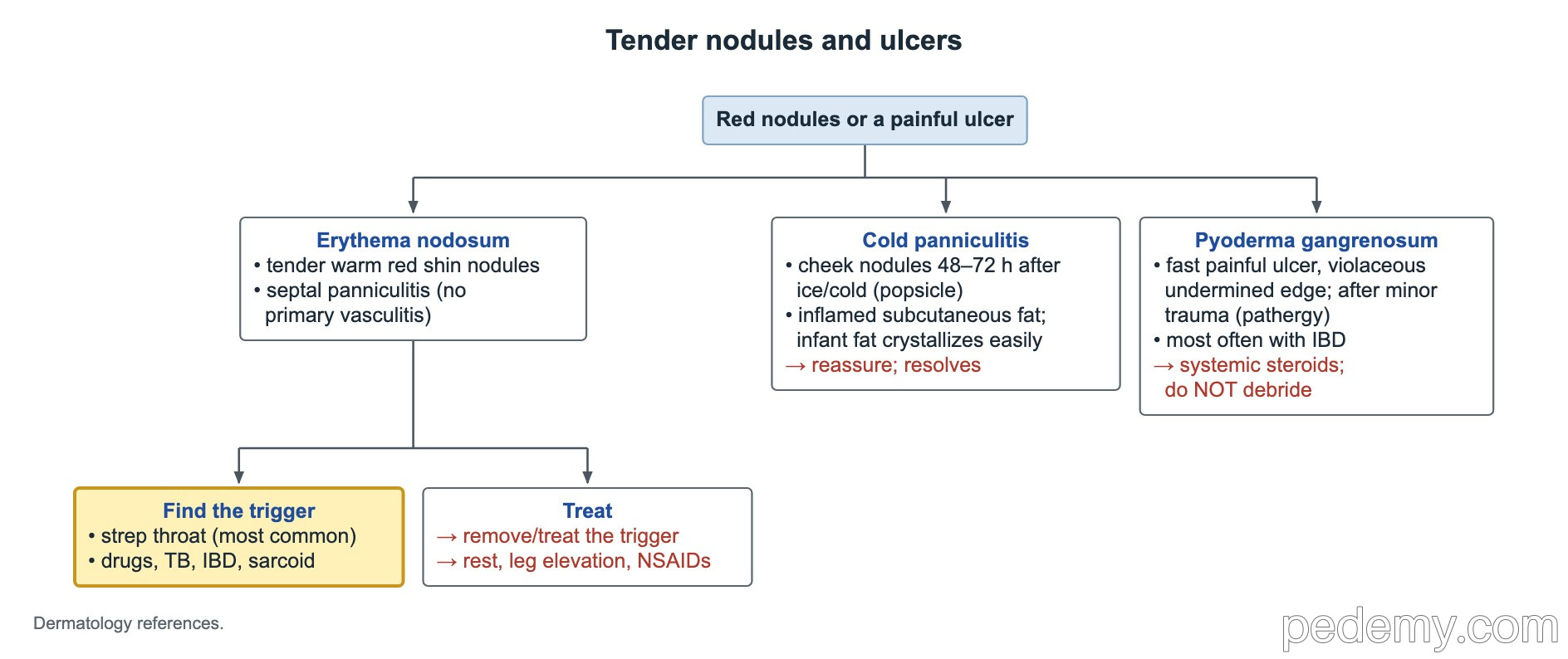
Erythema nodosum (tender red shin nodules) is a reaction pattern, so look for the trigger: strep throat most often in children, then drugs, TB, inflammatory bowel disease and sarcoidosis.
A teen with dry cough, uveitis, firm lymph nodes and erythema nodosum suggests sarcoidosis. Chest imaging for hilar nodes and a tissue biopsy confirm it. A high ACE level supports the diagnosis but is not specific enough to diagnose or exclude it.
Behçet disease
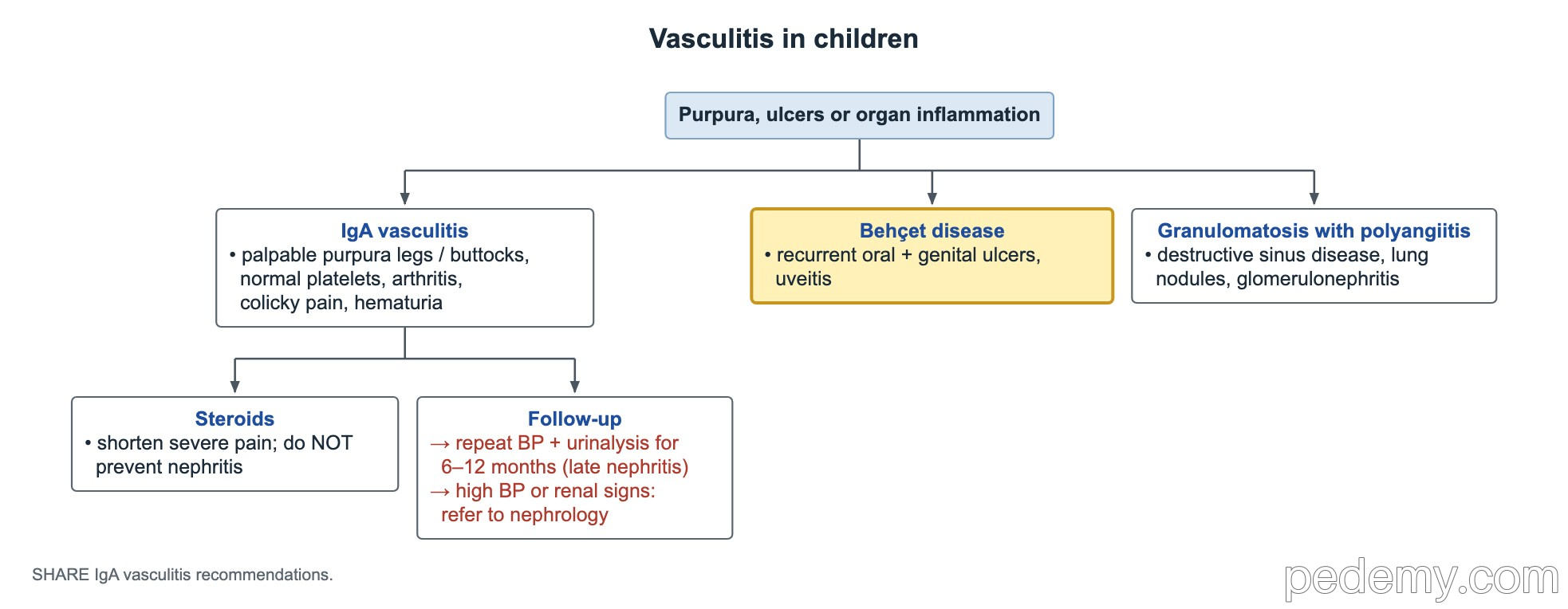
Behçet disease is a variable-vessel vasculitis most common in people with roots along the old Silk Road (Turkey, the Middle East, East Asia). Recurrent oral aphthous ulcers are the usual first sign.
The pediatric classification (PEDBD) needs 3 of 6 categories: recurrent oral ulcers, genital ulcers, skin lesions (erythema nodosum, folliculitis), eye disease (uveitis), neurologic signs and vascular disease (thrombosis). A pathergy reaction (a pustule at a needle stick) is a supportive clue.
Common questions
What conditions are linked to alopecia areata in children?
Autoimmune thyroid disease, vitiligo, atopic disease and Down syndrome. Many children are screened for thyroid disease when there is a suggestive history.
How long after an illness does telogen effluvium start?
Shedding starts about 2–3 months after the trigger, such as a high fever or surgery, and hair regrows within 6–12 months without treatment.
How do you tell trichotillomania from alopecia areata?
Trichotillomania leaves irregular patches with broken hairs of different lengths. Alopecia areata gives smooth, round, completely bald patches with exclamation-mark hairs at the edge.
Study this topic
Sources
- ATS 2020: Diagnosis and detection of sarcoidosis (Am J Respir Crit Care Med)
- PEDBD 2016: Classification criteria for pediatric Behçet disease (Ann Rheum Dis)
- Langerhans cell histiocytosis: evaluation and treatment guidelines (Pediatr Blood Cancer 2013)
- Alopecia areata (Nat Rev Dis Primers 2017)
Written for Pedemy in our own words from the sources above; checked November 2026. For education only, not medical advice; check doses and local protocols before treating a patient. Pedemy is independent and not affiliated with or endorsed by the organizations cited.