

Main decision tree
Jaundice in an infant: conjugated or not?
Key points
- Jaundice past 2 weeks, pale stools or dark urine: check total and direct (conjugated) bilirubin now.
- Biliary atresia: confirm with intraoperative cholangiogram and liver biopsy; Kasai portoenterostomy works best before 45–60 days.
- Choledochal cyst: ultrasound, MRCP confirms; complete excision with Roux-en-Y hepaticojejunostomy because of cancer risk.
- Wilson disease: low ceruloplasmin, high 24-hour urine copper, Kayser–Fleischer rings; dysarthria is the commonest neurologic sign.
- MASLD screening: ALT from age 10 in children with BMI ≥95th percentile, or ≥85th with risk factors.
- Acute pancreatitis: 2 of 3 of typical pain, lipase or amylase ≥3× normal, and imaging findings; feed early.
- Gallstones in teens: obesity and rapid weight loss; pigment stones with chronic hemolysis.
Jaundice in an infant: conjugated or not?
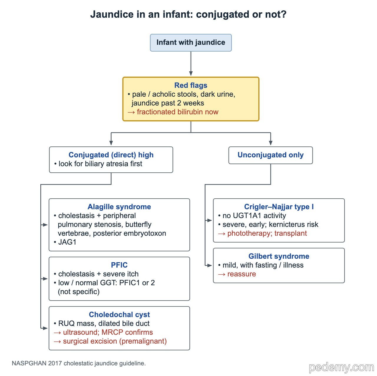
Any infant still jaundiced after 2 weeks of age, or with pale (acholic) stools or dark urine, needs a fractionated bilirubin right away. A raised direct (conjugated) bilirubin, above 1.0 mg/dL, is always abnormal and needs prompt workup for cholestasis, starting with biliary atresia.
| Pattern | Condition | Clues |
|---|---|---|
| Conjugated | Biliary atresia | Pale stools, dark urine, hepatomegaly by 2–8 weeks |
| Conjugated | Alagille syndrome (JAG1) | Peripheral pulmonary stenosis, butterfly vertebrae, posterior embryotoxon |
| Conjugated | PFIC | Severe itch; low or normal GGT suggests PFIC1 or 2 but is not specific |
| Conjugated | Choledochal cyst | Dilated bile duct, sometimes a right upper quadrant mass |
| Unconjugated | Crigler–Najjar type I | Absent UGT1A1 activity; severe early jaundice, kernicterus risk; phototherapy, liver transplant |
| Unconjugated | Gilbert syndrome | Mild jaundice with fasting or illness; normal enzymes, no hemolysis; reassure |
Biliary atresia
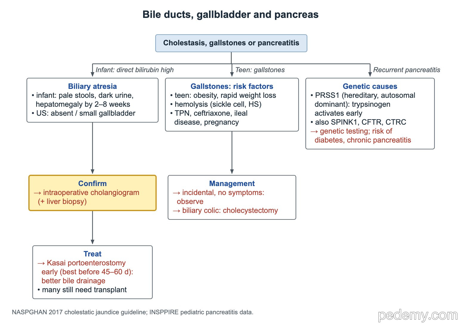
- Typical infant: well at first, then pale stools, dark urine and hepatomegaly in the first 2–8 weeks.
- Ultrasound may show an absent or small gallbladder, but a normal scan does not exclude it.
- Definitive test: intraoperative cholangiogram with liver biopsy.
- Treatment: Kasai portoenterostomy, best before 45–60 days of life, because earlier surgery gives a better chance of bile drainage.
- Many children still need a liver transplant later; biliary atresia is the leading reason for pediatric liver transplant.
Choledochal cyst
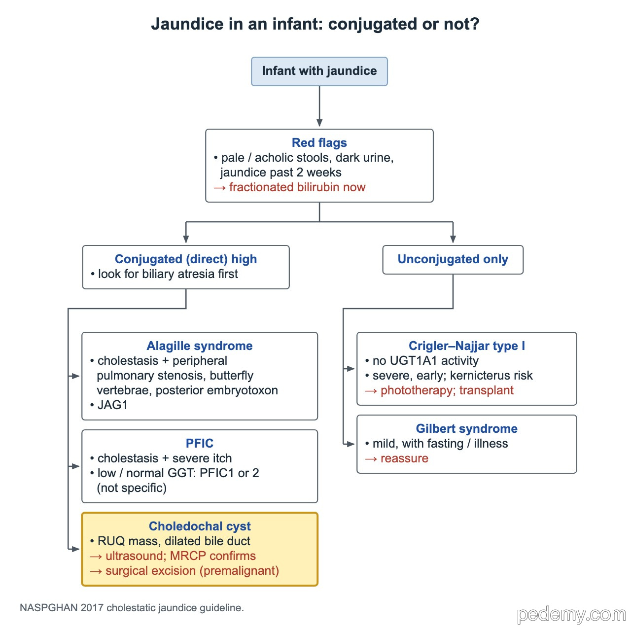
A choledochal cyst is a cystic dilation of the bile ducts. The classic triad is jaundice, abdominal pain and a right upper quadrant mass, but infants often present with cholestasis alone or an incidental finding.
- Type I (extrahepatic dilation) is the most common; Caroli disease (type V) is multiple intrahepatic duct dilations only.
- Ultrasound first; MRCP confirms the anatomy before surgery.
- A cholestatic infant with a cystic duct and patent intrahepatic ducts on cholangiogram has a choledochal cyst, not biliary atresia.
- Treatment: complete cyst excision with Roux-en-Y hepaticojejunostomy. Refer promptly even when found by chance; asymptomatic cysts are usually removed after about 6 months of age, symptomatic ones at any age.
- Long-term follow-up for cholangiocarcinoma, a risk that persists after excision.
Jaundice or high liver enzymes in an older child
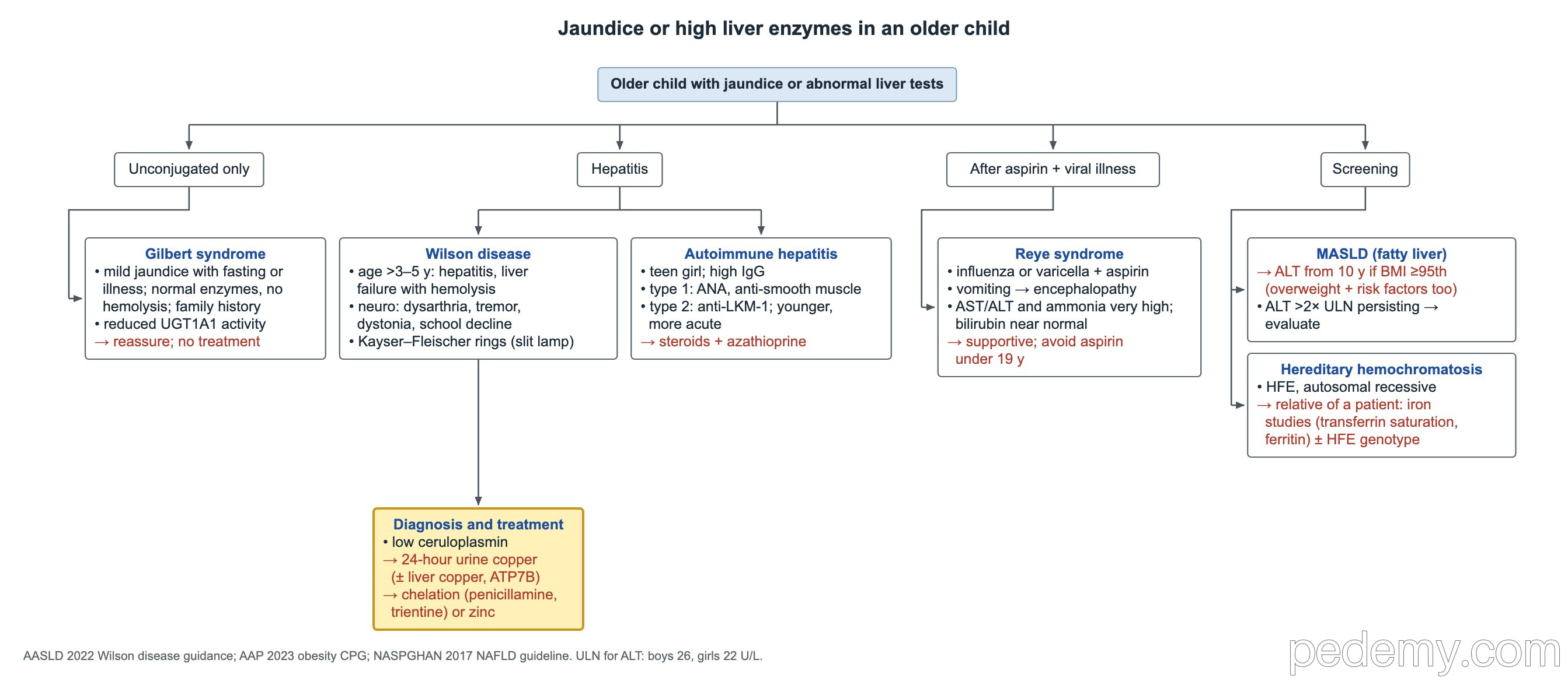
- Wilson disease is copper overload from ATP7B mutations: copper is not excreted into bile and builds up in liver, brain and eye.
- Kayser–Fleischer rings (golden-brown corneal copper ring on slit lamp) may be absent when disease is only in the liver.
- Wilson workup: low ceruloplasmin, then 24-hour urine copper (± liver copper, ATP7B testing). Treatment: chelation with penicillamine or trientine, or zinc.
- MASLD (formerly NAFLD): screen with ALT from age 10 if BMI ≥95th percentile, or ≥85th percentile with risk factors.
| Condition | Clues | Next step |
|---|---|---|
| Gilbert syndrome | Mild unconjugated jaundice with fasting or illness; normal enzymes; family history; reduced UGT1A1 | Reassure; no treatment |
| Wilson disease | Age over 3–5 years; hepatitis, liver failure with hemolysis; dysarthria, tremor, dystonia, school decline | Ceruloplasmin, 24-hour urine copper, slit lamp |
| Reye syndrome | Aspirin during influenza or varicella; vomiting then encephalopathy; very high AST/ALT and ammonia, near-normal bilirubin | Supportive care; no aspirin under 19 years |
| MASLD (fatty liver) | Obesity, acanthosis nigricans | ALT screening; ALT persistently >2× normal → evaluate |
| HFE hemochromatosis | Autosomal recessive; a parent or sibling is affected | Transferrin saturation and ferritin, ± HFE genotype |
| Acetaminophen toxicity | Acute liver failure, high acetaminophen level | The top known cause of acute liver failure in teens |
Autoimmune hepatitis
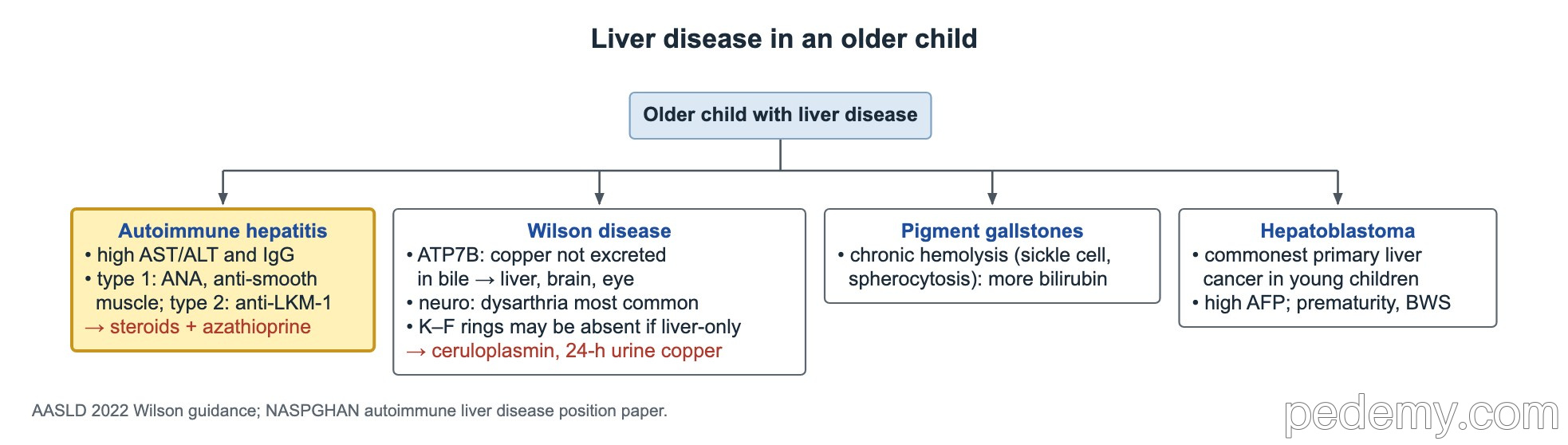
- High AST/ALT and IgG; often a teen girl with negative viral tests.
- Type 1: ANA and/or anti-smooth muscle antibody. Type 2: anti-LKM-1, younger children, more acute.
- Treatment: corticosteroids plus azathioprine.
- Hepatoblastoma is the commonest primary liver cancer in young children: high AFP; linked to prematurity and Beckwith–Wiedemann syndrome.
Gallstones
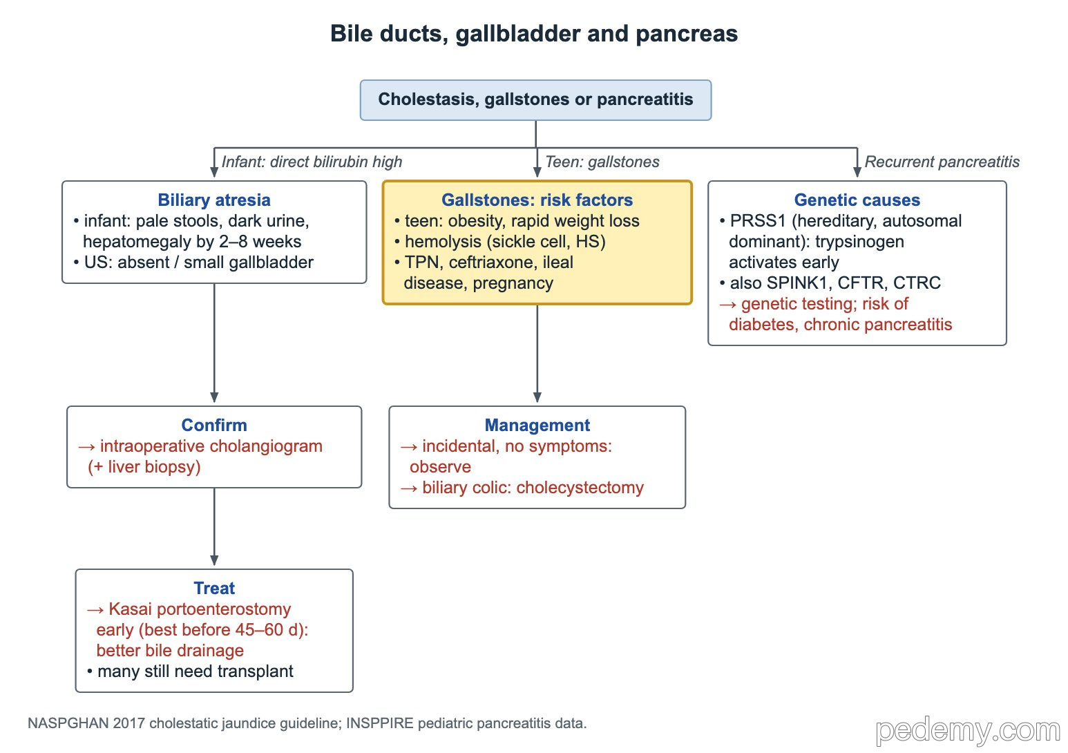
- Teen without hemolysis: obesity and rapid weight loss are the main risk factors.
- Chronic hemolysis (sickle cell disease, hereditary spherocytosis) raises bilirubin turnover and causes pigment stones.
- Other risks: TPN, ceftriaxone, ileal disease, pregnancy.
- Incidental stone, no symptoms: observe. Biliary colic or other symptoms: laparoscopic cholecystectomy.
- A stone in the common bile duct can cause acute (gallstone) pancreatitis.
Acute pancreatitis
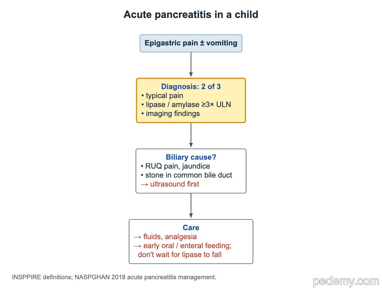
Diagnose acute pancreatitis when at least 2 of 3 are present: compatible abdominal pain, serum lipase or amylase at least 3 times the upper limit of normal, and imaging findings of pancreatitis.
- Epigastric or right upper quadrant pain with jaundice and a high lipase: abdominal ultrasound first to look for gallstones.
- Care: crystalloid fluids, especially in the first 24 hours, and analgesia.
- Start oral or enteral feeding early as tolerated; do not keep the child NPO until the lipase falls.
- Recurrent pancreatitis with relatives who had pancreatitis and later diabetes: hereditary pancreatitis from PRSS1 (cationic trypsinogen, autosomal dominant). Also test SPINK1, CFTR and CTRC.
Common questions
When is infant jaundice a concern for biliary atresia?
When it lasts beyond 2 weeks, or at any age with pale stools or dark urine. Check a direct bilirubin; a level above 1.0 mg/dL needs prompt evaluation.
Why must the Kasai operation be done early?
The earlier it is done, ideally before 45–60 days of life, the better the chance that bile drains and the native liver survives.
How is pediatric acute pancreatitis diagnosed?
With at least 2 of 3: typical abdominal pain, lipase or amylase at least 3 times normal, and imaging findings.
Study this topic
Sources
- NASPGHAN/ESPGHAN 2017: Evaluation of cholestatic jaundice in infants (JPGN)
- AASLD 2022: Practice guidance on Wilson disease (Hepatology)
- NASPGHAN 2018: Management of acute pancreatitis in children (JPGN)
- NASPGHAN 2017: Diagnosis and treatment of NAFLD in children (JPGN)
- ESPGHAN 2018: Pediatric autoimmune liver disease position statement (JPGN)
Written for Pedemy in our own words from the sources above; checked November 2026. For education only, not medical advice; check doses and local protocols before treating a patient. Pedemy is independent and not affiliated with or endorsed by the organizations cited.