

Main decision tree
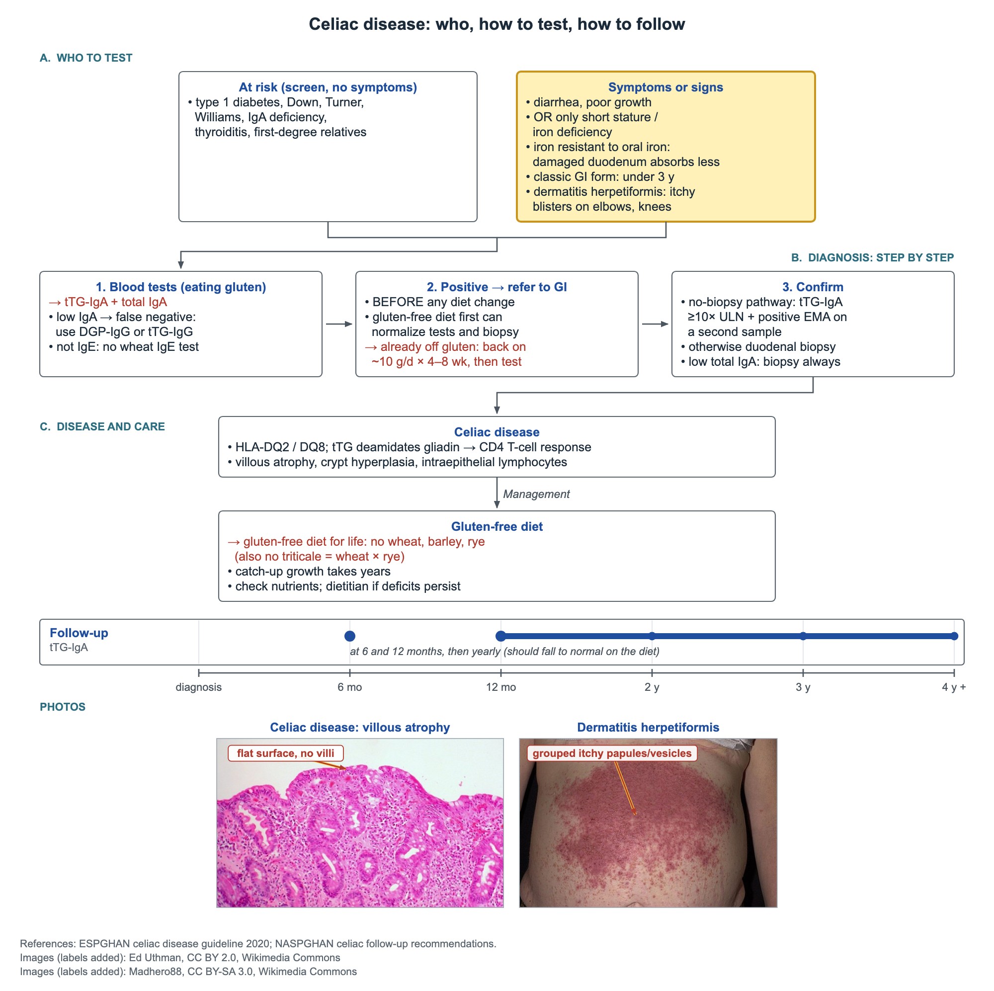
Celiac disease: who, how to test, how to follow
Key points
- Celiac first test (child eating gluten): tTG-IgA plus total serum IgA; low total IgA → DGP-IgG or tTG-IgG.
- No-biopsy celiac diagnosis: tTG-IgA ≥10× the upper limit of normal plus positive EMA-IgA on a second blood sample.
- Low total IgA or tTG-IgA below 10× ULN → duodenal biopsies while still eating gluten.
- After diagnosis, recheck tTG-IgA at 6 and 12 months, then yearly; it should normalize within 12–24 months.
- Crohn disease: mouth to anus, skip lesions, transmural; ulcerative colitis: rectum upward, continuous, mucosal.
- Stool osmotic gap = 290 − 2 × (stool Na + K): >100 osmotic, <50 secretory.
- CF enzymes: ≤2,500 lipase units/kg/meal and ≤10,000 units/kg/day to avoid fibrosing colonopathy.
Celiac disease: who to test
Celiac disease is an immune reaction to gluten in people who carry HLA-DQ2 or HLA-DQ8. Tissue transglutaminase (tTG) deamidates gliadin, which then binds DQ2 or DQ8 and activates CD4 T cells. The result is villous atrophy, crypt hyperplasia and more intraepithelial lymphocytes.
The classic picture of diarrhea, a distended belly and poor weight gain is most common under 3 years. Older children often have only one clue.
- Short stature or delayed puberty with no other cause.
- Iron deficiency, especially when it does not respond to oral iron: the damaged duodenum absorbs iron poorly.
- Dermatitis herpetiformis: very itchy grouped vesicles on the elbows and knees, with granular IgA in the dermal papillae on biopsy. Treat with a gluten-free diet, with dapsone for fast itch control.
- Higher-risk groups to test: type 1 diabetes, autoimmune thyroiditis, Turner and Williams syndromes, IgA deficiency and first-degree relatives.
- Down syndrome also carries a high risk; the AAP Down syndrome guidance asks about celiac symptoms at each visit and tests when they appear.
Celiac disease: testing and confirming the diagnosis
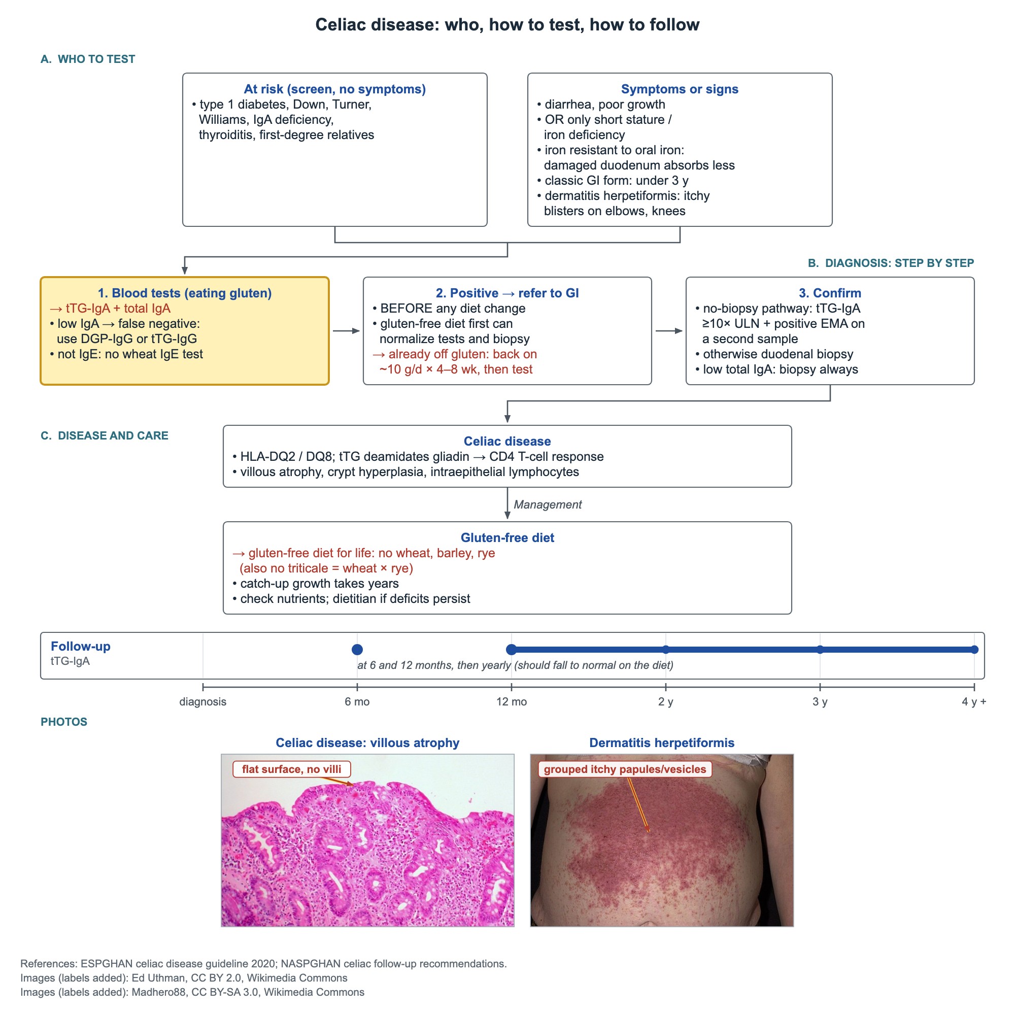
Test while the child is still eating gluten. A gluten-free diet started before testing heals the gut and lowers the antibodies, so both blood tests and biopsy can turn falsely normal.
- First tests: tTG-IgA plus total serum IgA.
- Why total IgA: IgA deficiency gives a false-negative tTG-IgA. If total IgA is low, use IgG tests (DGP-IgG or tTG-IgG).
- Do not order wheat-specific IgE: celiac disease is not IgE-mediated.
- Positive test → refer to pediatric gastroenterology before any diet change.
- Already off gluten: restart about 10 g of gluten a day (about 2 slices of bread) for 4–8 weeks, then test.
- No-biopsy pathway (specialist decision, family agrees): tTG-IgA ≥10× ULN plus a positive EMA-IgA on a second sample.
- All others, including every child with low total IgA and a positive IgG test, need upper endoscopy with duodenal biopsies on a gluten diet.
Gluten-free diet and follow-up
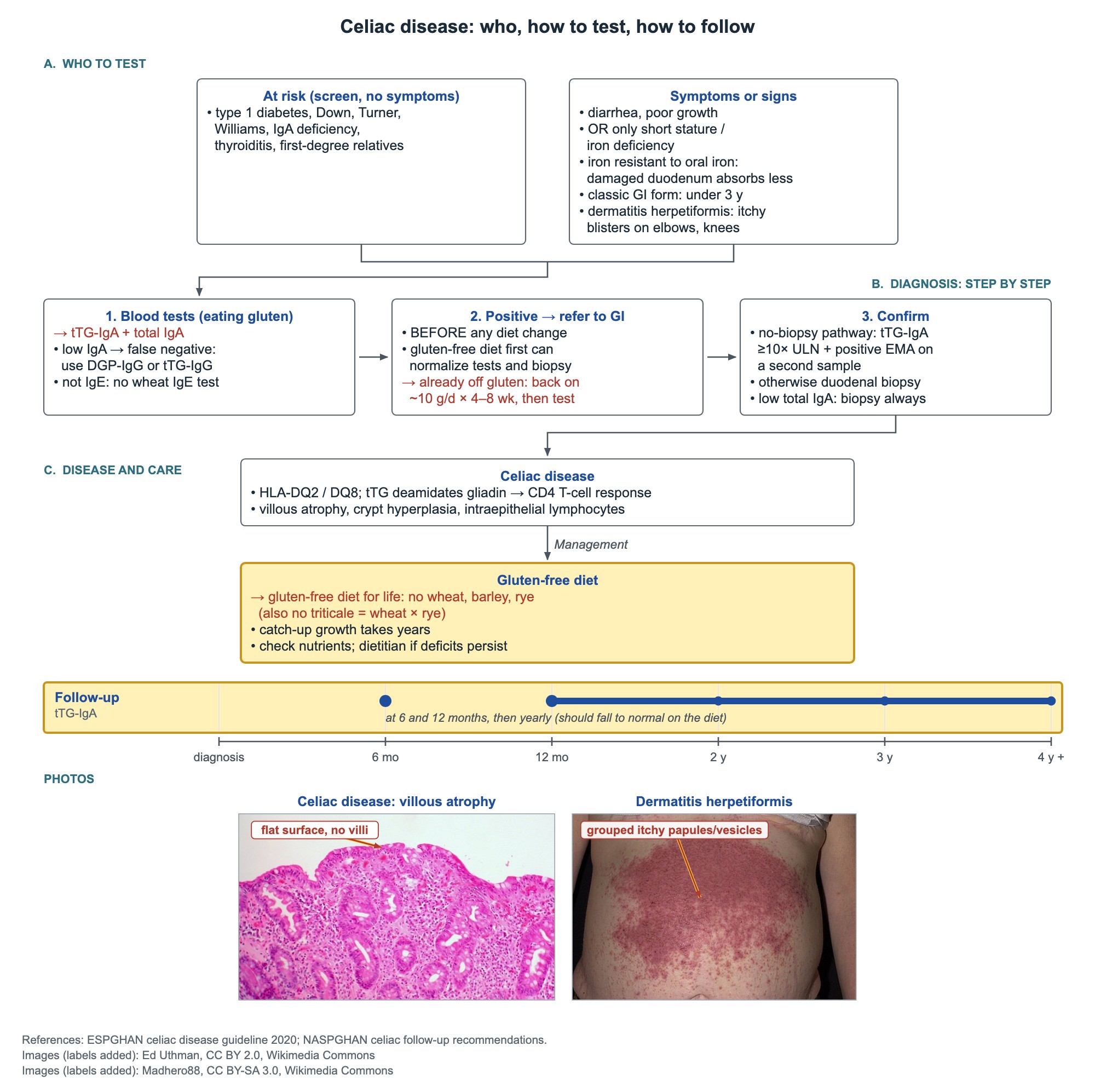
- Treatment is a strict gluten-free diet for life: no wheat, barley or rye.
- Triticale is not allowed: it is a wheat–rye hybrid and contains gluten.
- Catch-up growth is gradual and takes several years.
- Check tTG-IgA at 6 and 12 months, then yearly. It should fall to normal within 12–24 months on the diet.
- Persistent micronutrient deficits on a strict diet → reassess with gastroenterology and a dietitian.
Crohn disease vs ulcerative colitis
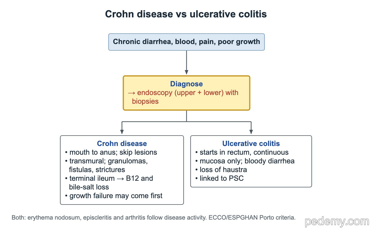
Think of IBD in a child with chronic diarrhea, blood in the stool, abdominal pain or poor growth. In Crohn disease, slow growth and delayed puberty can come before gut symptoms. A teen with poor growth, a right lower quadrant mass and heme-positive stool fits Crohn disease.
Diagnose with upper endoscopy plus ileocolonoscopy with biopsies; small-bowel imaging (MR enterography or capsule) completes the work-up unless the picture is typical ulcerative colitis.
| Feature | Crohn disease | Ulcerative colitis |
|---|---|---|
| Extent | Anywhere, mouth to anus; skip lesions | Starts in the rectum, continuous up the colon |
| Depth | Transmural; granulomas, fistulas, strictures | Mucosal; bloody diarrhea |
| Other clues | Terminal ileum disease → B12 and bile-salt loss | Loss of haustra; linked to PSC |
IBD outside the gut
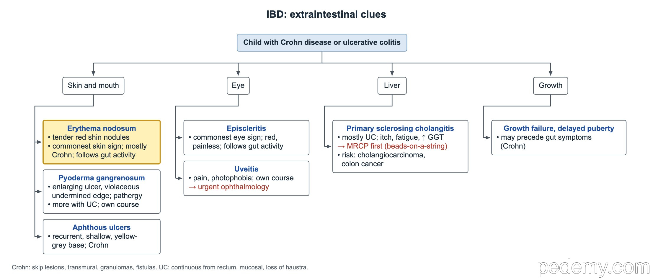
- Erythema nodosum: tender red shin nodules; the commonest skin sign, mostly Crohn disease; follows gut activity.
- Pyoderma gangrenosum: an enlarging painful ulcer with a violaceous, undermined edge; more often ulcerative colitis; runs its own course. Ask about bloody diarrhea, cramping and tenesmus.
- Aphthous mouth ulcers: recurrent, shallow, yellow-grey base with a red halo; Crohn disease.
- Episcleritis: red, painless eye; the commonest eye sign; follows gut activity.
- Uveitis: eye pain and photophobia; threatens vision and runs its own course → urgent ophthalmology.
- Primary sclerosing cholangitis: mostly ulcerative colitis; itch, fatigue and a raised GGT → MRCP first. It raises the risk of cholangiocarcinoma and colon cancer.
Chronic diarrhea, lactose and short bowel
Chronic diarrhea in children means diarrhea lasting more than 2 weeks (stool output above 10 mL/kg/day). Alarm features that need a work-up: stools at night, blood in the stool, weight loss or growth faltering, and unexplained fever.
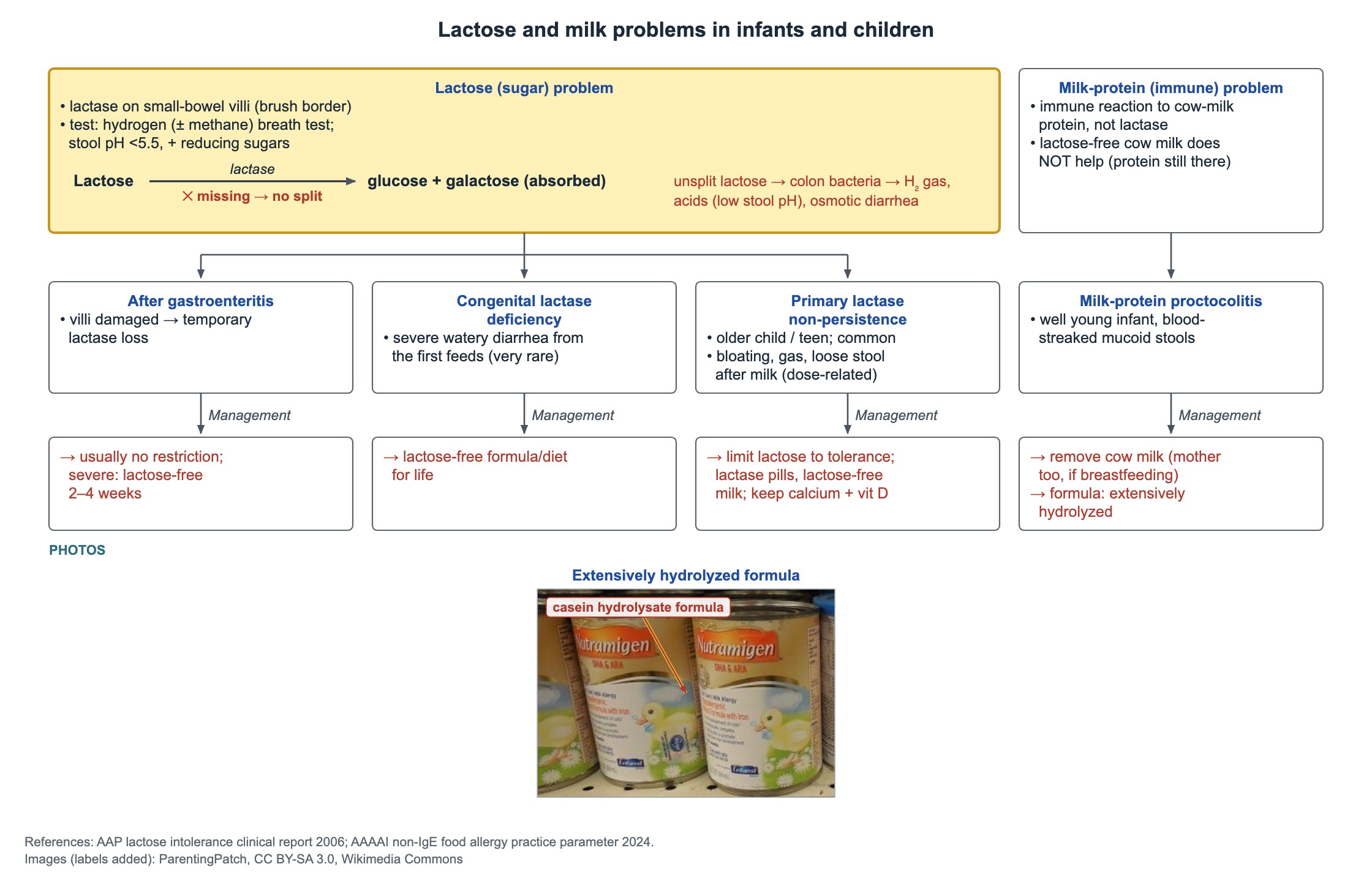
Lactase sits on the brush border of the small-bowel villi. Unsplit lactose reaches the colon, where bacteria ferment it into gas and acids: the stool turns acidic and the diarrhea is osmotic.
- Osmotic gap = 290 − 2 × (stool Na + stool K).
- Carbohydrate malabsorption: stool pH <5.5 and positive reducing substances; a hydrogen breath test (often with methane) after a lactose load supports lactose malabsorption.
- Primary lactase non-persistence: common in people of Asian, African or Native American ancestry, usually after age 5. Bloating and diarrhea after milk or ice cream, but yogurt and hard cheese are tolerated. Limit lactose to tolerance, use lactase pills or lactose-free milk, and keep calcium and vitamin D intake up.
- After gastroenteritis: lactase loss is temporary. Most children need no restriction; if intolerance is significant, use lactose-free feeds for about 2–4 weeks. Lifelong avoidance is not needed.
- Congenital lactase deficiency (very rare): severe watery diarrhea from the first milk feeds → lactose-free diet for life.
- Congenital chloride diarrhea: watery diarrhea from birth, hypochloremic metabolic alkalosis and stool chloride >90 mEq/L.
- Short bowel syndrome: after jejunal loss the ileum adapts well. After ileal loss, B12 and bile salts are lost, so fat and fat-soluble vitamins are malabsorbed; loss of the ileocecal valve adds small-bowel bacterial overgrowth.
| Type | Fasting | Stool osmotic gap | Examples |
|---|---|---|---|
| Osmotic | Stops | >100 mOsm/kg | Lactose or other carbohydrate malabsorption |
| Secretory | Continues | <50 mOsm/kg | Congenital chloride diarrhea, microvillus inclusion disease |
Cystic fibrosis: the gut
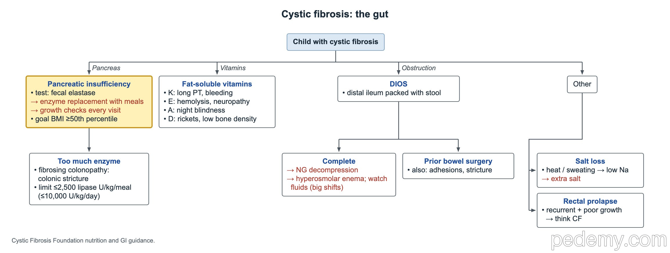
- Test every new diagnosis for pancreatic insufficiency with fecal elastase (below 200 µg/g suggests insufficiency).
- Treat with pancreatic enzymes with meals and snacks; keep lipase ≤2,500 units/kg/meal and ≤10,000 units/kg/day.
- Long-term high enzyme doses can cause fibrosing colonopathy, a colonic stricture that mimics obstruction.
- Check growth at every visit; aim for BMI at or above the 50th percentile from age 2 to 20 years.
- Fat-soluble vitamins: K (long PT, bruising), E (hemolysis, ataxia or neuropathy), A (night blindness), D (rickets, low bone density).
- Salt loss: heavy sweating in hot weather causes hyponatremia from high sweat sodium chloride; give extra salt.
- Distal intestinal obstruction syndrome (DIOS): stool packs the distal ileum. For complete obstruction, decompress with an NG sump on low intermittent suction and give a hyperosmolar enema; watch fluids closely because large fluid shifts cause hypovolemia and electrolyte problems.
- After previous bowel surgery, also think of adhesions or an anastomotic stricture.
Common questions
What is the best blood test for celiac disease in children?
tTG-IgA together with total serum IgA, done while the child is eating gluten. If total IgA is low, an IgG-based test (DGP-IgG or tTG-IgG) is used instead.
Can celiac disease be diagnosed without a biopsy?
In selected children, yes: a tTG-IgA of 10 times the upper limit of normal or more, plus a positive EMA-IgA on a second blood sample, decided with a pediatric gastroenterologist. Children with low total IgA always need biopsies.
How long should a child eat gluten before celiac testing?
A child who has already cut out gluten should eat about 10 g a day, roughly 2 slices of bread, for several weeks (about 4–8) before testing.
Does lactose intolerance after a stomach bug last forever?
No. Lactase loss after gastroenteritis is temporary. Most children need no change; a few need lactose-free feeds for about 2–4 weeks.
Study this topic
Sources
- ESPGHAN 2020: Guidelines for diagnosing coeliac disease (JPGN)
- NASPGHAN 2016: Clinical report on the diagnosis and treatment of gluten-related disorders (JPGN)
- ESPGHAN revised Porto criteria for the diagnosis of pediatric IBD (JPGN 2014)
- CF Foundation: Nutrition and pancreatic enzyme recommendations (J Am Diet Assoc 2008)
Written for Pedemy in our own words from the sources above; checked November 2026. For education only, not medical advice; check doses and local protocols before treating a patient. Pedemy is independent and not affiliated with or endorsed by the organizations cited.