

Main decision tree
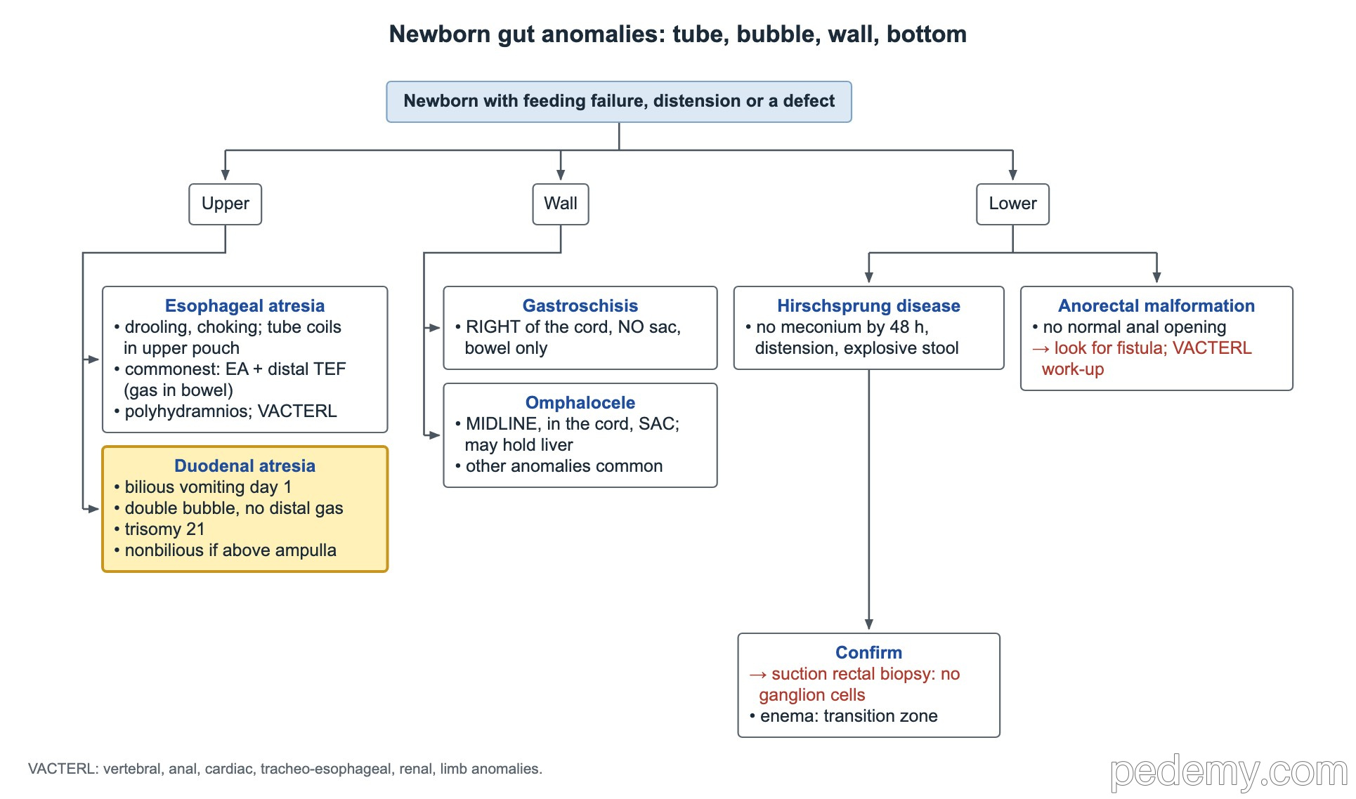
Newborn gut anomalies: tube, bubble, wall, bottom
Key points
- Bilious vomiting in an infant is malrotation with volvulus until proven otherwise: urgent upper GI series, or straight to surgery if in shock.
- Duodenal atresia: double bubble with no distal gas; linked to trisomy 21.
- Gastroschisis: right of the cord, no sac, rarely chromosomal; omphalocele: midline sac, often with other anomalies.
- Pyloric stenosis: 3–8 weeks, projectile nonbilious vomiting, hypochloremic hypokalemic metabolic alkalosis; fix fluids and alkalosis before pyloromyotomy.
- Intussusception: ultrasound first (target sign); enema reduction if stable without peritonitis, perforation or shock.
- Appendicitis imaging: ultrasound first, MRI if unclear, CT last; risk scores perform poorly under 5 years.
- No meconium by 48 hours → think of Hirschsprung disease or meconium ileus.
Newborn obstruction: atresias, Hirschsprung and anorectal malformations
- Esophageal atresia: drooling and choking with the first feed; the tube stops at about 10–12 cm or coils in the upper pouch. The fetus could not swallow, so expect polyhydramnios. The commonest type is atresia with a distal tracheoesophageal fistula (gas in the bowel). Look for VACTERL anomalies.
- Duodenal atresia: vomiting on day 1 and a double bubble with little or no distal gas; associated with trisomy 21. Vomiting is usually bilious but can be nonbilious when the block is above the ampulla.
- Jejunoileal atresia: bilious vomiting; blind bowel ends from an in-utero vascular accident (maternal cocaine, vasoconstrictors or smoking).
- No meconium by 48 hours: worry about bowel obstruction such as Hirschsprung disease or meconium ileus.
- Hirschsprung disease: distension and explosive stool; a contrast enema shows a transition zone; suction rectal biopsy showing no ganglion cells confirms it.
- Anorectal malformation: no normal anal opening; look for a fistula and do a VACTERL work-up.
Abdominal wall defects and inguinal hernia
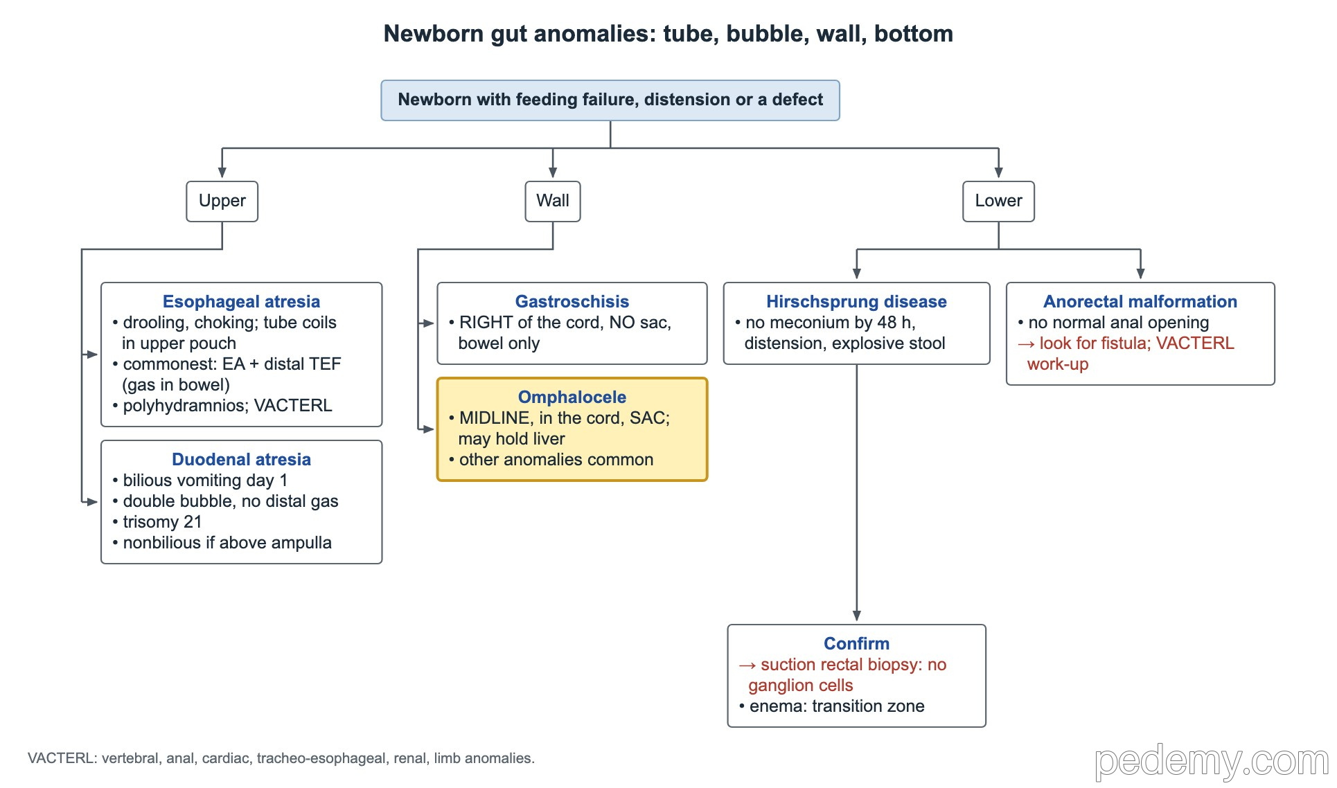
- Small omphaloceles that hold only bowel carry the highest aneuploidy risk.
- Gastroschisis first steps without a pediatric surgeon: put the bowel in a sterile bowel bag (or wrap it), lay the baby on the right side, then NG tube, IV fluids and warmth before transfer.
- Inguinal hernia in children is indirect, from a patent processus vaginalis. Risk rises the more preterm the baby.
- Reducible, symptom-free hernia → prompt referral for elective repair.
- Incarcerated hernia (hard, painful bulge that will not reduce, often with vomiting) → urgent pediatric surgery to prevent strangulation, ischemia and bowel necrosis.
| Feature | Omphalocele | Gastroschisis |
|---|---|---|
| Site | Midline, through the umbilical cord | Right of a normal cord insertion |
| Covering | Sac; may contain liver | No sac; bowel only |
| Associations | Heart defects, trisomy 13 and 18, Beckwith–Wiedemann | Chromosomal anomalies rare |
Pyloric stenosis
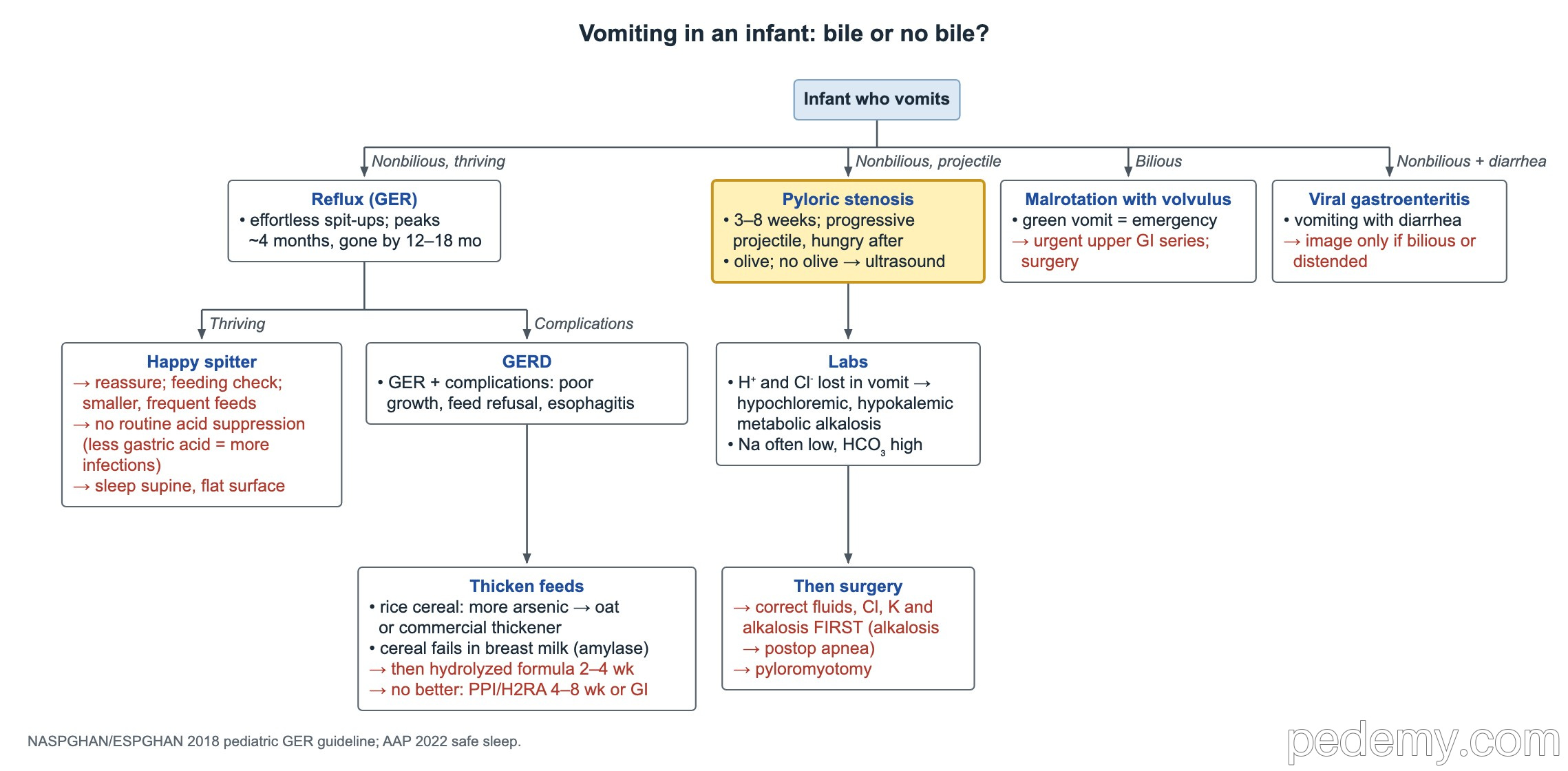
Hypertrophic pyloric stenosis causes progressive, projectile, nonbilious vomiting after feeds in an infant 3–8 weeks old who stays hungry. A firm epigastric olive may be felt; if not, pyloric ultrasound confirms it (muscle about 3 mm thick or more and 15 mm long or more).
- Risk factors: male sex, first-born, preterm birth, an affected parent (especially the mother), maternal smoking, and oral erythromycin or azithromycin in the first 2 weeks of life.
- Labs: vomiting loses gastric hydrogen and chloride → hypochloremic, hypokalemic metabolic alkalosis. Bicarbonate is high; sodium is normal or low.
- Correct dehydration, chloride, potassium and alkalosis first: persistent alkalosis raises the risk of postoperative apnea or hypoventilation.
- Then pyloromyotomy.
Bilious vomiting: malrotation and SMA syndrome
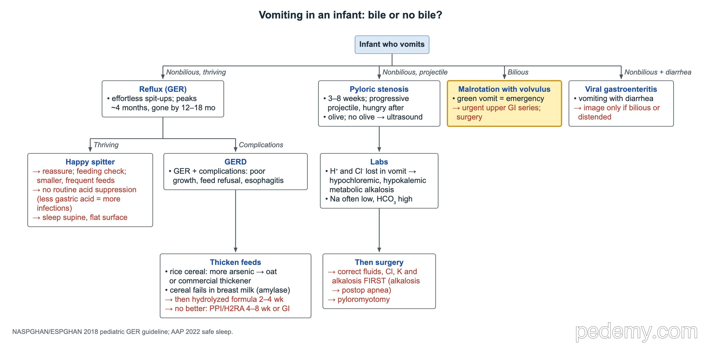
- Green vomit in a young infant is an emergency: exclude malrotation with midgut volvulus.
- Stable infant → urgent upper GI series.
- Shock or peritonitis → straight to laparotomy, without waiting for imaging.
- Superior mesenteric artery syndrome: bilious vomiting with a cut-off at the third part of the duodenum after rapid weight loss (anorexia) or after spinal fusion or body casting. Treat with nutritional rehabilitation (often nasojejunal feeds) and left-side or prone positioning.
Acute abdomen and intussusception
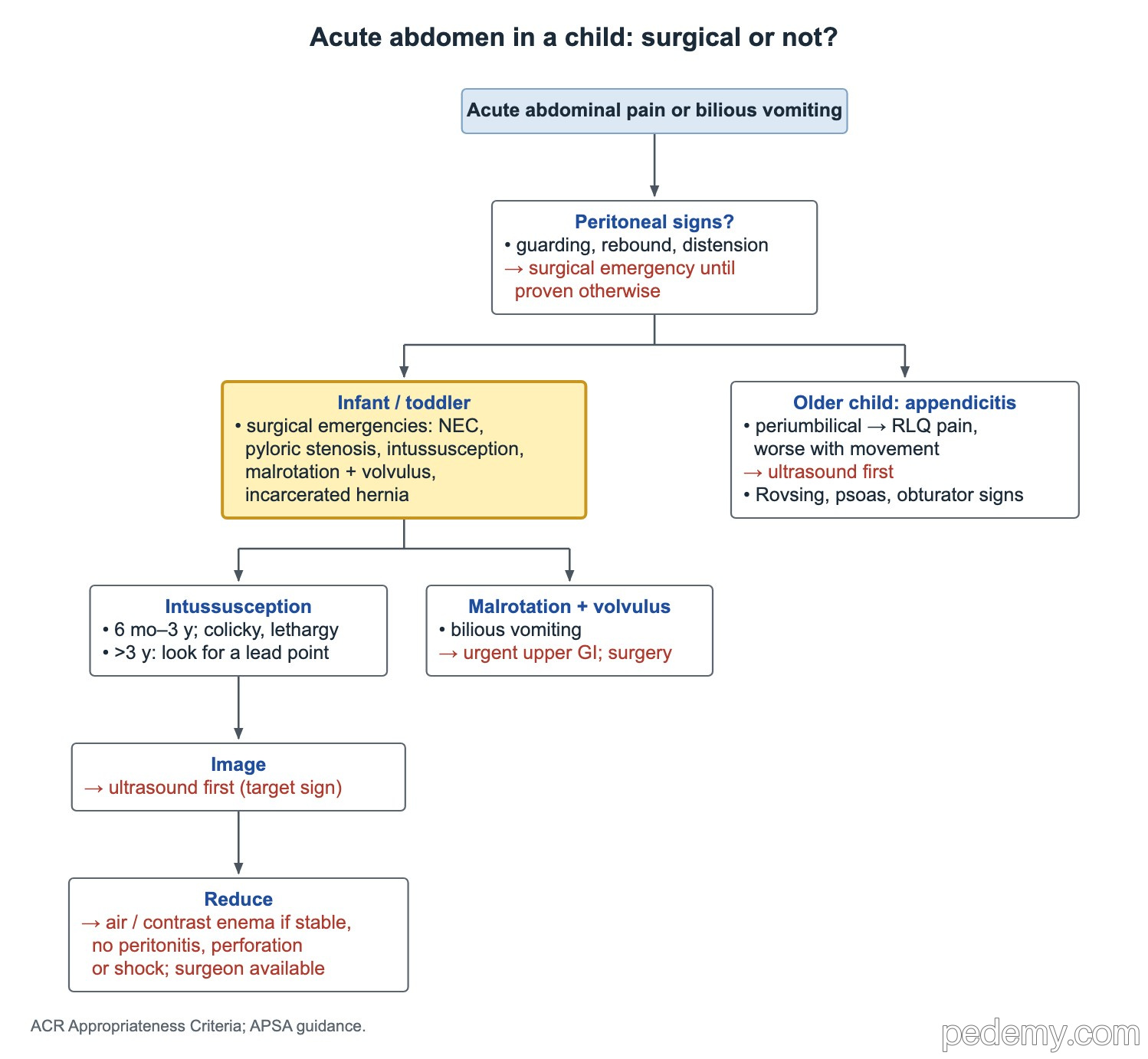
Guarding, rebound or distension mean a surgical emergency until proven otherwise. In infants and toddlers the main surgical causes are NEC, pyloric stenosis, intussusception, malrotation with volvulus and incarcerated hernia.
- Intussusception: most common from 6 months to 3 years; colicky pain with legs drawn up, lethargy, a sausage-shaped right upper quadrant mass and currant-jelly stool late.
- Ultrasound first; it shows a target sign.
- Air or contrast enema reduction if the child is stable with no peritonitis, perforation or shock, and a surgeon is available.
- Older than 3 years: look for a pathologic lead point (for example a Peutz–Jeghers polyp or a Meckel diverticulum).
Appendicitis
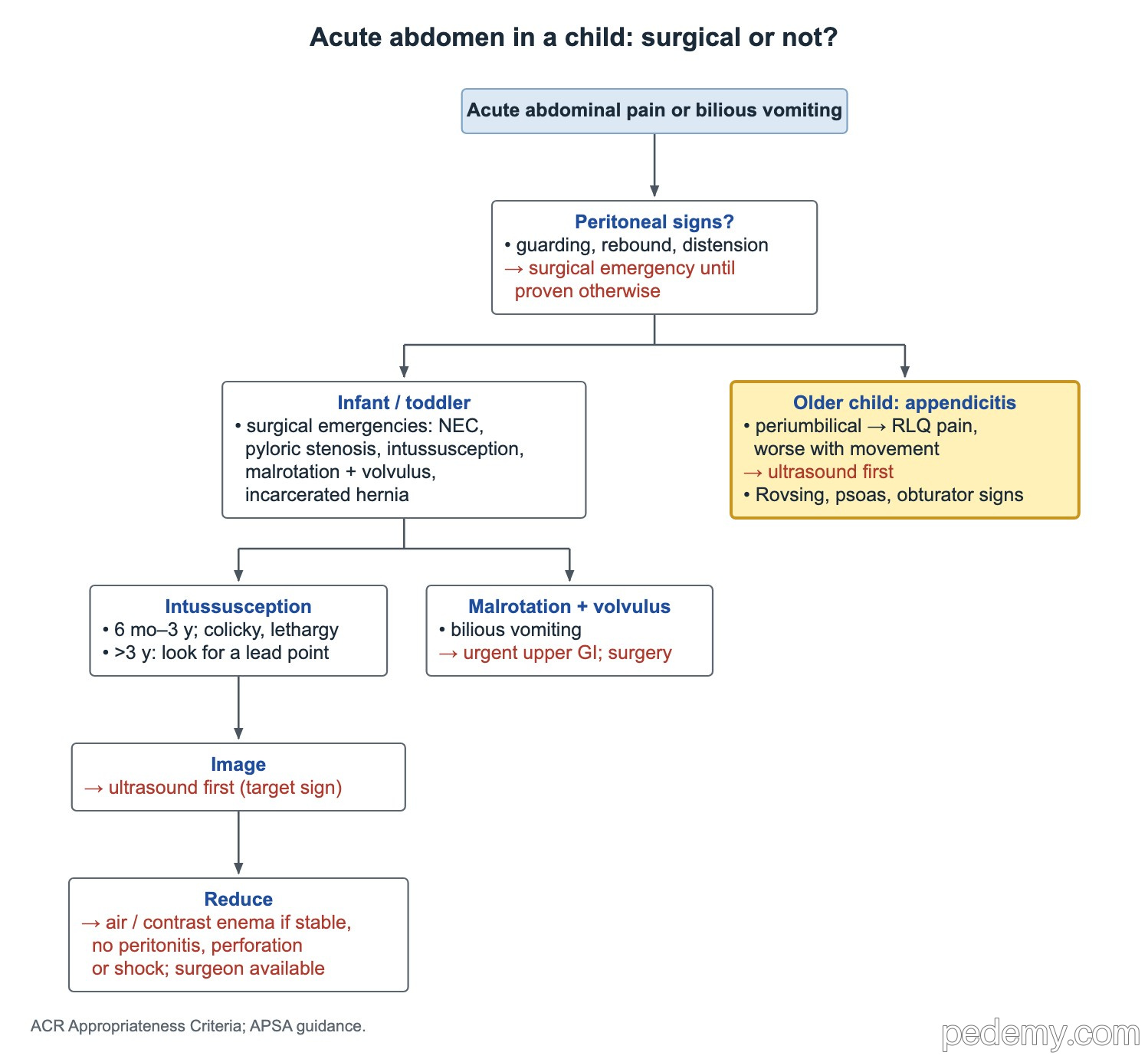
- Classic course: periumbilical pain that moves to the right lower quadrant and is worse with movement.
- Rovsing sign: right lower quadrant pain when the left lower quadrant is pressed. Psoas sign: pain with right hip extension.
- An inflamed appendix next to the bladder causes dysuria and sterile pyuria (no nitrites or bacteria); a pelvic appendix irritating the sigmoid causes frequent small loose stools.
- A boy with fever, dysuria and severe pain on moving is more likely to have appendicitis than a UTI.
- Raised WBC and CRP support risk scoring but do not confirm the diagnosis; imaging does.
- Imaging: ultrasound first, MRI if ultrasound is unclear, CT last.
- Risk scores (PAS, Alvarado, pARC, AIR) perform poorly in children under 5 years.
Painless rectal bleeding: Meckel diverticulum and polyps
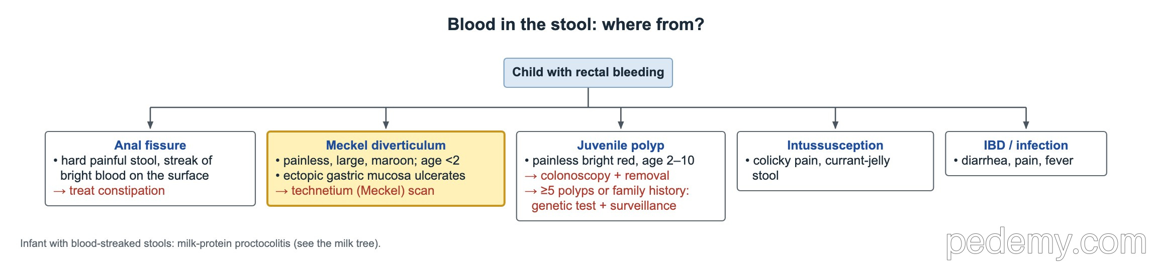
- Meckel diverticulum: large, painless, maroon bleeding with anemia, usually under 2 years; ectopic gastric mucosa ulcerates nearby bowel.
- Confirm with a technetium-99m pertechnetate (Meckel) scan. A negative scan does not rule it out.
- Juvenile polyp: painless bright red bleeding at age 2–10 → colonoscopy and removal. A single juvenile polyp with no family history needs no follow-up.
- 5 or more juvenile polyps or a family history → genetic testing and surveillance colonoscopy.
- Peutz–Jeghers syndrome: dark freckles on the lips and buccal mucosa; hamartomatous small-bowel polyps cause bleeding and recurrent intussusception. Start upper endoscopy, colonoscopy and small-bowel capsule around age 8.
- Gardner syndrome (familial adenomatous polyposis, APC): hundreds of colonic adenomas plus jaw osteomas, extra teeth and retinal pigment spots.
Common questions
What is the first test for suspected appendicitis in a child?
Abdominal ultrasound. MRI is used if ultrasound is unclear, and CT is the last choice because of radiation.
Why must alkalosis be corrected before pyloromyotomy?
Persistent metabolic alkalosis raises the risk of apnea and hypoventilation after anesthesia, so fluids, chloride and potassium are corrected first.
How do you tell gastroschisis from omphalocele?
Gastroschisis lies to the right of a normal cord with no sac and rarely has chromosomal problems. Omphalocele is midline through the cord, covered by a sac, and often comes with heart defects or trisomy 13 or 18.
Study this topic
Sources
- AAP 2012: Assessment and management of inguinal hernia in infants (Pediatrics)
- ESPGHAN 2019: Management of Peutz–Jeghers syndrome in children (JPGN)
- ESPGHAN 2019: Management of familial adenomatous polyposis in children (JPGN)
Written for Pedemy in our own words from the sources above; checked November 2026. For education only, not medical advice; check doses and local protocols before treating a patient. Pedemy is independent and not affiliated with or endorsed by the organizations cited.