

Main decision tree
Brain and spinal cord tumors
Key points
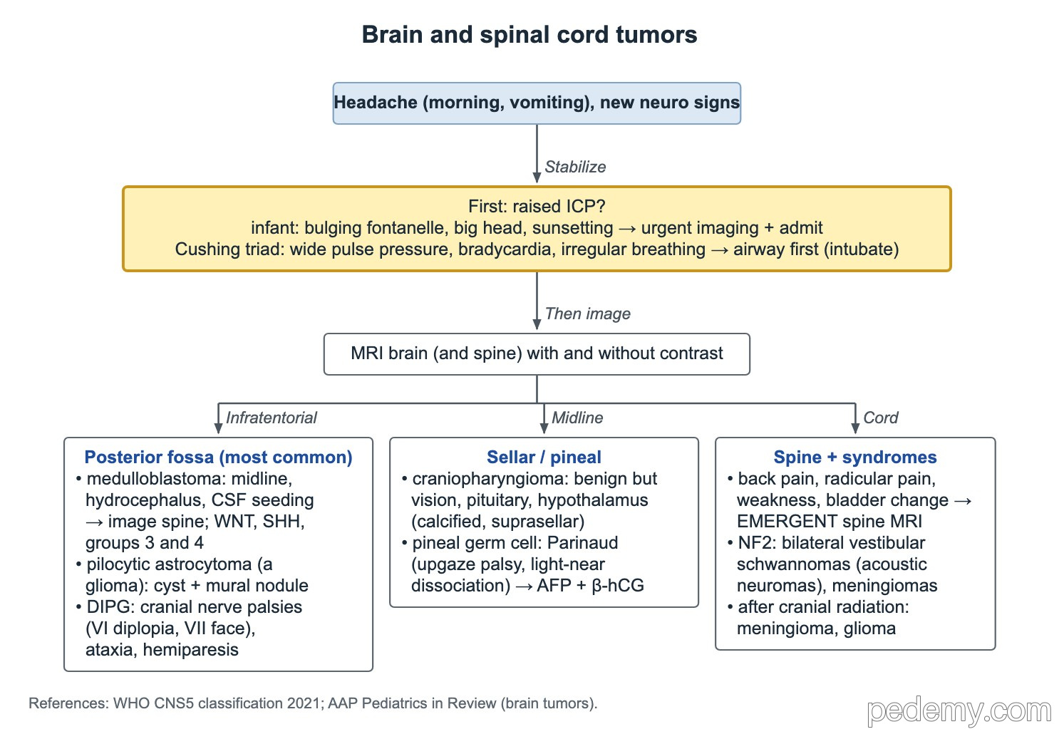
- Cushing triad = hypertension with wide pulse pressure, bradycardia and irregular breathing: secure the airway (intubate) first.
- Infant with bulging fontanelle, vomiting and sunsetting eyes: urgent head imaging and admission.
- Stable child with a possible brain tumor: MRI brain with and without gadolinium; add the whole spine for staging.
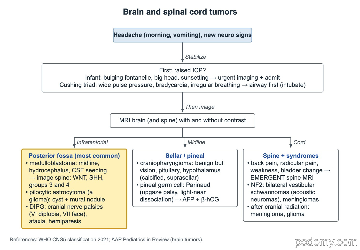
- Posterior fossa mass + obstructive hydrocephalus → medulloblastoma is the likely malignant tumor; image the spine for drop metastases.
- Cerebellar cyst with an enhancing mural nodule → pilocytic astrocytoma, a low-grade glioma.
- CN VI and VII palsies with ataxia and a diffuse non-enhancing pontine lesion → diffuse midline (intrinsic pontine) glioma.
- Back pain with new weakness: emergent MRI of the spine.
Raised intracranial pressure comes first
Brain tumors often present with morning headache, vomiting and new neurologic signs. A posterior fossa mass blocks CSF outflow at the fourth ventricle and causes obstructive hydrocephalus.
The Cushing triad (hypertension with a wide pulse pressure, bradycardia and irregular breathing) means impending herniation. Secure the airway with endotracheal intubation first, then treat the pressure and get neurosurgery involved.
- Infant: bulging fontanelle, growing head circumference, vomiting and sunsetting eyes → urgent head imaging and admission.
- Older child: morning headache with vomiting, papilledema, new cranial nerve palsy or ataxia.
Which imaging?
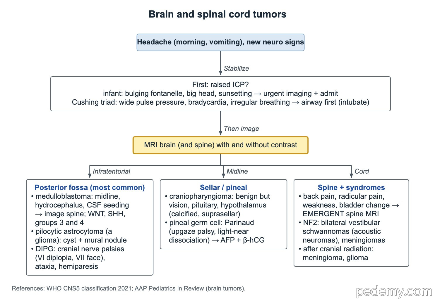
In a stable child, MRI of the brain with and without gadolinium is the best study to characterize a possible tumor. CT is faster and is used when the child is unstable or MRI is not available.
For staging, image the brain and the whole spine with and without gadolinium. Tumors that seed the CSF, such as medulloblastoma, can drop metastases down the spinal canal.
Posterior fossa tumors
- Medulloblastoma predisposition: Gorlin syndrome, Turcot syndrome and Li–Fraumeni syndrome.
- WNT-activated medulloblastoma has the best outcome.
| Tumor | Clues | Note |
|---|---|---|
| Medulloblastoma | Midline cerebellar mass, obstructive hydrocephalus | Malignant embryonal tumor; seeds CSF → image the spine. Groups: WNT, SHH, group 3, group 4 |
| Pilocytic astrocytoma | Well-circumscribed cerebellar cyst with an enhancing mural nodule | Low-grade glioma; astrocytoma is a type of glioma |
| Diffuse midline glioma of the pons (DIPG) | Diplopia (CN VI), facial weakness (CN VII), ataxia, long-tract signs | Diffuse non-enhancing pontine lesion on MRI; diagnosed by imaging |
Sellar and pineal region tumors
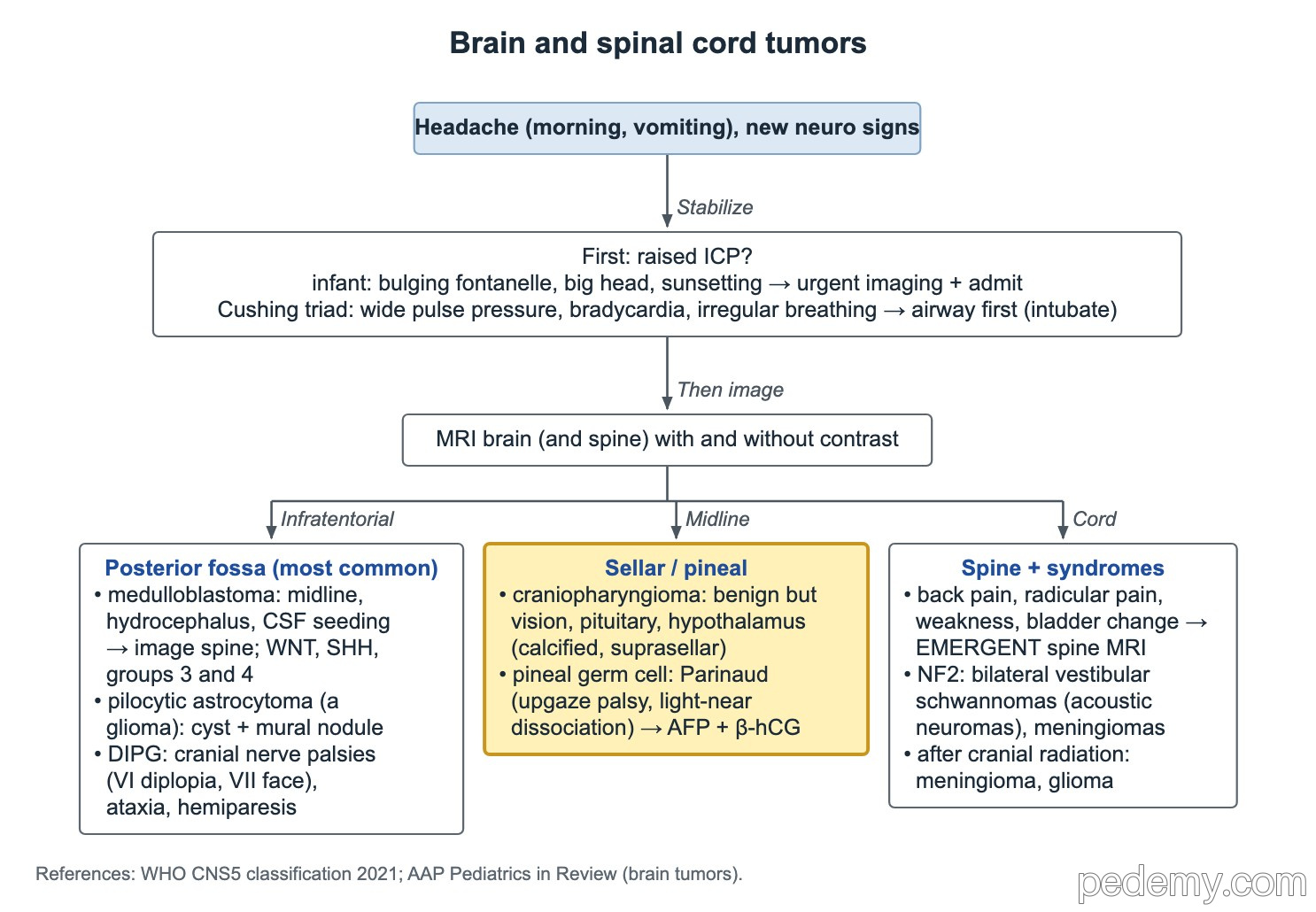
Craniopharyngioma is histologically benign but sits in the sellar and suprasellar region, often calcified. It threatens vision (bitemporal field loss), pituitary function (growth failure, diabetes insipidus) and the hypothalamus (obesity).
A pineal mass presses on the dorsal midbrain and causes Parinaud syndrome: impaired upgaze, light-near dissociation of the pupils and convergence-retraction nystagmus. Pineal germ cell tumors are midline tumors, so send AFP and β-hCG in both serum and CSF.
Spinal cord compression and tumor syndromes
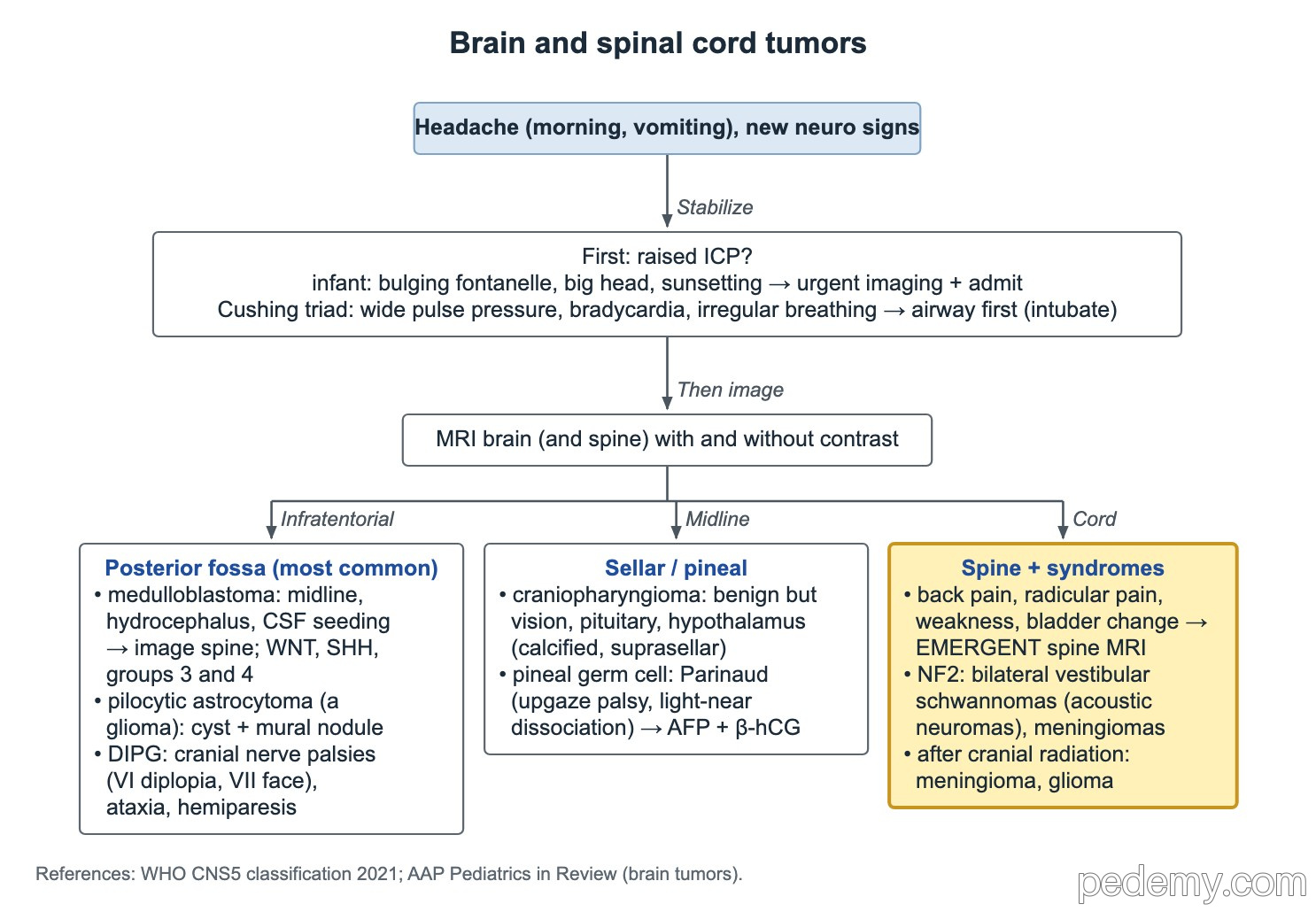
Back pain in a child with cancer, radicular pain, new weakness or bladder change needs emergent MRI of the spine. Spinal cord compression gives brisk reflexes below the level; cauda equina compression gives leg weakness with absent knee and ankle reflexes and normal arms.
- NF2-related schwannomatosis (formerly NF2): bilateral vestibular schwannomas (acoustic neuromas) and meningiomas.
- After cranial radiation: later meningioma and glioma.
Common questions
What is the most common malignant brain tumor in children?
Medulloblastoma, a posterior fossa embryonal tumor that often presents with obstructive hydrocephalus.
What are the warning signs of a brain tumor in a child?
Morning headache with vomiting, new squint or double vision, unsteady walking, head tilt, a growing head in an infant, vision loss, growth failure or new seizures.
What is the Cushing triad?
Hypertension with a wide pulse pressure, bradycardia and irregular breathing. It is a late sign of raised intracranial pressure and impending herniation.
Study this topic
Sources
- The 2021 WHO classification of tumors of the central nervous system: a summary (Neuro Oncol)
- Updated diagnostic criteria for NF2 and schwannomatosis, 2022 (Genet Med)
- NCI PDQ: Childhood brain and spinal cord tumors treatment overview
Written for Pedemy in our own words from the sources above; checked November 2026. For education only, not medical advice; check doses and local protocols before treating a patient. Pedemy is independent and not affiliated with or endorsed by the organizations cited.