

Main decision tree
Hemolysis: is the Coombs test positive?
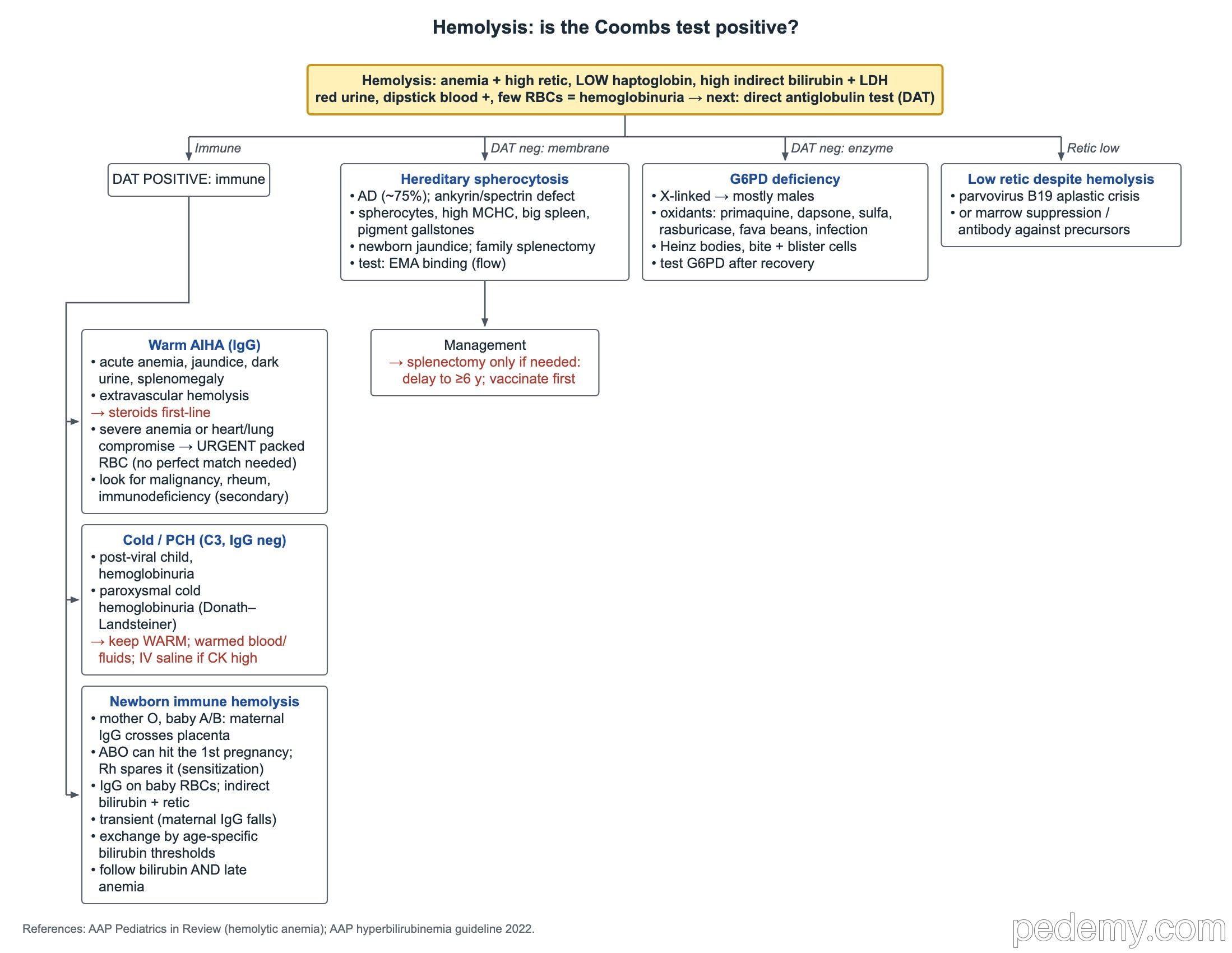
Key points
- Hemolysis labs: anemia, high reticulocytes, low haptoglobin, high LDH and high unconjugated bilirubin.
- Red urine, strongly positive dipstick for blood, few RBCs on microscopy = hemoglobinuria (intravascular hemolysis).
- Hereditary spherocytosis: autosomal dominant in about 75%, high MCHC, DAT negative; EMA binding test; delay splenectomy to ≥6 years.
- G6PD deficiency is X-linked; oxidants cause bite and blister cells and Heinz bodies; the level can look normal during acute hemolysis.
- Warm AIHA: DAT positive for IgG, steroids first-line. PCH: DAT C3 positive, IgG negative, keep the child warm.
- ABO hemolytic disease (group O mother, A or B baby) can occur in the first pregnancy; Rh disease usually spares the first baby.
- β-thalassemia major: no HbA, mostly HbF, severe microcytic anemia at 6–12 months; transfusions lead to iron overload and chelation.
Confirm hemolysis, then do the DAT
Suspect hemolysis with pallor, jaundice, dark urine or a big spleen. The labs show anemia with a high reticulocyte count, low haptoglobin, high LDH and high unconjugated (indirect) bilirubin.
Red urine with a strongly positive dipstick for heme but few or no red cells on microscopy is hemoglobinuria, a sign of intravascular hemolysis. The next test is the DAT.
- DAT positive: immune hemolysis (warm AIHA, cold AIHA or PCH, newborn alloimmune hemolysis).
- DAT negative: think of a membrane defect (hereditary spherocytosis) or an enzyme defect (G6PD deficiency).
- Low reticulocyte count despite hemolysis: parvovirus B19 aplastic crisis, other marrow suppression, or antibodies that also destroy reticulocytes.
- Parvovirus B19 stops red cell production for about a week; in a child with chronic hemolysis this causes a sudden, severe drop in hemoglobin.
Hereditary spherocytosis
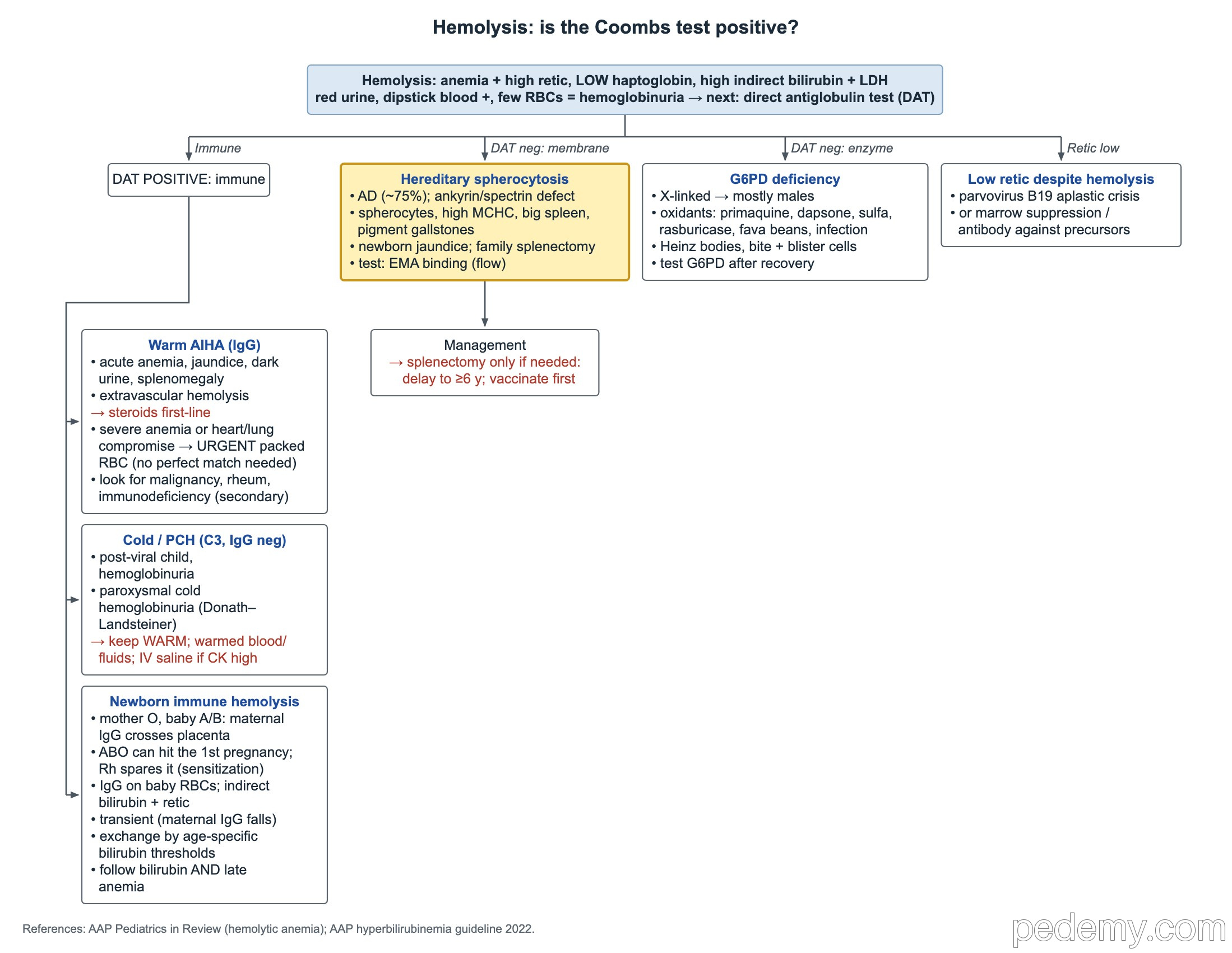
A defect in the red cell membrane skeleton makes cells lose membrane and become spheres, which the spleen removes. Look for a family history of anemia, jaundice, gallstones or splenectomy.
- Proteins: ankyrin and spectrin most often; also band 3 and protein 4.2.
- Inheritance: autosomal dominant in about 75%.
- Clues: spherocytes on the smear, high MCHC, splenomegaly, pigment gallstones.
- Newborn: severe indirect jaundice with a negative DAT and a parent who had a splenectomy.
- Testing: eosin-5-maleimide (EMA) binding by flow cytometry is useful when the diagnosis is uncertain; osmotic fragility is older and less specific.
- Splenectomy only when needed for severe disease; delay it until at least 6 years and give pneumococcal, meningococcal and Hib vaccines first.
- After splenectomy, Howell–Jolly bodies appear on the smear.
G6PD deficiency
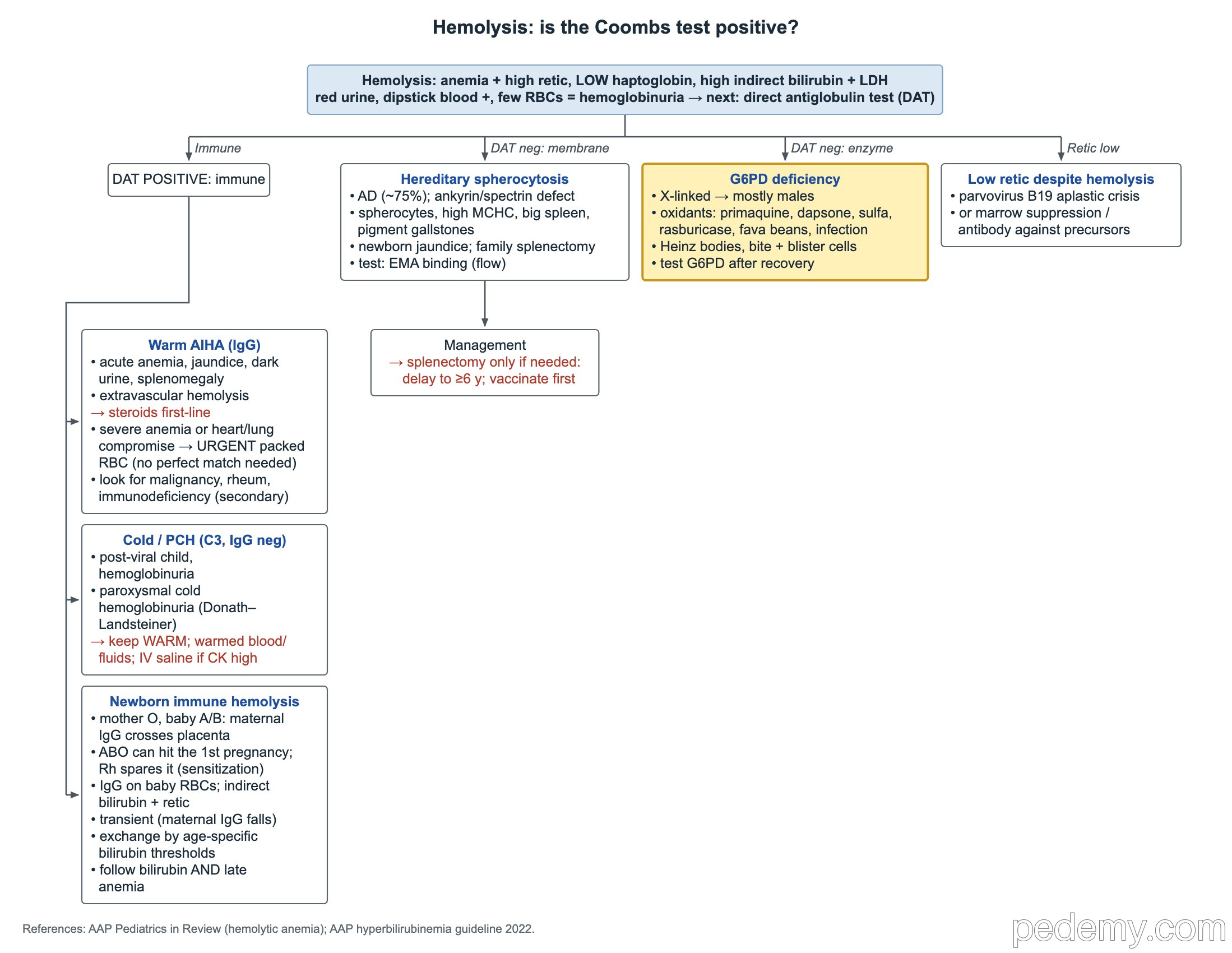
G6PD protects red cells from oxidant damage. Without it, an oxidant stress causes sudden hemolysis with pallor, jaundice and dark urine, usually 1–3 days after the trigger.
- Inheritance: X-linked, so most affected patients are male.
- Triggers: primaquine, tafenoquine, dapsone, sulfonamides, rasburicase, fava beans and infection.
- Routine smear: bite cells and blister cells. Heinz bodies need a supravital stain.
- During or just after a hemolytic episode the G6PD level can be falsely normal, because young cells have more enzyme. Recheck in 2–3 months.
- Quantitative G6PD testing is required before primaquine or tafenoquine for malaria.
- This is an idiosyncratic, genetically determined drug reaction, not an IgE-mediated allergy; record it as G6PD deficiency.
Warm autoimmune hemolytic anemia
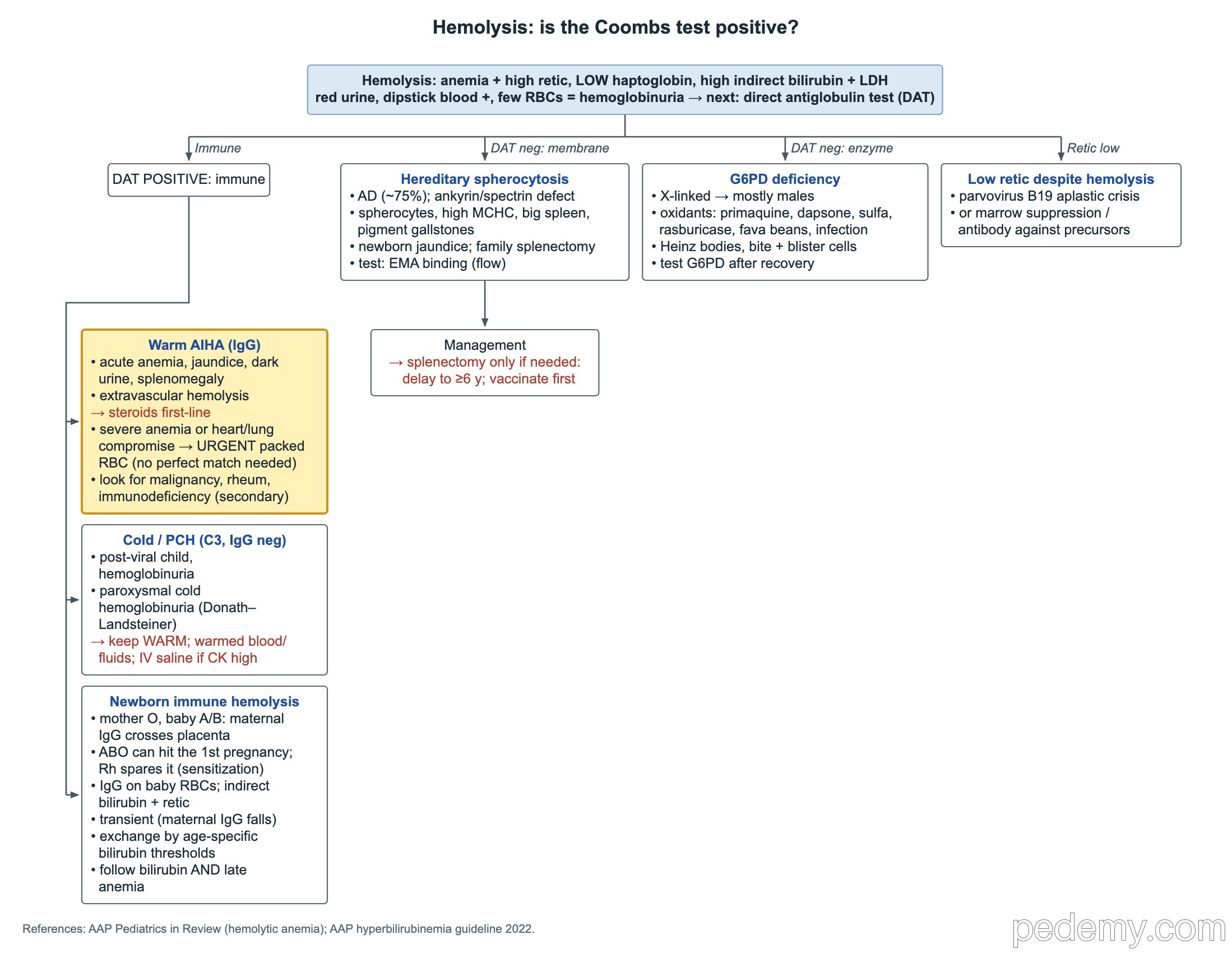
A typical case is a previously well school-age child who, after a viral illness, has pallor, jaundice, dark urine and a big spleen. The DAT is positive for IgG, and destruction is mostly in the spleen (extravascular).
- Treatment: glucocorticoids are first-line.
- Severe anemia or heart or lung compromise: give packed red cells urgently; do not delay for a perfectly matched unit.
- Look for an underlying cause: malignancy, rheumatologic disease such as lupus, and immunodeficiency.
- Spherocytes on the smear are common in warm AIHA as well as in hereditary spherocytosis; the DAT tells them apart.
Cold hemolysis and paroxysmal cold hemoglobinuria
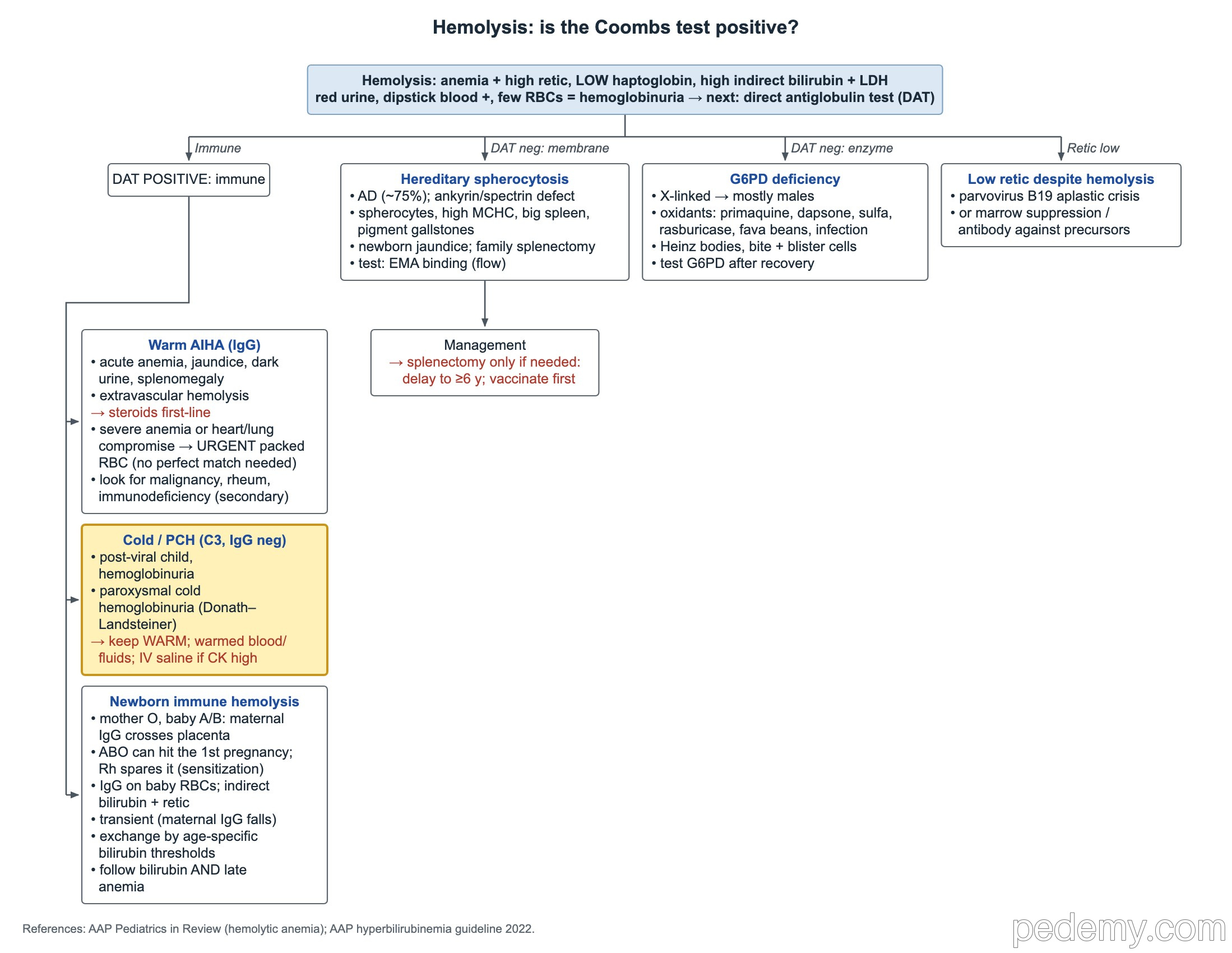
Paroxysmal cold hemoglobinuria (PCH) is the classic cold hemolysis of young children. After a viral illness, a Donath–Landsteiner IgG antibody binds red cells in the cold and fixes complement, which destroys them inside the vessels when they warm up.
- DAT: positive for C3, negative for IgG.
- Clue: sudden pallor with hemoglobinuria after a viral infection.
- Keep the child warm and give needed blood and IV fluids through a warmer.
- Hemoglobinuria with a raised CK: give careful IV saline to protect the kidneys.
- PCH is usually self-limited over days to weeks.
Immune hemolysis in the newborn
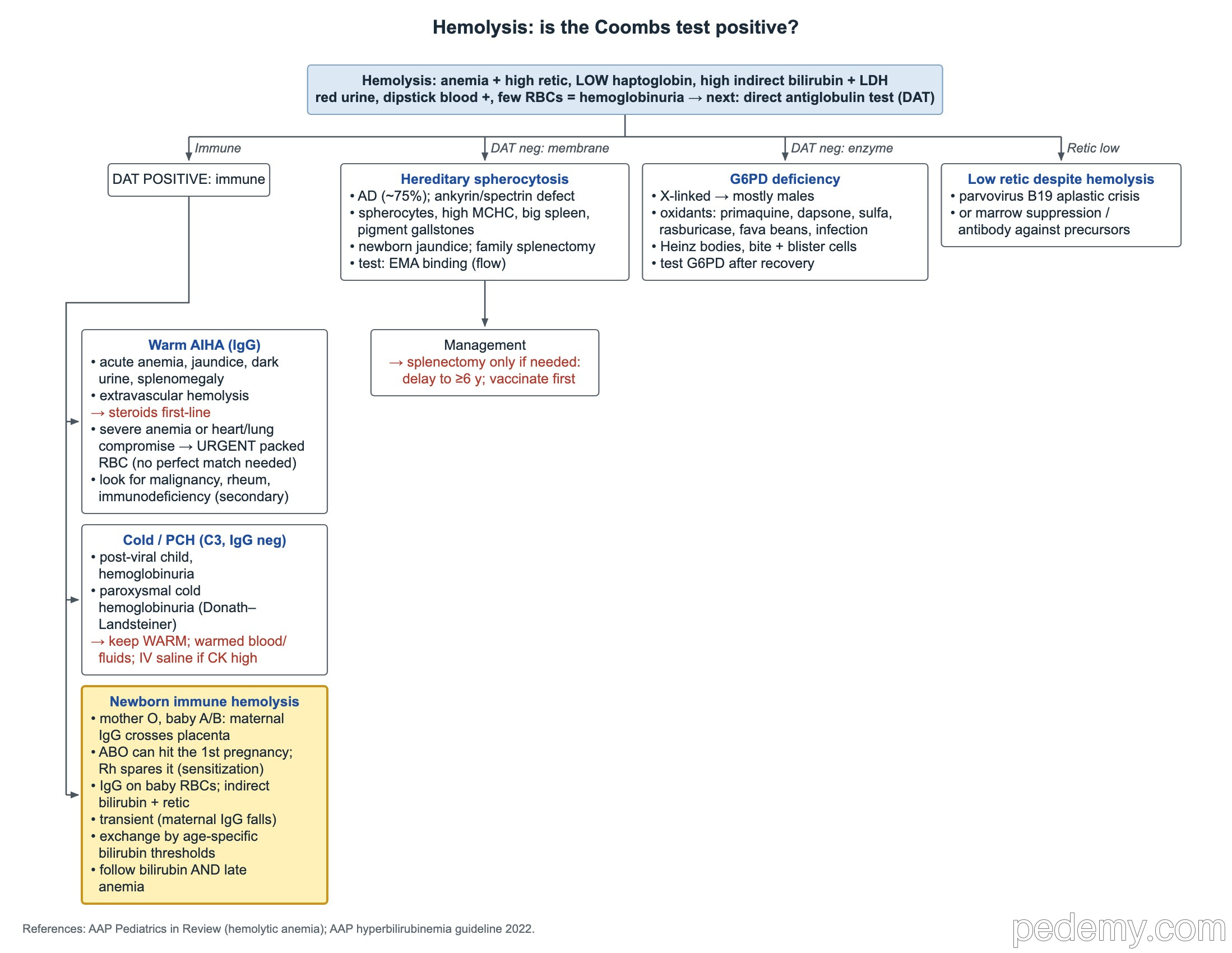
Maternal IgG antibodies cross the placenta and coat the baby's red cells. A positive DAT for IgG detects that coating. The baby has a rising unconjugated bilirubin and a reticulocyte response.
- ABO disease: group O mother with a group A or B baby; anti-A or anti-B IgG crosses the placenta. It can occur in the first pregnancy because no prior sensitization is needed.
- Rh disease: the mother is usually sensitized at the first delivery, so the first Rh-positive baby is usually spared. Anti-D prophylaxis prevents most cases.
- Severe disease despite anti-D: antibodies to minor antigens such as Kell, c or E.
- Isoimmune hemolysis is a risk factor in the 2022 AAP hyperbilirubinemia guideline: it lowers phototherapy and exchange thresholds.
- Exchange transfusion is decided by hour-specific, gestation-specific bilirubin thresholds and neurotoxicity risk factors.
- A 1-week-old with marked anemia, indirect hyperbilirubinemia and a positive IgG DAT needs immediate emergency department evaluation.
- The problem fades as maternal IgG clears over weeks, but follow both bilirubin and hemoglobin: late anemia can appear weeks after birth.
Hemoglobinopathies and thalassemia
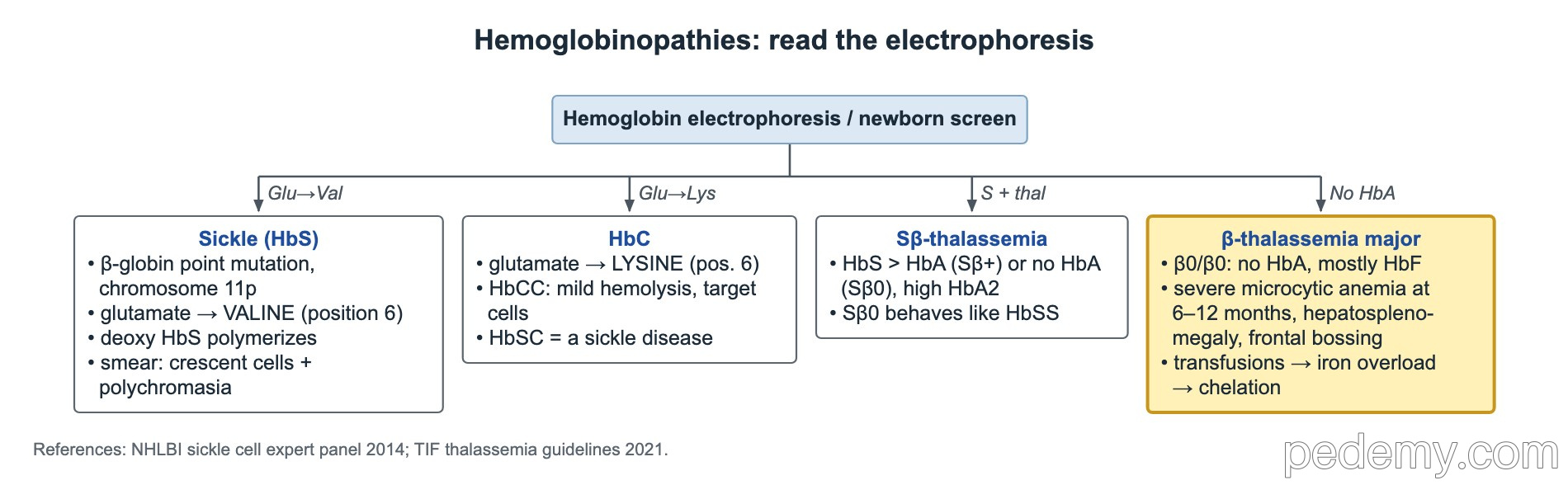
Hemoglobin electrophoresis or the newborn screen shows which hemoglobins are present. Abnormal β-chains (HbS, HbC) or too little β-chain (β-thalassemia) usually show up after 6 months, when HbF falls.
- β-thalassemia major: severe microcytic anemia at 6–12 months with hepatosplenomegaly.
- Without regular transfusion, marrow expansion causes frontal bossing and maxillary overgrowth.
- Repeated transfusions add iron the body cannot excrete, so transfusion-dependent patients need iron chelation.
| Result | Meaning |
|---|---|
| HbS: glutamate → valine at position 6 | Sickle hemoglobin; polymerizes when deoxygenated |
| HbC: glutamate → lysine at position 6 | HbCC = mild hemolysis with target cells, not sickle disease; HbSC = a form of sickle cell disease |
| HbS > HbA with raised HbA2 | HbS β+-thalassemia |
| HbS, no HbA, raised HbA2 | HbS β0-thalassemia; behaves like HbSS |
| No HbA, mostly HbF | Severe β-thalassemia (β0/β0, thalassemia major) |
Common questions
What labs confirm hemolysis?
Anemia with a high reticulocyte count, low haptoglobin, high LDH and high unconjugated bilirubin. The direct antiglobulin test then shows whether the cause is immune.
Why can a G6PD test be normal right after hemolysis?
The oldest, most enzyme-poor cells have been destroyed and young cells have more G6PD. Repeat the test 2–3 months after the episode.
Can ABO incompatibility affect a first baby?
Yes. Group O mothers already have IgG anti-A and anti-B, so ABO hemolytic disease can occur in the first pregnancy, unlike Rh disease.
Study this topic
Sources
- AAP 2022: Clinical practice guideline revision, hyperbilirubinemia in the newborn ≥35 weeks (Pediatrics)
- CPIC 2022 guideline for G6PD genotype and drug use (Clinical Pharmacology & Therapeutics)
- GeneReviews: Beta-thalassemia (NCBI Bookshelf)
Written for Pedemy in our own words from the sources above; checked November 2026. For education only, not medical advice; check doses and local protocols before treating a patient. Pedemy is independent and not affiliated with or endorsed by the organizations cited.