

Main decision tree
Bone and soft-tissue tumors on x-ray: LCH
Key points
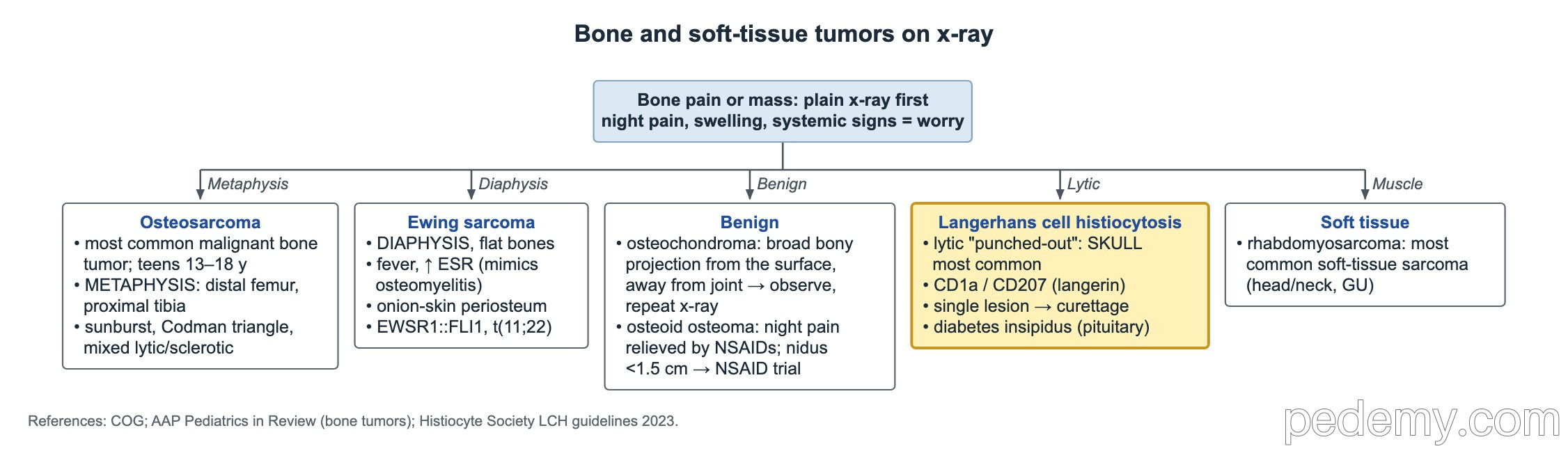
- The skull is the most common site of lytic bone lesions in LCH.
- Lesions look punched out on x-ray, without a sclerotic rim.
- Biopsy confirms LCH: CD1a and/or CD207 (langerin) staining in lesional cells, read with morphology.
- Polyuria with dilute urine in a child with LCH → central diabetes insipidus (AVP deficiency).
- A single bone lesion is usually treated with curettage, with or without an intralesional steroid.
- Multisystem disease needs systemic chemotherapy, usually vinblastine plus prednisone.
Recognize LCH
LCH comes from abnormal myeloid dendritic cells, most often driven by BRAF V600E or other MAPK pathway mutations. It can be a single bone lesion or involve skin, bone marrow, liver, spleen, lungs and the pituitary.
Bone is the most common site, and the skull is the most common bone. The lesion is lytic and punched out. A scaly or seborrheic-like rash in an infant, ear discharge and a skull defect together strongly suggest LCH.
- Diagnosis needs a biopsy: CD1a and/or CD207 (langerin) positive cells.
- Staging: blood count, liver tests, urine osmolality and a skeletal survey or PET-CT.
Central diabetes insipidus
LCH can infiltrate the pituitary stalk. New polyuria, thirst and dilute urine mean central diabetes insipidus, now also called AVP deficiency. It can appear before, with or after the bone lesions, and it is usually permanent.
Skull base, orbit, mastoid and sphenoid lesions carry a higher risk of central nervous system and pituitary involvement.
- Check serum sodium and osmolality with urine osmolality.
- Get a brain MRI looking at the pituitary stalk.
- Treat with desmopressin; look for other pituitary hormone deficiencies, especially growth hormone.
Treatment
- Single bone lesion: curettage, with or without an intralesional steroid; some heal after biopsy alone.
- Multifocal bone, high-risk sites or multisystem disease: vinblastine plus prednisone for 12 months.
- Risk organs (bone marrow, liver, spleen) predict a worse outcome.
- BRAF or MEK inhibitors are options for refractory disease.
Common questions
What is the most common bone involved in Langerhans cell histiocytosis?
The skull. The lesion is lytic and punched out on x-ray.
Why does LCH cause diabetes insipidus?
LCH can infiltrate the pituitary stalk and posterior pituitary, so less vasopressin (AVP) is released. The child passes large amounts of dilute urine.
Study this topic
Sources
- LCH guidelines for patients up to 18 years (Pediatr Blood Cancer 2013)
- Histiocyte Society consensus recommendations for LCH in adults (Blood 2022)
- NCI PDQ: Langerhans cell histiocytosis treatment
Written for Pedemy in our own words from the sources above; checked November 2026. For education only, not medical advice; check doses and local protocols before treating a patient. Pedemy is independent and not affiliated with or endorsed by the organizations cited.