

Main decision tree
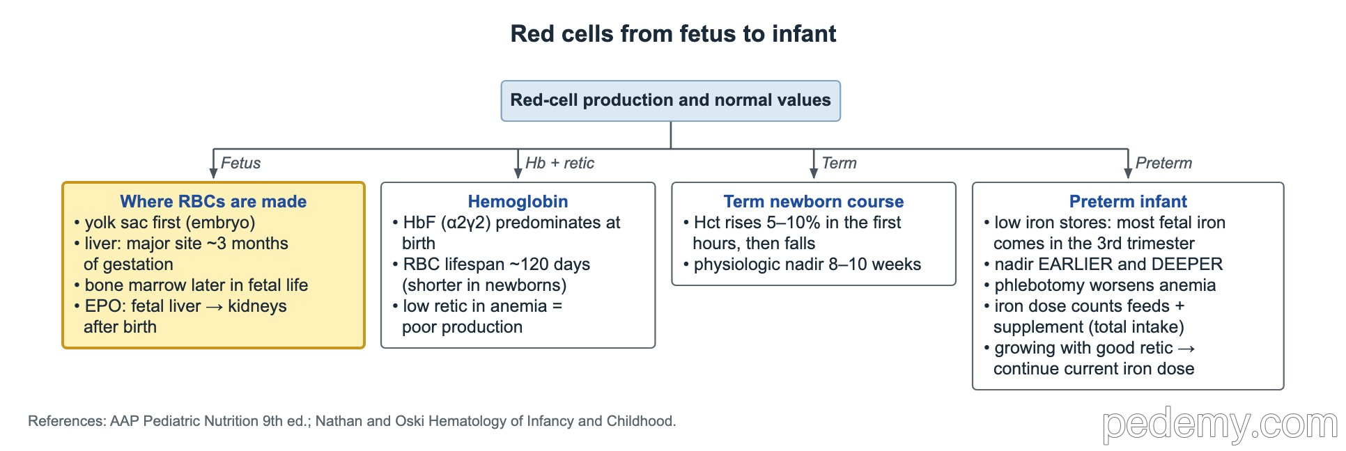
Red cells from fetus to infant: where RBCs are made
Key points
- Red cells are made first in the yolk sac, then mainly in the liver (major site at about 3 months of gestation), then in the bone marrow.
- Erythropoietin comes mainly from the fetal liver before birth and from the kidneys after birth.
- HbF (α2γ2) predominates at birth; a normal red cell lives about 120 days, less in newborns.
- A low reticulocyte count in anemia means the marrow is not making enough red cells.
- Term infants: hematocrit rises in the first hours, then falls to a physiologic nadir at about 8–12 weeks.
- Preterm infants reach their nadir earlier and lower; repeated blood draws make it worse.
- Hypersegmented neutrophils suggest folate or B12 deficiency; parasites are the commonest cause of eosinophilia worldwide.
Where red cells are made
- Yolk sac: primitive embryonic red cells, in the first weeks of gestation.
- Liver: the major site of red cell production by about 3 months of gestation.
- Bone marrow: takes over later in fetal life and is the main site at birth.
- Erythropoietin: made mainly by the fetal liver before birth and by the kidneys after birth.
Hemoglobin, red cell lifespan and reticulocytes
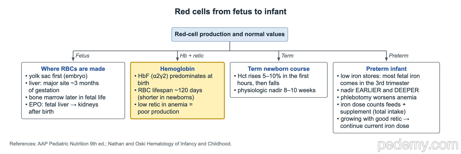
- HbF (alpha2-gamma2) is the main hemoglobin at birth. It falls over the first year as adult HbA (alpha2-beta2) takes over.
- This switch is why β-chain disorders such as sickle cell disease and β-thalassemia usually show symptoms after about 6 months.
- A normal mature red cell lives about 120 days; newborn red cells live a shorter time.
- Reticulocytes show marrow output. In anemia, a low reticulocyte count means poor production; a high count means blood loss or hemolysis.
Physiologic anemia of the term infant
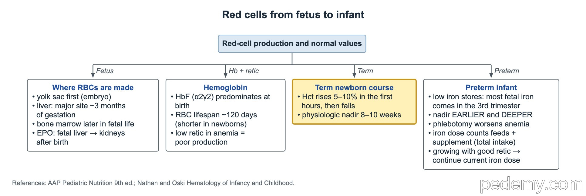
After birth, plasma volume shifts and the hematocrit rises 5–10% over the first hours. Then oxygen levels rise, erythropoietin falls and red cell production slows, so hemoglobin drifts down.
- Lowest hemoglobin (physiologic nadir) in a term infant: about 8–12 weeks.
- This is normal and needs no treatment in a well, growing infant.
Anemia of prematurity
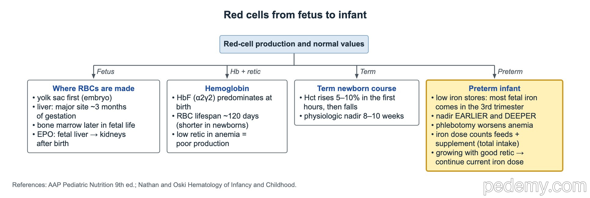
- Preterm infants reach their hemoglobin nadir earlier and lower than term infants.
- Causes: low erythropoietin response, shorter red cell lifespan, rapid growth and low iron stores, since most fetal iron arrives in the third trimester.
- Repeated laboratory blood draws in the NICU make anemia of prematurity worse; limit sampling.
- Iron dosing counts total daily intake from feeds plus supplement.
White cell clues on the smear
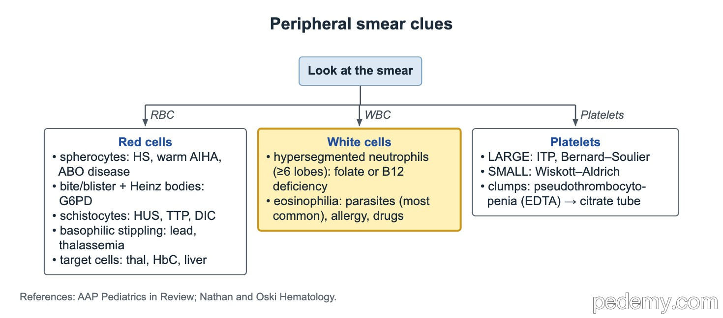
- Hypersegmented neutrophils (6 or more lobes): folate or vitamin B12 deficiency, often with a high MCV.
- Eosinophilia: parasitic infection is the most common cause in children worldwide; allergy and drugs are other causes.
Common questions
When is hemoglobin lowest in a term baby?
At about 8–12 weeks of age. This physiologic anemia needs no treatment in a well infant.
Which hemoglobin is highest at birth?
Fetal hemoglobin, HbF (alpha2-gamma2). Adult HbA takes over during the first year.
Where are red blood cells made in the fetus?
First in the yolk sac, then mainly in the liver from about 3 months of gestation, and later in the bone marrow.
Study this topic
Sources
- AAP Clinical Report: Diagnosis and prevention of iron deficiency in infants and young children (Pediatrics 2010)
- Red blood cell transfusion thresholds for anemia of prematurity (NeoReviews 2023)
- Erythropoiesis in the mammalian embryo (Experimental Hematology 2024)
Written for Pedemy in our own words from the sources above; checked November 2026. For education only, not medical advice; check doses and local protocols before treating a patient. Pedemy is independent and not affiliated with or endorsed by the organizations cited.