

Main decision tree
Neural tube defects
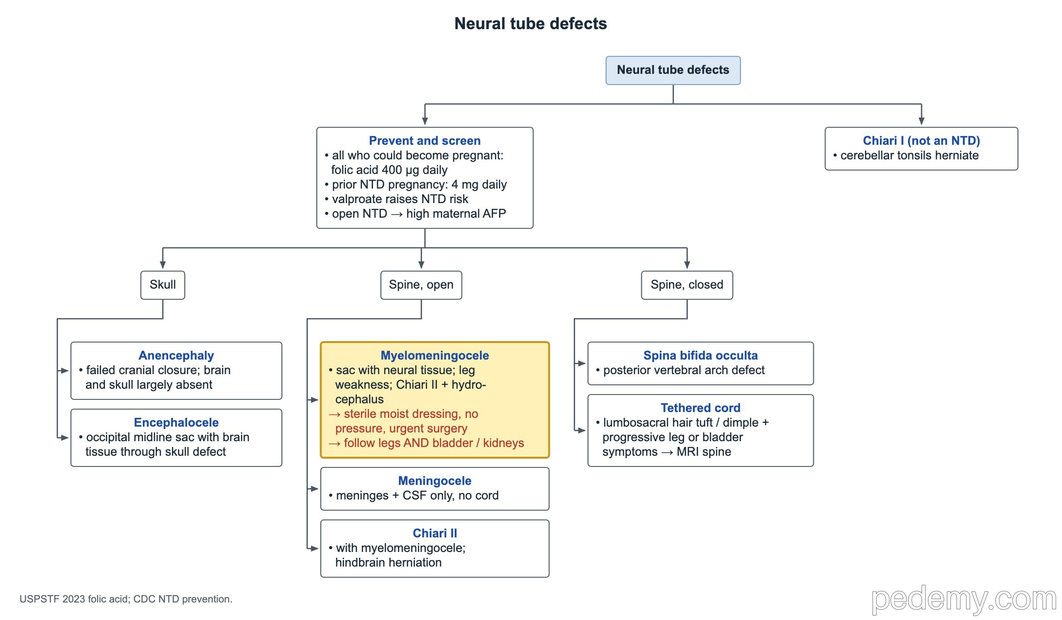
Key points
- Myelomeningocele: sac with neural tissue, leg weakness, Chiari II and hydrocephalus in most; most need a shunt or ETV.
- Open lesion at birth: sterile moist dressing, no pressure on the sac, urgent neurosurgery.
- Everyone who could become pregnant: folic acid 400 µg (0.4 mg) daily, starting at least 1 month before conception.
- A previous pregnancy with a neural tube defect: 4 mg folic acid daily; recurrence risk is about 2–3%.
- Valproate in pregnancy raises the neural tube defect risk; open defects raise maternal serum AFP.
- Lumbosacral hair tuft or dimple with progressive leg or bladder symptoms: tethered cord → lumbar spine MRI.
- Chiari I: tonsils herniate; cough-induced occipital headache; image the whole spine for a syrinx.
Myelomeningocele and Chiari II
The tree splits neural tube defects into skull, open spine and closed spine. Myelomeningocele is the key open spinal defect: a lumbosacral sac that contains spinal cord or nerve roots, with weakness and sensory loss below the level.
- Chiari II malformation (hindbrain herniation) occurs with myelomeningocele.
- Hydrocephalus develops in most, from the Chiari II malformation; most need a VP shunt or endoscopic third ventriculostomy (ETV).
- At birth: cover with a sterile moist (saline) dressing, nurse prone or on the side with no pressure on the sac, and arrange urgent neurosurgical closure.
- Lifelong follow-up of legs AND bladder and kidneys: neurogenic bladder needs regular urologic and renal checks.
- Watch for later shunt failure, tethered cord, scoliosis and symptomatic Chiari II (stridor, apnea, swallowing problems).
Other neural tube defects
| Defect | What it is |
|---|---|
| Anencephaly | Failed closure at the cranial end; most of the brain and skull are absent; not compatible with long survival |
| Encephalocele | Sac with brain tissue through a skull defect, usually occipital and midline |
| Meningocele | Spinal sac with meninges and CSF only, no cord tissue; usually little or no deficit |
| Myelomeningocele | Spinal sac with neural tissue; deficits below the level |
| Spina bifida occulta | Bony defect of the posterior vertebral arches, skin closed; often an incidental finding |
Occult spinal dysraphism and tethered cord
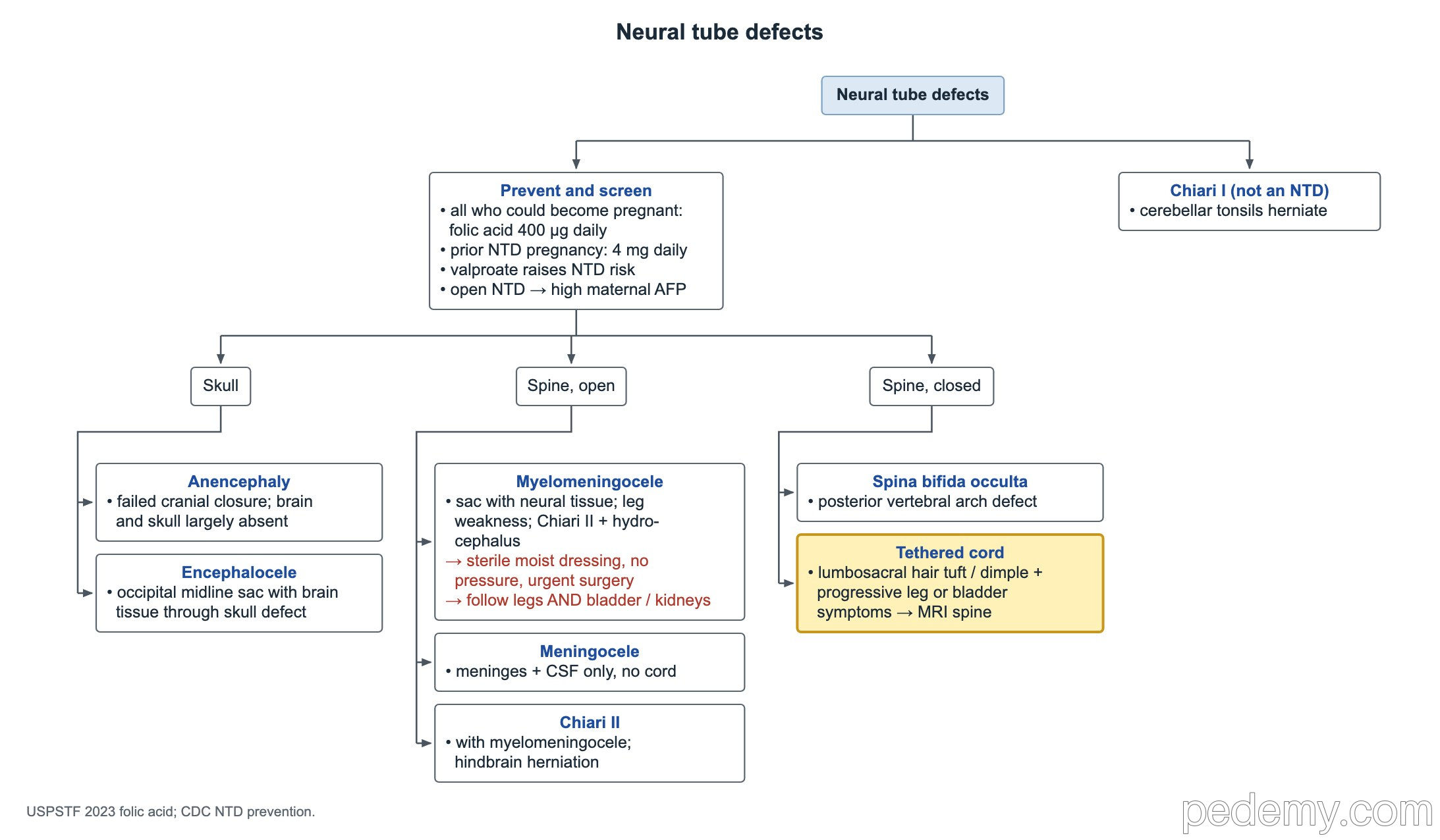
Closed defects hide under intact skin. Skin markers over the lower spine are the clue: a hair tuft, a dimple above the gluteal crease, a lipoma, a hemangioma or a skin tag.
- Tethered cord: the cord is pulled down and stretched as the child grows.
- Symptoms: progressive leg weakness, foot deformity, back or leg pain, new bladder or bowel problems.
- Best test: MRI of the lumbar spine; surgical untethering stops progression.
Chiari I malformation
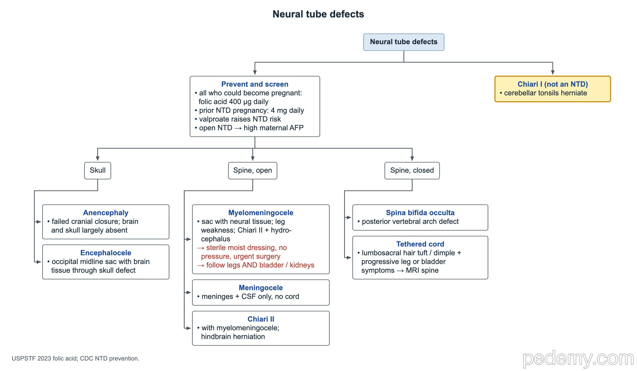
Chiari I is downward displacement of the cerebellar tonsils through the foramen magnum. It is not a neural tube defect, and it is often found by chance on MRI.
- Typical symptom: occipital headache brought on by coughing, straining or laughing.
- Other signs: unsteady gait, neck pain, lower cranial nerve signs, scoliosis.
- Image the whole spine: an associated syrinx (syringomyelia) changes management.
- Symptomatic cases are treated with posterior fossa decompression.
Prevention and prenatal screening
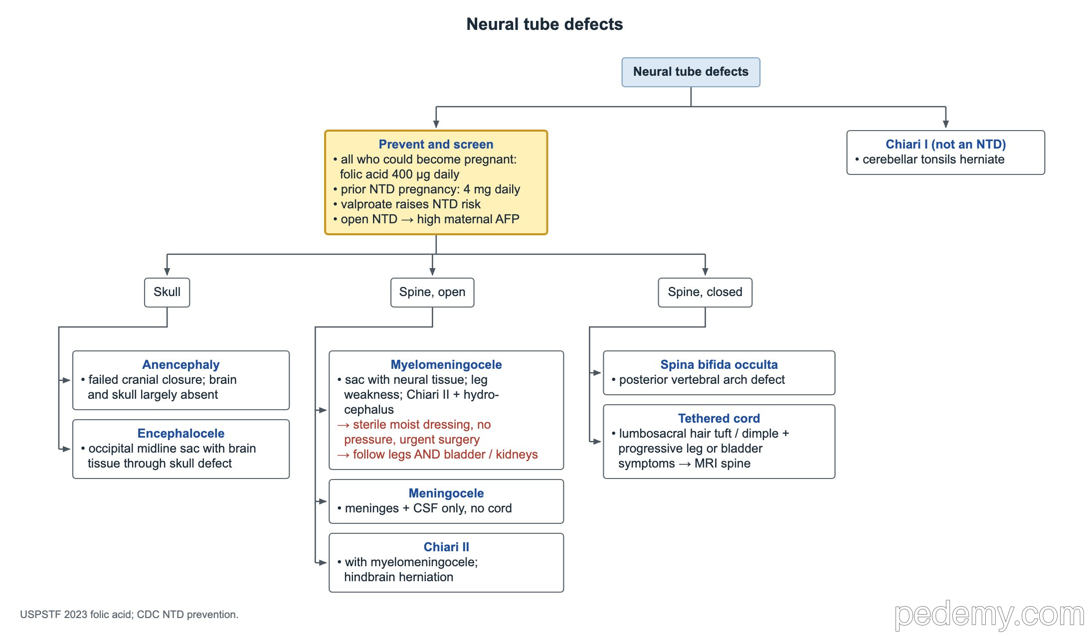
- All who could become pregnant: folic acid 400 µg (0.4 mg) daily; USPSTF advises 0.4–0.8 mg daily, starting at least 1 month before conception and continuing through the first 2–3 months of pregnancy.
- Previous pregnancy with a neural tube defect: 4 mg daily from at least 1 month before conception; recurrence risk is about 2–3%.
- Valproate is the antiseizure medicine most strongly linked to neural tube defects.
- Open neural tube defects raise maternal serum alpha-fetoprotein (AFP); detailed ultrasound confirms.
Brain malformations on MRI
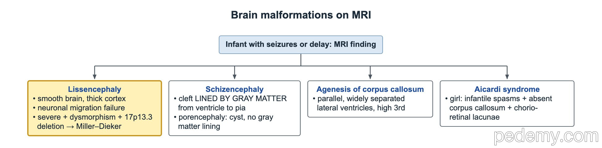
These usually present in infancy with seizures, developmental delay or an abnormal head size.
| Malformation | MRI clue | Note |
|---|---|---|
| Lissencephaly | Smooth brain surface, thick cortex | Failure of neuronal migration; severe form + dysmorphism + 17p13.3 deletion = Miller–Dieker syndrome |
| Schizencephaly | Cleft lined by gray matter, from ventricle to pia | The gray-matter lining separates it from porencephaly |
| Porencephaly | CSF-filled cyst without gray-matter lining | Usually from earlier destructive injury, such as stroke |
Agenesis of the corpus callosum and Aicardi syndrome
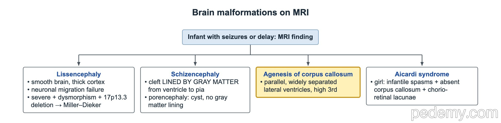
- Agenesis of the corpus callosum: widely separated, parallel lateral ventricles and a high-riding third ventricle.
- It can be isolated, with normal development, or part of a syndrome.
- Aicardi syndrome: a girl with infantile spasms, absent corpus callosum and chorioretinal lacunae.
Common questions
How much folic acid prevents neural tube defects?
400 µg (0.4 mg) daily for everyone who could become pregnant (USPSTF: 0.4–0.8 mg). After a previous affected pregnancy, 4 mg daily starting at least 1 month before conception.
What is the difference between Chiari I and Chiari II?
Chiari I is tonsillar herniation, often found by chance, without a neural tube defect. Chiari II is hindbrain herniation that comes with myelomeningocele and causes hydrocephalus.
What does a hair tuft on the lower back mean?
It can mark occult spinal dysraphism. With progressive leg or bladder symptoms, think of tethered cord and order a lumbar spine MRI.
Study this topic
Sources
- USPSTF: Folic acid supplementation to prevent neural tube defects, reaffirmation (JAMA 2023)
- Congress of Neurological Surgeons guidelines for patients with Chiari malformation: surgical interventions (Neurosurgery 2023)
Written for Pedemy in our own words from the sources above; checked November 2026. For education only, not medical advice; check doses and local protocols before treating a patient. Pedemy is independent and not affiliated with or endorsed by the organizations cited.