

Main decision tree
Acute demyelination: which one?
Key points
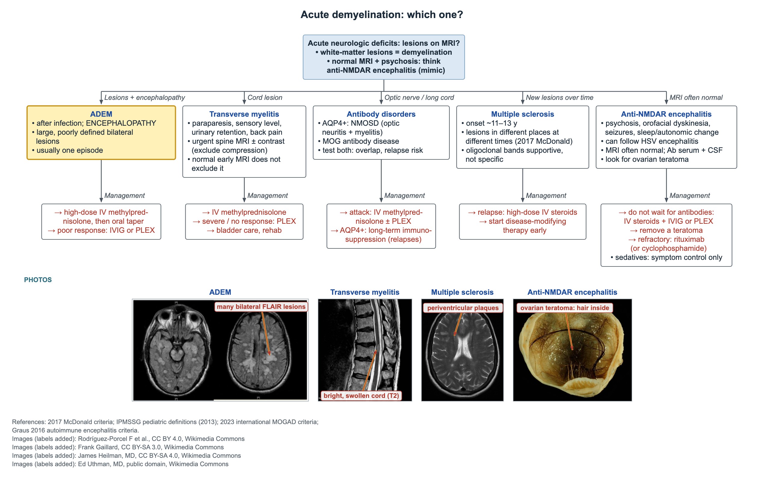
- ADEM: after an infection, encephalopathy plus large, poorly defined bilateral white-matter lesions; usually one episode.
- Acute weakness with a sensory level: emergency spine MRI with and without contrast to exclude compression.
- Transverse myelitis: CSF mild lymphocytic pleocytosis and raised protein; a normal early MRI does not exclude it.
- Test aquaporin-4 IgG (NMOSD) and MOG antibodies: they predict relapse and change long-term treatment.
- Pediatric MS usually starts around 11–14 years; oligoclonal bands support but do not prove MS.
- Guillain–Barré: ascending weakness, areflexia, CSF high protein with few cells; give IVIG or plasma exchange and monitor breathing.
- Acute cerebellar ataxia: toddler, days after varicella or a viral illness, alert with normal reflexes; it resolves.
ADEM
The tree sorts acute neurologic deficits by MRI: lesions with encephalopathy (ADEM), a cord lesion (transverse myelitis), optic nerve or long cord lesions (antibody disorders), new lesions over time (MS), and a normal MRI with psychosis (anti-NMDA receptor encephalitis).
ADEM typically follows an infection by days to weeks and is most common in young children.
- Encephalopathy (confusion, irritability, drowsiness) is required and separates ADEM from transverse myelitis or a first MS attack.
- Multifocal deficits: ataxia, weakness, cranial nerve palsies, seizures.
- MRI: large, poorly defined, bilateral white-matter lesions, often also deep gray matter.
- Treatment: high-dose IV methylprednisolone, then an oral taper; IVIG or plasma exchange if the response is poor.
- Usually a single episode; test MOG antibodies, since MOG-positive children are more likely to relapse.
Transverse myelitis
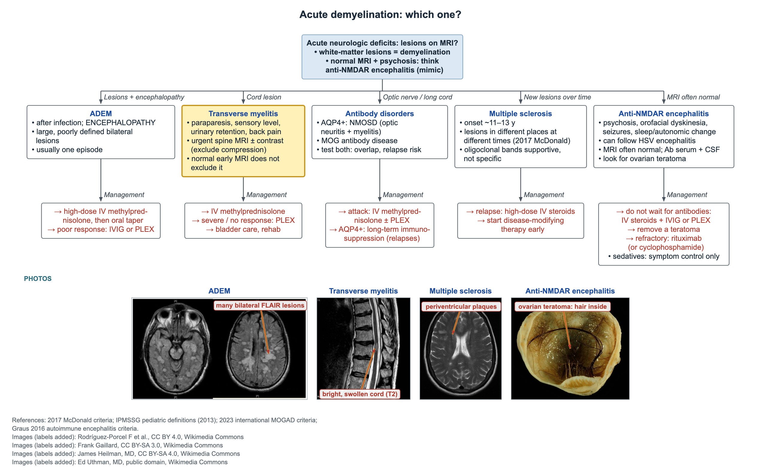
Picture: back pain, then paraparesis, a truncal sensory level (for example T7) and urinary retention or other sphincter problems over hours to days.
- First step: emergency spine MRI with and without contrast, to exclude cord compression (tumor, abscess, hematoma), which needs surgery.
- MRI: swollen, T2-bright cord segment; a normal early MRI does not exclude myelitis when the clinical picture fits; repeat it.
- CSF: mild lymphocytic pleocytosis and mildly raised protein.
- Treatment: high-dose IV methylprednisolone; plasma exchange for severe disease or no response.
- Supportive care: bladder catheterization, bowel care, early rehabilitation.
NMOSD and MOG antibody disease
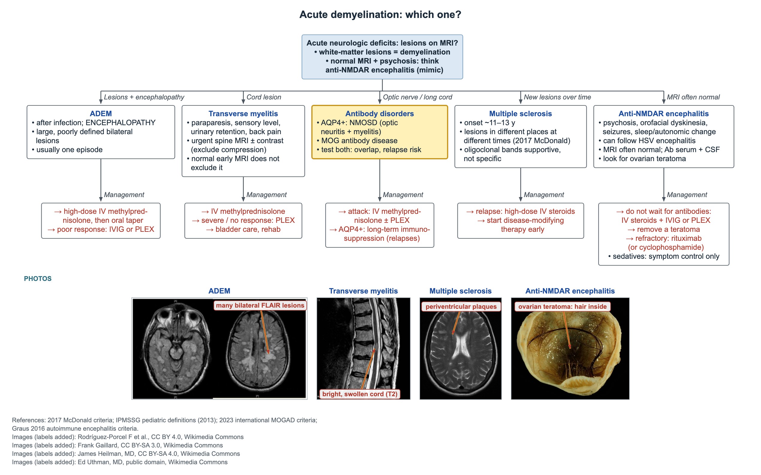
- Optic neuritis plus longitudinally extensive myelitis: test serum aquaporin-4 IgG; a positive result confirms NMOSD.
- MOG antibody-associated disease (MOGAD): ADEM, optic neuritis or myelitis with MOG antibodies; common in children.
- Test both antibodies in children with myelitis or optic neuritis: the features overlap, and the results predict relapse and chronic disease.
- Attack: IV methylprednisolone, with plasma exchange if severe.
- Aquaporin-4-positive NMOSD needs long-term immunosuppression to prevent relapses; MS disease-modifying drugs can worsen it.
Pediatric multiple sclerosis
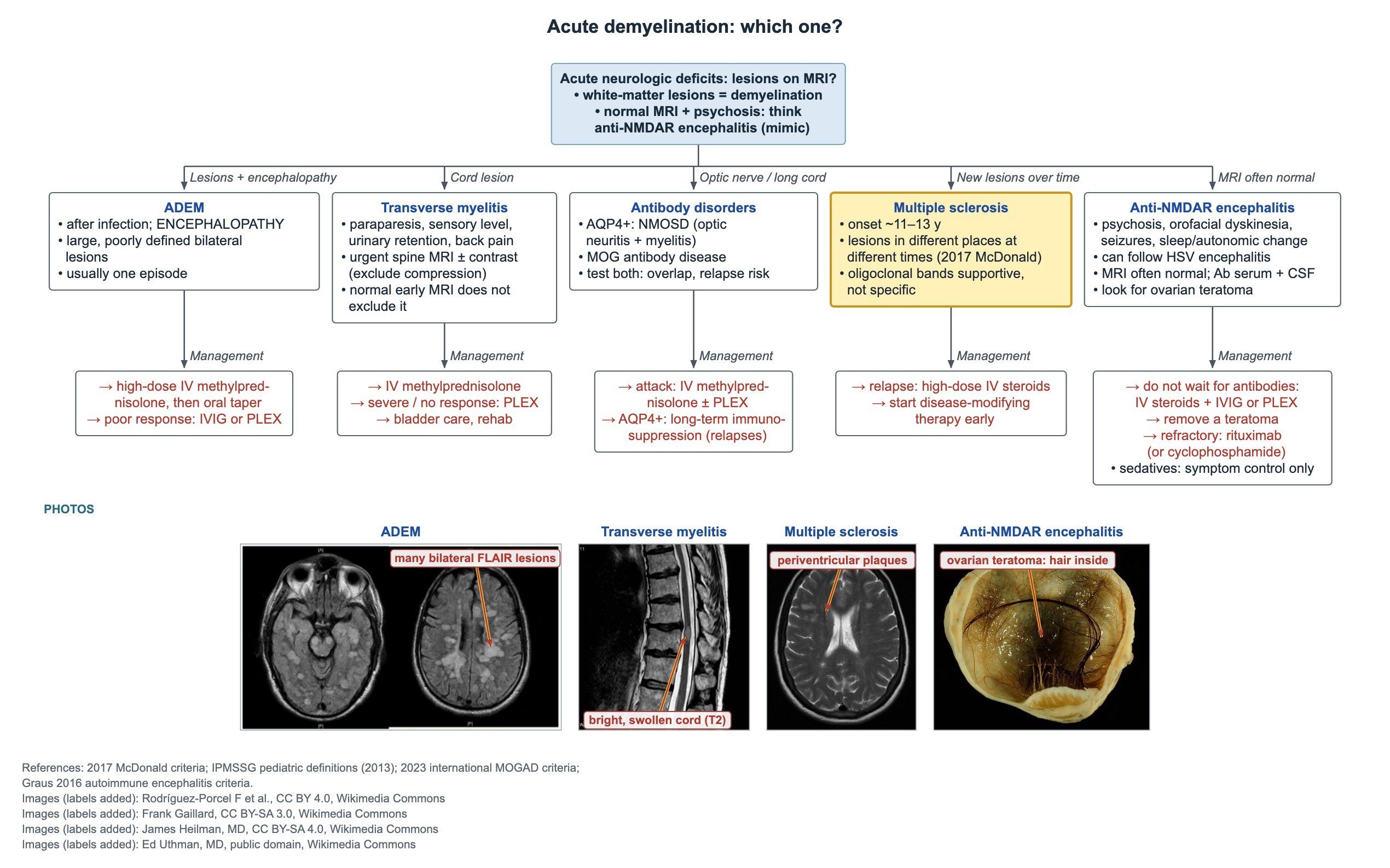
Think of MS in a teen with a past attack, such as optic neuritis, who now has a new deficit and an MRI with both old and new lesions. Onset is usually around 11–14 years.
- McDonald criteria idea: lesions in separate CNS areas (dissemination in space), plus separate times or supportive CSF evidence.
- The 2024 McDonald revision adds the optic nerve as a fifth area, so dissemination in space means typical lesions in at least 2 of 5 areas (periventricular, cortical or juxtacortical, infratentorial, spinal cord, optic nerve).
- CSF oligoclonal bands are supportive, not specific; they alone do not establish MS.
- Relapse: high-dose IV methylprednisolone.
- Start disease-modifying therapy early to reduce relapses and disability.
Guillain–Barré syndrome vs spinal cord disease
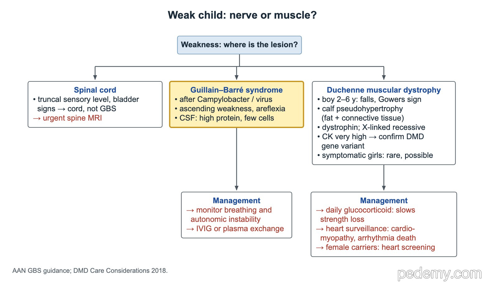
Guillain–Barré syndrome (GBS) often follows Campylobacter jejuni diarrhea or a viral illness. Weakness ascends from the legs, reflexes are lost, and back or leg pain is common in children.
- CSF: high protein with normal or few white cells (albuminocytologic dissociation); protein may be normal in the first week.
- A distinct truncal sensory level or early bladder dysfunction suggests spinal cord disease, not typical GBS: get an urgent spine MRI.
- Monitor closely: breathing (vital capacity, negative inspiratory force) and autonomic instability (arrhythmia, blood pressure swings).
- Treatment for a child who cannot walk or is getting worse: IVIG (2 g/kg over 2–5 days) or plasma exchange.
- Corticosteroids do not help GBS.
Acute ataxia
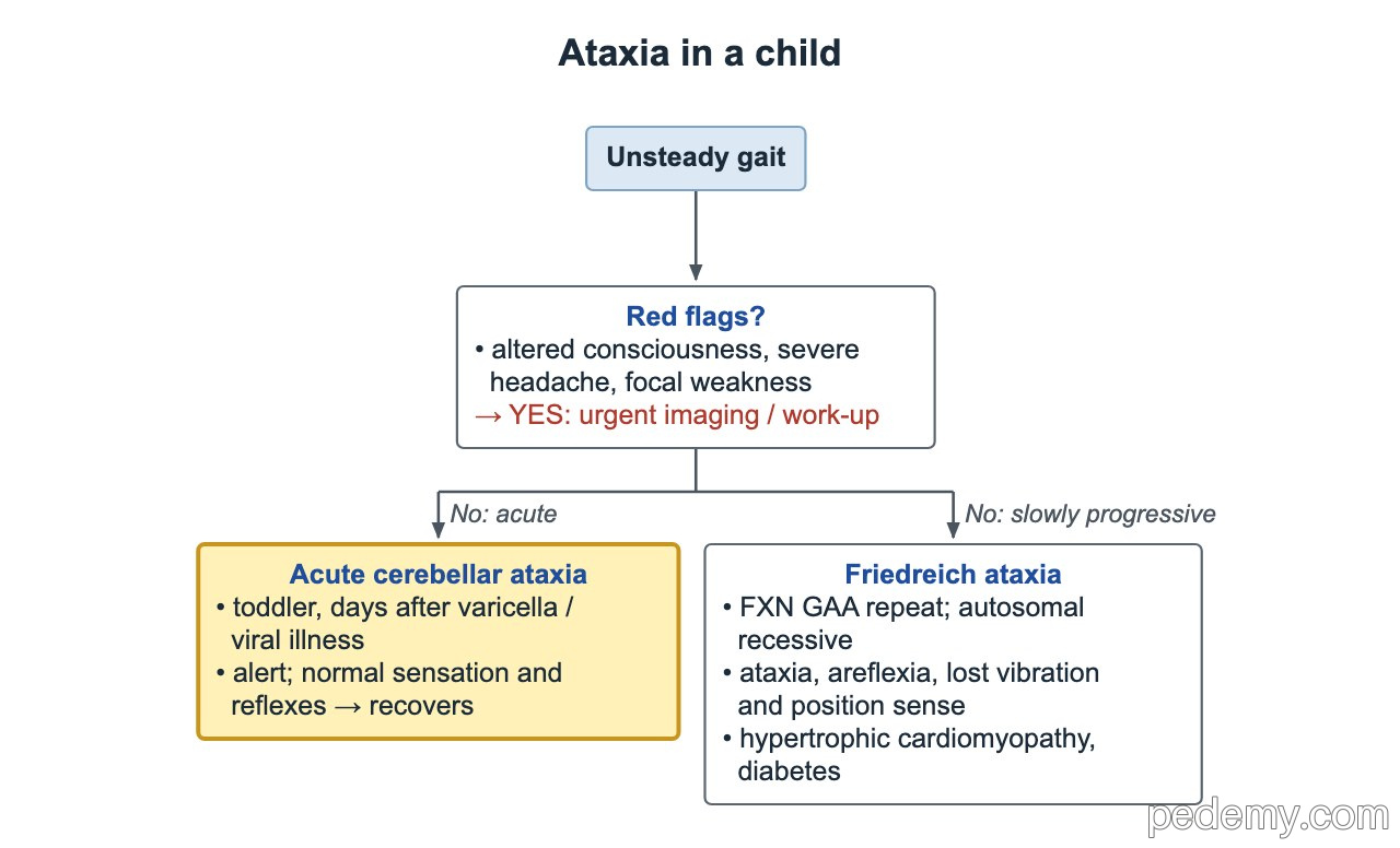
Acute cerebellar ataxia is the most common cause of acute ataxia in toddlers. It follows varicella or another viral illness by days to a few weeks.
- Wide-based, unsteady gait, often truncal ataxia, sometimes nystagmus; the child is alert, with normal sensation and reflexes.
- Recovery over weeks is the rule; no specific treatment.
- Red flags needing urgent imaging and work-up: altered consciousness, severe headache, focal weakness, papilledema or progressive course.
- Also consider ingestion, posterior fossa tumor, stroke, opsoclonus-myoclonus (neuroblastoma) and GBS variants.
- Slowly progressive ataxia with areflexia and lost vibration and position sense: Friedreich ataxia.
Common questions
How do you tell ADEM from a first attack of MS?
ADEM causes encephalopathy with large, poorly defined bilateral lesions and is usually a single episode. MS shows lesions in separate places and over time without encephalopathy.
What does CSF show in Guillain–Barré syndrome?
High protein with normal or few white cells. Protein can still be normal in the first week.
Is acute cerebellar ataxia after chickenpox dangerous?
Usually not. The child stays alert and recovers over weeks. Altered consciousness, severe headache or focal weakness need urgent imaging.
Study this topic
Sources
- Montalban X et al. Diagnosis of multiple sclerosis: 2024 revisions of the McDonald criteria (Lancet Neurol 2025)
- Krupp LB et al. International Pediatric MS Study Group criteria for pediatric MS and immune-mediated CNS demyelinating disorders (Mult Scler 2013)
- Leonhard SE et al. Diagnosis and management of Guillain–Barré syndrome in ten steps (Nat Rev Neurol 2019)
Written for Pedemy in our own words from the sources above; checked November 2026. For education only, not medical advice; check doses and local protocols before treating a patient. Pedemy is independent and not affiliated with or endorsed by the organizations cited.