

Main decision tree
Eye emergencies and eyelid lumps
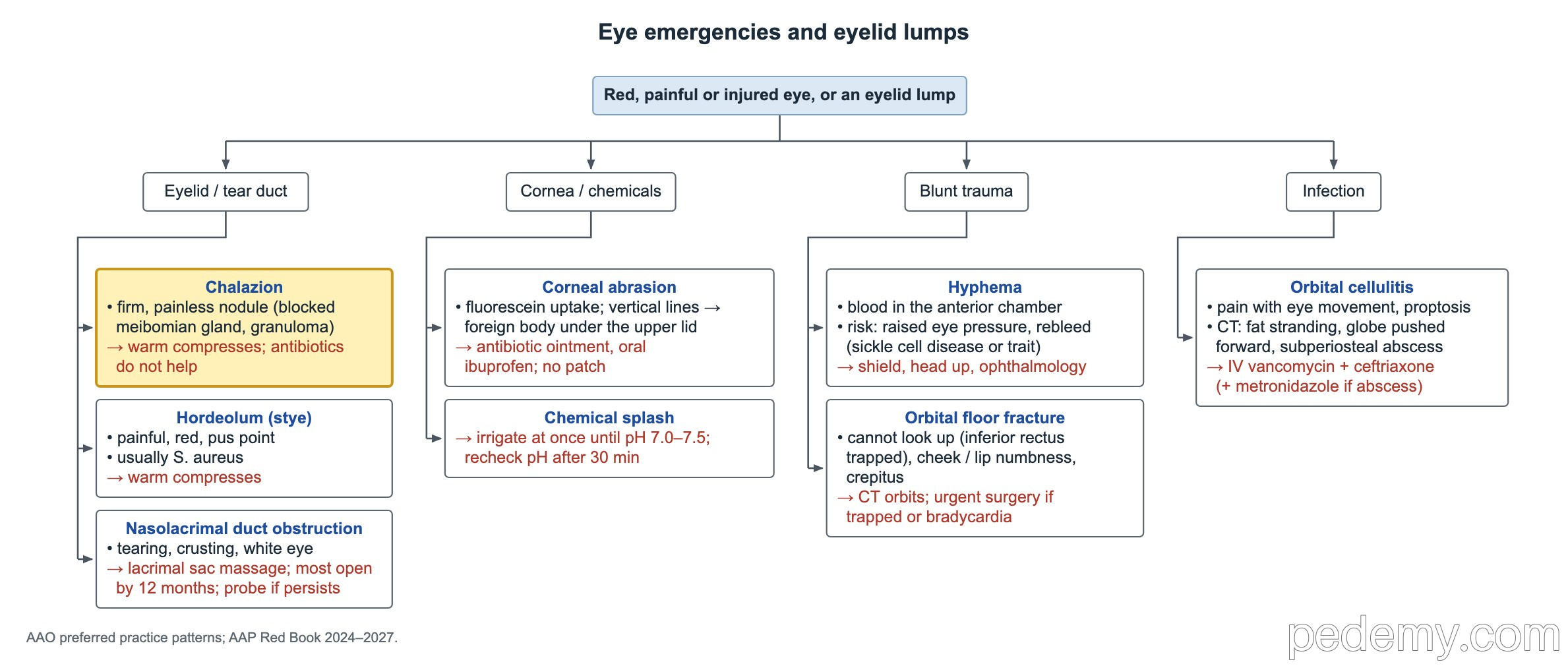
Key points
- Chalazion is firm and painless; hordeolum is painful and usually Staphylococcus aureus. Both get warm compresses.
- Infant tearing plus photophobia, blepharospasm or a cloudy, enlarged cornea means congenital glaucoma: urgent referral.
- Pain with eye movement, limited movement, proptosis or reduced vision means orbital cellulitis: admit, IV antibiotics, CT.
- Chemical splash: irrigate at once until the pH is 7.0–7.5, then recheck after 30 minutes. Alkali is worse than acid.
- Corneal abrasion: antibiotic ointment and oral pain relief, no patch; contact lens wearers need antipseudomonal drops.
- Hyphema: shield, head up 30°, same-day ophthalmology, sickle cell screen, no aspirin or NSAIDs.
- Open globe: rigid shield, IV antibiotics, antiemetic, head up, CT of the orbits; no pressure, drops, fluorescein or ultrasound.
Eyelid lumps and the tear sac
Start with the lid. Ask whether the lump is painful, where it sits and whether the child has fever.
- Chronic blepharitis (crusted, inflamed lid margins) predisposes to both hordeola and chalazia.
- Lid hygiene and warm compresses treat blepharitis and help prevent new lumps.
| Lesion | Clues | Treatment |
|---|---|---|
| Hordeolum (stye) | Acute, painful, red lump at the lid margin, often with a pus point; usually S. aureus | Warm compresses |
| Chalazion | Firm, painless nodule in the lid for weeks; a blocked meibomian gland forms a granuloma | Warm compresses; antibiotic or steroid drops do not help |
| Acute dacryocystitis | Tender red swelling at the inner corner of the eye (medial canthus), often with fever | Systemic antibiotics |
The tearing infant: blocked duct or glaucoma?
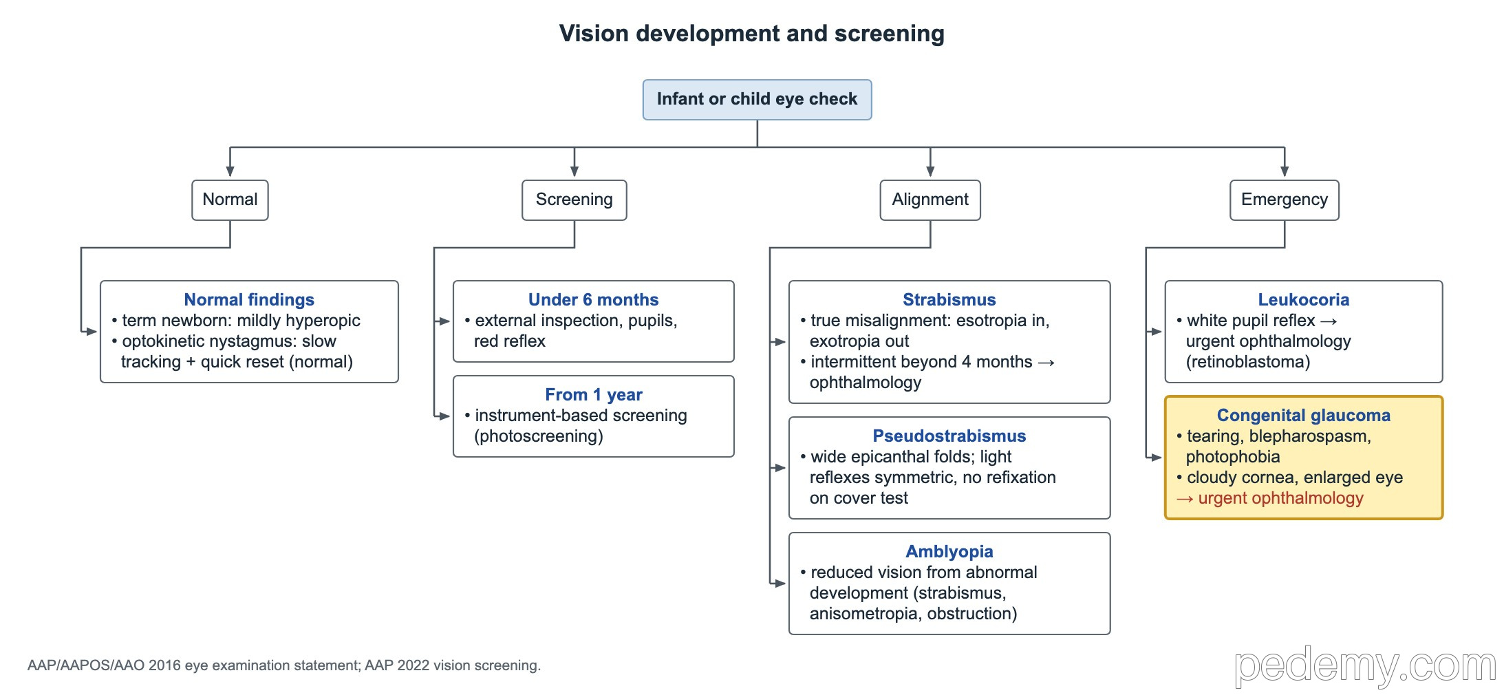
Persistent tearing with lash crusting and a white, quiet eye is congenital nasolacrimal duct obstruction. Treat with lacrimal sac (Crigler) massage. Most ducts open by about 12 months; refer for probing if it persists at about 1 year.
Tearing with photophobia or blepharospasm points to primary congenital glaucoma instead. High pressure stretches the infant eye, so the cornea becomes hazy and enlarged. This needs urgent ophthalmology referral.
| Feature | Blocked tear duct | Congenital glaucoma |
|---|---|---|
| Tearing | Yes, with mucus or crusting | Yes |
| Light sensitivity, squeezing the lids | No | Yes (photophobia, blepharospasm) |
| Cornea | Clear, normal size | Cloudy, enlarged |
| Cause | Membrane blocking the lower end of the duct | Abnormal trabecular meshwork blocks aqueous outflow |
| Treatment | Massage; probing if it persists at ~12 months | Surgery (angle surgery), with drops as support |
Swollen eyelid: preseptal or orbital cellulitis
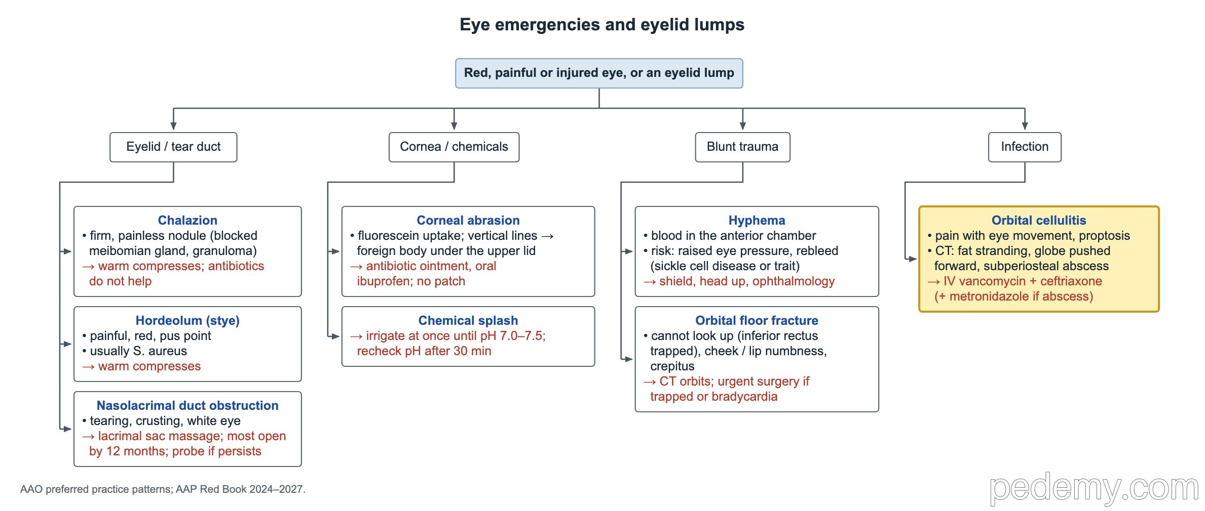
The orbital septum separates the lid from the orbit. Infection in front of it (preseptal) leaves the eye itself normal. Infection behind it (orbital) affects eye movement and vision.
- CT in orbital cellulitis shows fat stranding, a globe pushed forward and sometimes a subperiosteal abscess.
- Typical IV regimen: vancomycin plus ceftriaxone; add metronidazole for anaerobes when there is an abscess.
- Watery red eyes that spread from one eye to the other during a cold are viral conjunctivitis, most often adenovirus; the lids are not tender and eye movements are normal.
| Finding | Preseptal (periorbital) | Orbital |
|---|---|---|
| Typical story | Insect bite, skin break or stye; child looks well | Fever; usual source is ethmoid sinusitis |
| Eye movements | Full and painless | Painful and limited |
| Globe and vision | Normal | Proptosis; vision may be reduced |
| Management | Oral antibiotics in a well child | Admit, IV antibiotics, CT of the orbits, ophthalmology and ENT |
Corneal abrasion and chemical splash
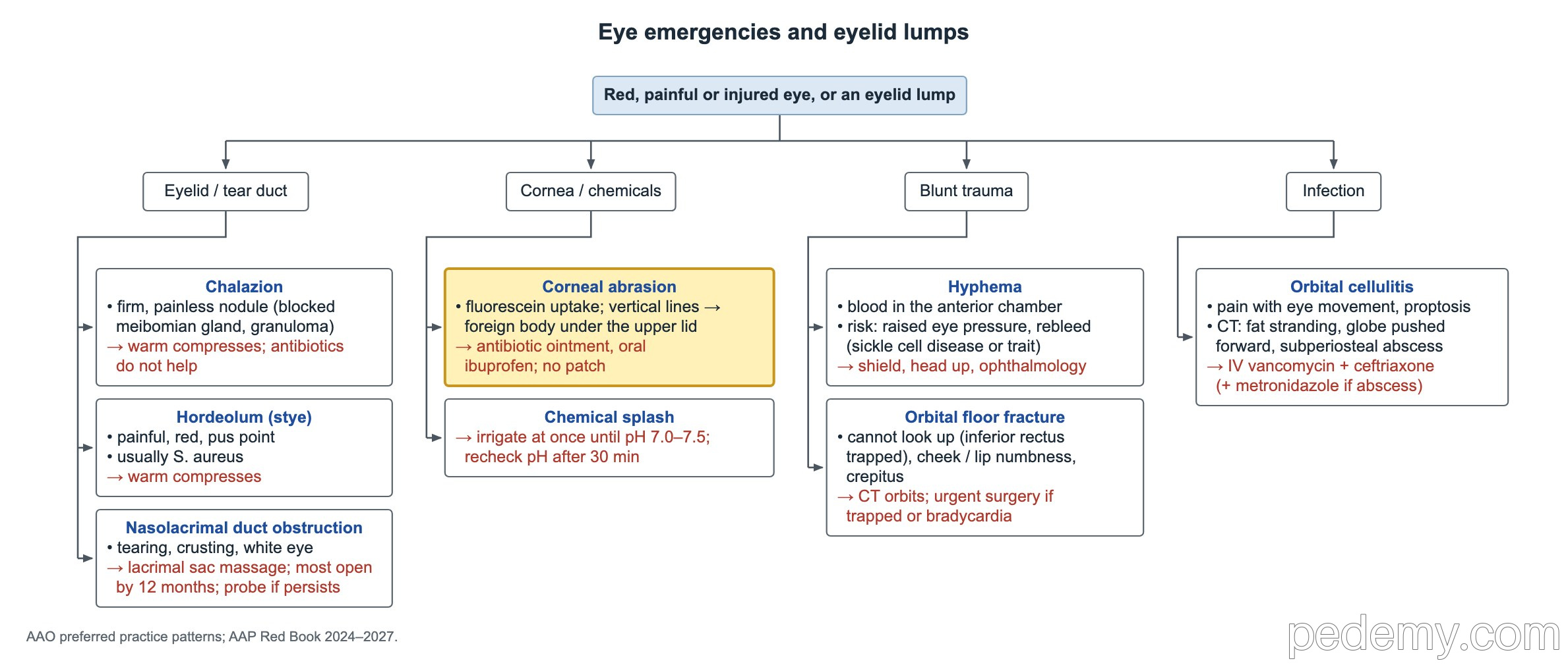
A scratched cornea causes pain, tearing and photophobia with normal vision. Fluorescein under blue light shows a bright green epithelial defect. Several vertical linear marks suggest a foreign body under the upper lid, so evert the lid.
- Treat with topical antibiotic ointment (for example erythromycin) and oral ibuprofen or acetaminophen.
- Do not patch, and do not send the child home with topical anesthetic drops.
- Contact lens wearers: stop lens wear, use a topical antipseudomonal antibiotic and recheck closely, because microbial keratitis is a risk.
- Chemical splash: irrigate at once, before the full exam, until the pH is neutral (7.0–7.5). Recheck the pH after 30 minutes.
- Alkali burns are worse than acid burns because alkali penetrates deeper into the eye.
Blunt trauma: hyphema and orbital floor fracture
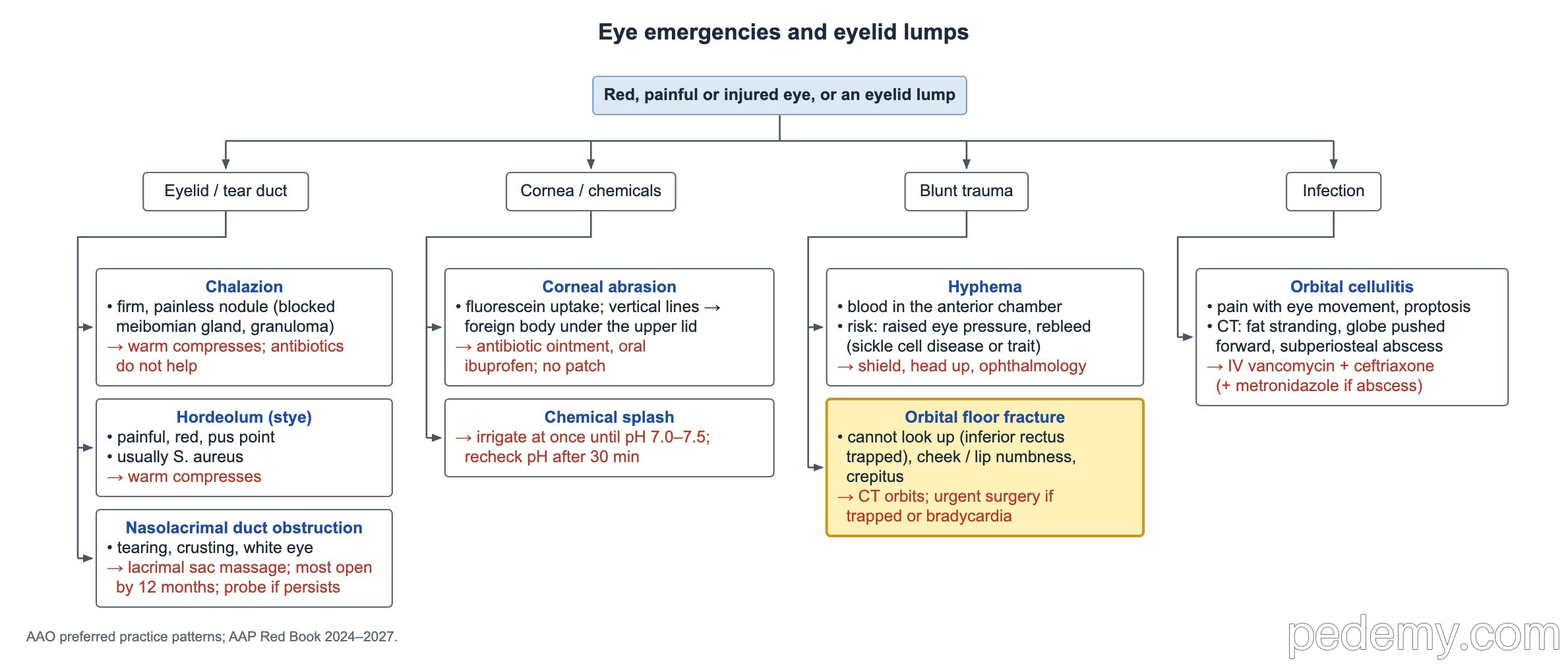
Hyphema is blood layering in the anterior chamber in front of the iris after blunt injury, such as a ball or hockey stick. The main early complication is raised eye pressure, because red cells block the trabecular meshwork; rebleeding is the other risk.
Orbital floor fracture traps the inferior rectus muscle. The child cannot look up and has cheek and upper-lip numbness (infraorbital nerve) and crepitus from air in the tissues.
- Hyphema: rigid shield, head of the bed up 30°, rest and same-day ophthalmology.
- Hyphema: screen for sickle cell disease or trait when status is unknown; sickled cells raise the pressure risk.
- Hyphema: avoid aspirin and NSAIDs.
- Floor fracture: CT of the orbits is the first study.
- Floor fracture with bradycardia, nausea or vomiting is the oculocardiac reflex from a trapped muscle and needs urgent surgery.
Open globe injury
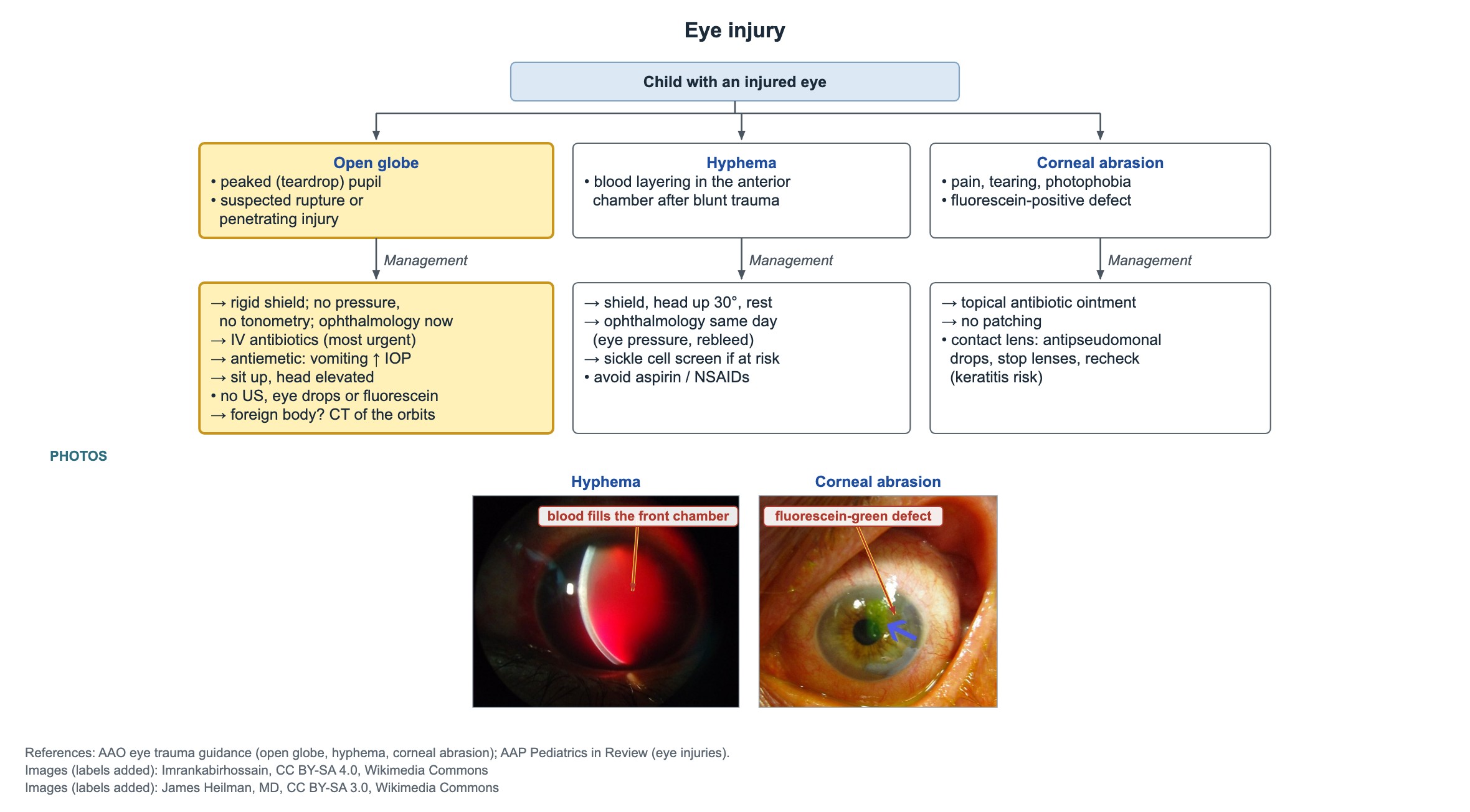
Suspect an open globe after a sharp or high-speed injury, such as an air gun, when the pupil is peaked or teardrop-shaped. The goal before the ophthalmologist arrives is to protect the eye and prevent infection.
- Place a rigid shield. Put no pressure on the eye and do no tonometry.
- Give IV antibiotics: this is the most urgent medication, to prevent endophthalmitis.
- Give an antiemetic, because vomiting raises eye pressure and can push eye contents out.
- Keep the child sitting up with the head elevated.
- Skip fluorescein, topical drops and bedside ocular ultrasound; leave the exam to ophthalmology.
- Possible intraocular foreign body: CT of the orbits.
Drooping eyelid (ptosis)
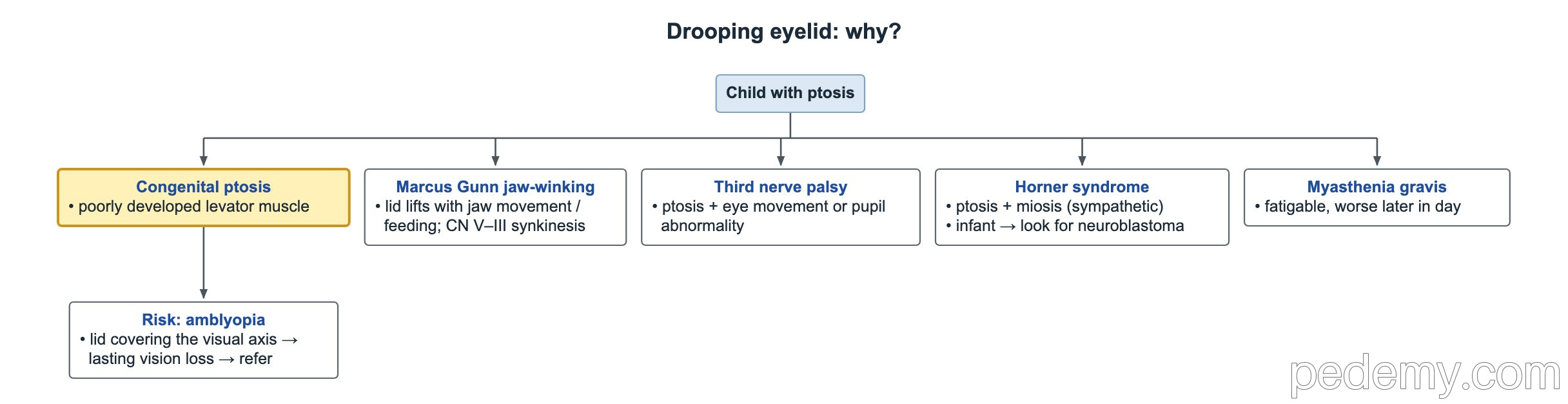
Ptosis present from birth is usually congenital ptosis from a poorly developed levator palpebrae muscle. A lid that covers the pupil can cause amblyopia, so refer.
| Cause | Key clue |
|---|---|
| Congenital ptosis | Present from birth; levator muscle dysgenesis |
| Marcus Gunn jaw-winking | Lid lifts with sucking or chewing; trigeminal (V) to oculomotor (III) miswiring |
| Third nerve (CN III) palsy | Ptosis with impaired eye movements, with or without a dilated pupil |
| Horner syndrome | Mild ptosis with a small pupil on the same side |
| Myasthenia gravis | Fatigable ptosis, worse by evening, better after rest; neuromuscular junction |
Common questions
How do you tell orbital from preseptal cellulitis?
Orbital cellulitis causes pain with eye movement, limited movement, proptosis or reduced vision. In preseptal cellulitis the lid is swollen but vision and eye movements are normal and painless.
Should a corneal abrasion be patched?
No. Use antibiotic ointment and oral pain relief. Contact lens wearers need an antipseudomonal antibiotic and no lens wear.
What is the first step for a chemical in the eye?
Irrigate right away and keep going until the pH is 7.0–7.5, then recheck after 30 minutes.
What should you not do with a suspected ruptured globe?
Do not press on the eye, measure pressure, put in drops or fluorescein, or use ultrasound. Shield the eye, give IV antibiotics and an antiemetic, and call ophthalmology.
Study this topic
Sources
- AAO 2022: Pediatric Eye Evaluations Preferred Practice Pattern (Ophthalmology)
- AAO 2024: Blepharitis Preferred Practice Pattern (Ophthalmology)
- AAO 2024: Bacterial Keratitis Preferred Practice Pattern (Ophthalmology)
- AAP 2016: Visual system assessment in infants, children and young adults (Pediatrics)
Written for Pedemy in our own words from the sources above; checked November 2026. For education only, not medical advice; check doses and local protocols before treating a patient. Pedemy is independent and not affiliated with or endorsed by the organizations cited.