

Main decision tree
Bone lesions in children
Key points
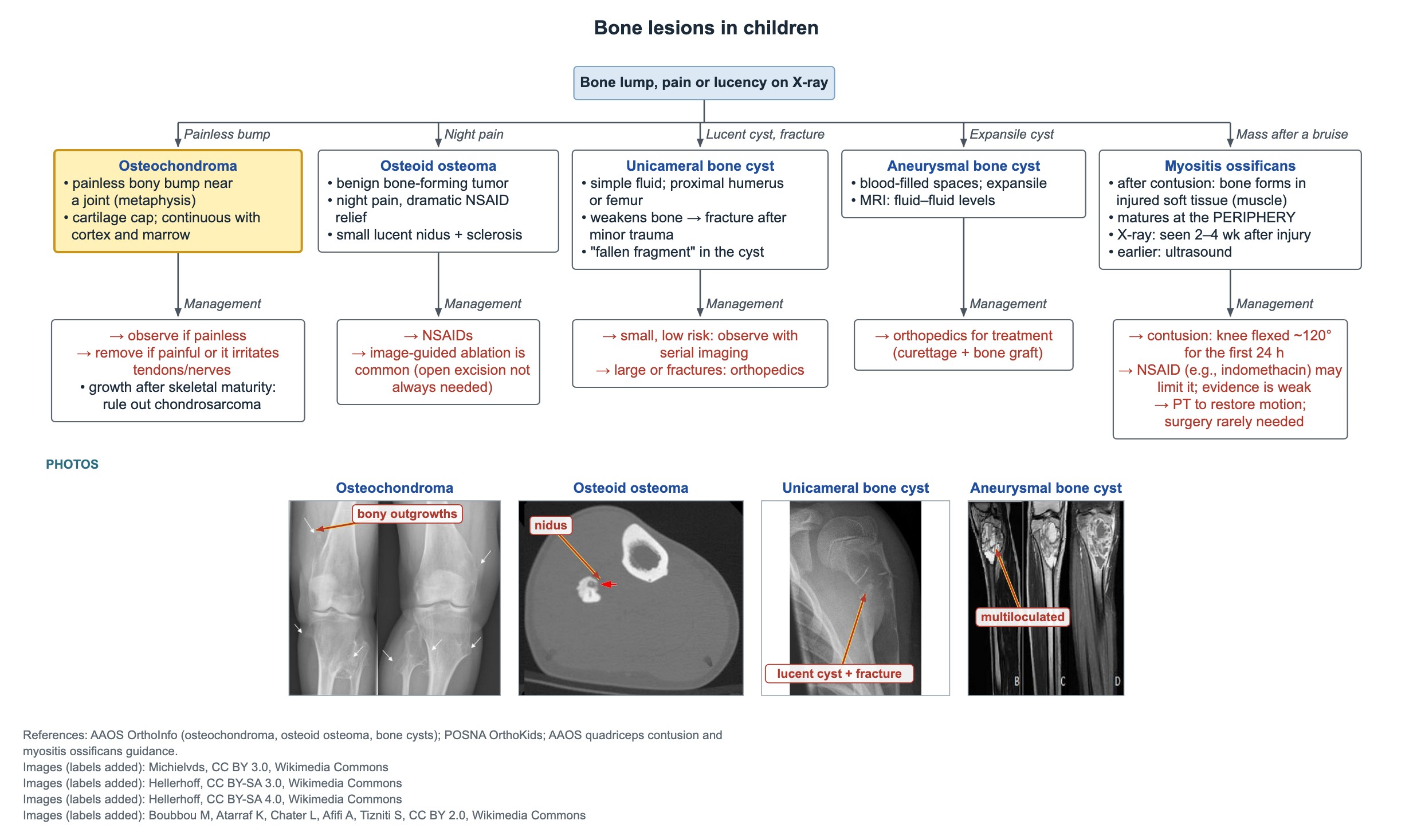
- Osteochondroma: painless metaphyseal bump with a cartilage cap, continuous with the cortex and marrow; observe if painless.
- Osteoid osteoma: benign bone-forming tumor; night pain with dramatic NSAID relief; small lucent nidus with reactive sclerosis.
- Osteoid osteoma treatment: NSAIDs, or image-guided ablation; open excision is not always needed.
- Unicameral bone cyst: simple fluid, proximal humerus or femur; weakens bone and fractures after minor trauma.
- Aneurysmal bone cyst: expansile, blood-filled spaces with fluid–fluid levels on MRI; refer to orthopedics.
- Myositis ossificans: bone forms in injured muscle; X-ray shows it 2–4 weeks after injury, ultrasound earlier; matures at the periphery.
- After a quadriceps contusion, hold the knee in about 120° of flexion for the first 24 hours.
Osteochondroma: the painless bump
Osteochondroma is the most common benign bone tumor in children. It is a bony outgrowth near a joint, at the metaphysis, capped with cartilage. The key X-ray sign is that its cortex and marrow are continuous with the parent bone.
A painless osteochondroma is observed. Remove it if it hurts, irritates a tendon, nerve or vessel, or limits motion.
- Multiple lesions suggest hereditary multiple osteochondromas (EXT1 or EXT2 genes).
- Growth or new pain after skeletal maturity raises concern for chondrosarcoma: image and refer.
Osteoid osteoma: night pain relieved by NSAIDs
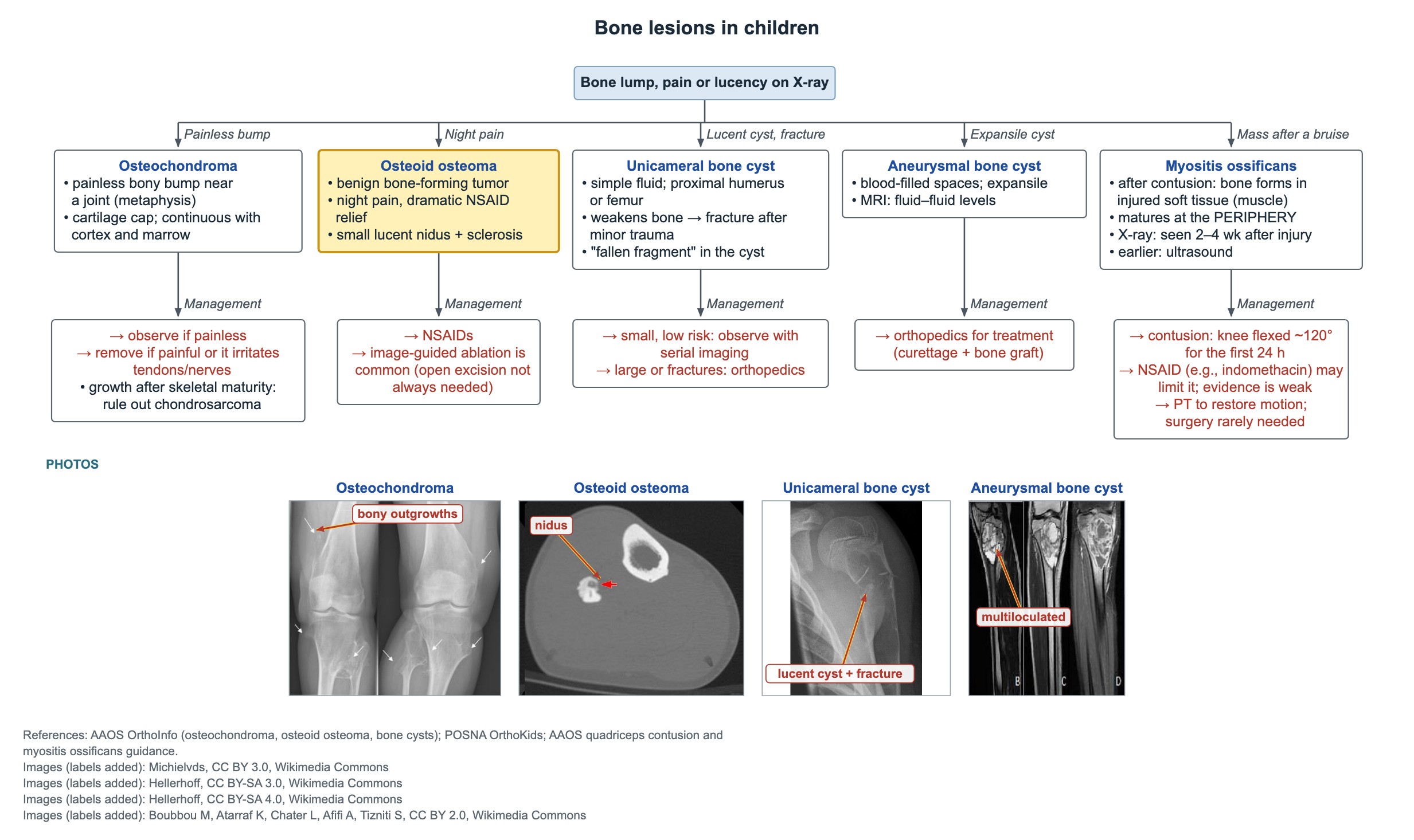
Osteoid osteoma is a benign bone-forming tumor, most common in older children and teens, often in the femur or tibia. The classic story is focal pain that is worse at night and dramatically relieved by NSAIDs, because the nidus makes prostaglandins.
Imaging shows a small lucent nidus, usually under 1.5 cm, surrounded by dense reactive sclerosis. CT shows the nidus best.
- In the spine it can cause painful scoliosis.
- Near a joint it can mimic arthritis with effusion and stiffness.
Treating osteoid osteoma
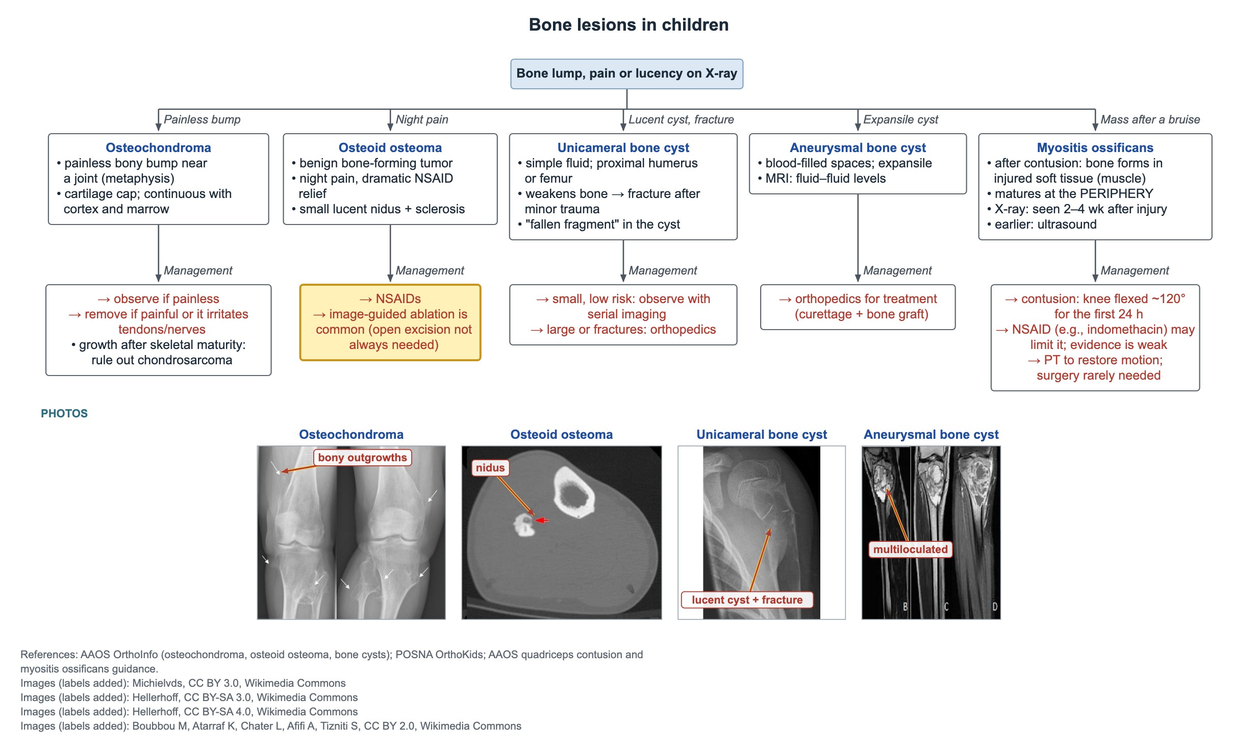
Open excision is not always required. Some children control symptoms with NSAIDs until the lesion burns out, which can take years.
Image-guided (CT-guided) radiofrequency ablation is a common definitive treatment, with high success and few complications. Surgery is kept for lesions that cannot be ablated safely.
Unicameral and aneurysmal bone cysts
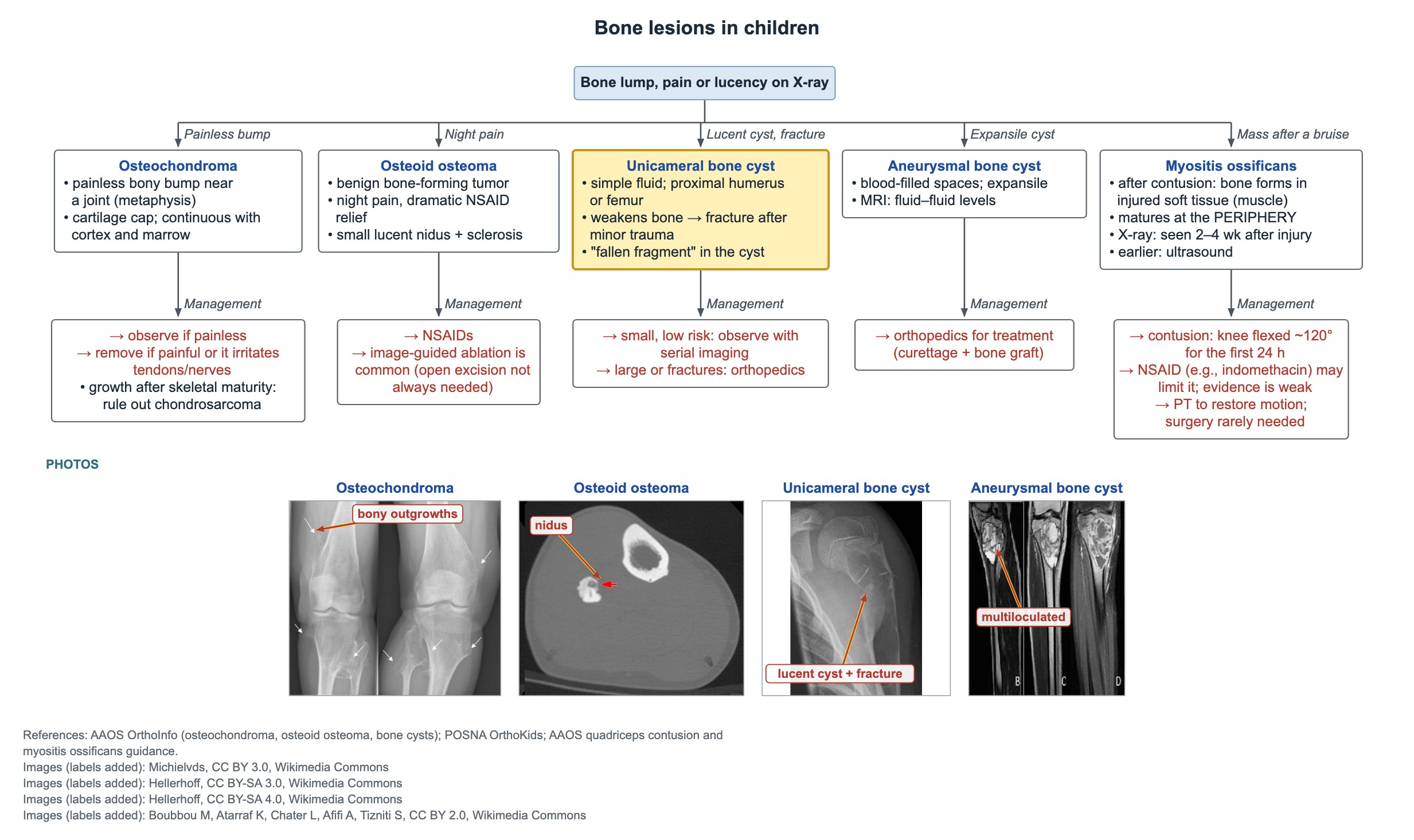
A unicameral (simple) bone cyst is a fluid-filled cavity, usually in the proximal humerus or proximal femur of a growing child. It is often painless and found on an X-ray or after a pathologic fracture: the thin cortex breaks after minor trauma. A bone chip lying in the bottom of the cyst is the fallen fragment sign.
A small, asymptomatic cyst with low fracture risk is observed with serial X-rays. Large cysts, cysts in the proximal femur and cysts that have fractured go to orthopedics.
Aneurysmal bone cyst
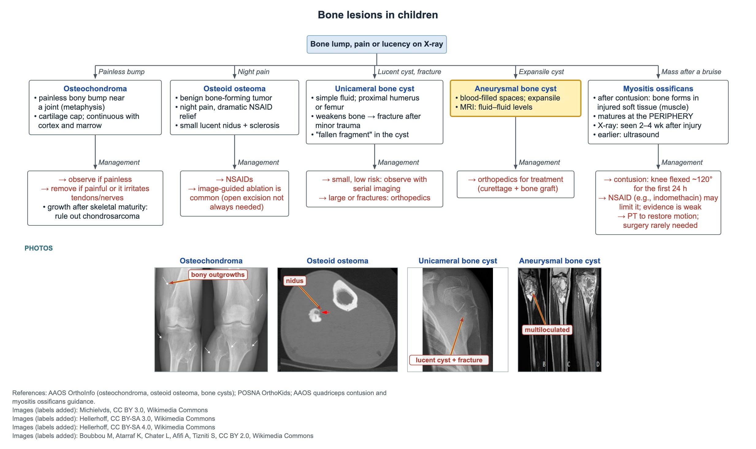
An expansile aneurysmal bone cyst needs orthopedic evaluation for lesion-directed treatment. Telangiectatic osteosarcoma can look similar, so tissue diagnosis is part of the work-up.
| Feature | Unicameral bone cyst | Aneurysmal bone cyst |
|---|---|---|
| Contents | Simple fluid | Blood-filled spaces |
| X-ray | Central lucency, thin cortex | Expansile, eccentric lucency |
| MRI | Single fluid cavity | Multiple fluid–fluid levels |
| Usual course | Small, low risk: observe | Grows; needs orthopedic treatment (often curettage and bone graft) |
Myositis ossificans after a thigh contusion
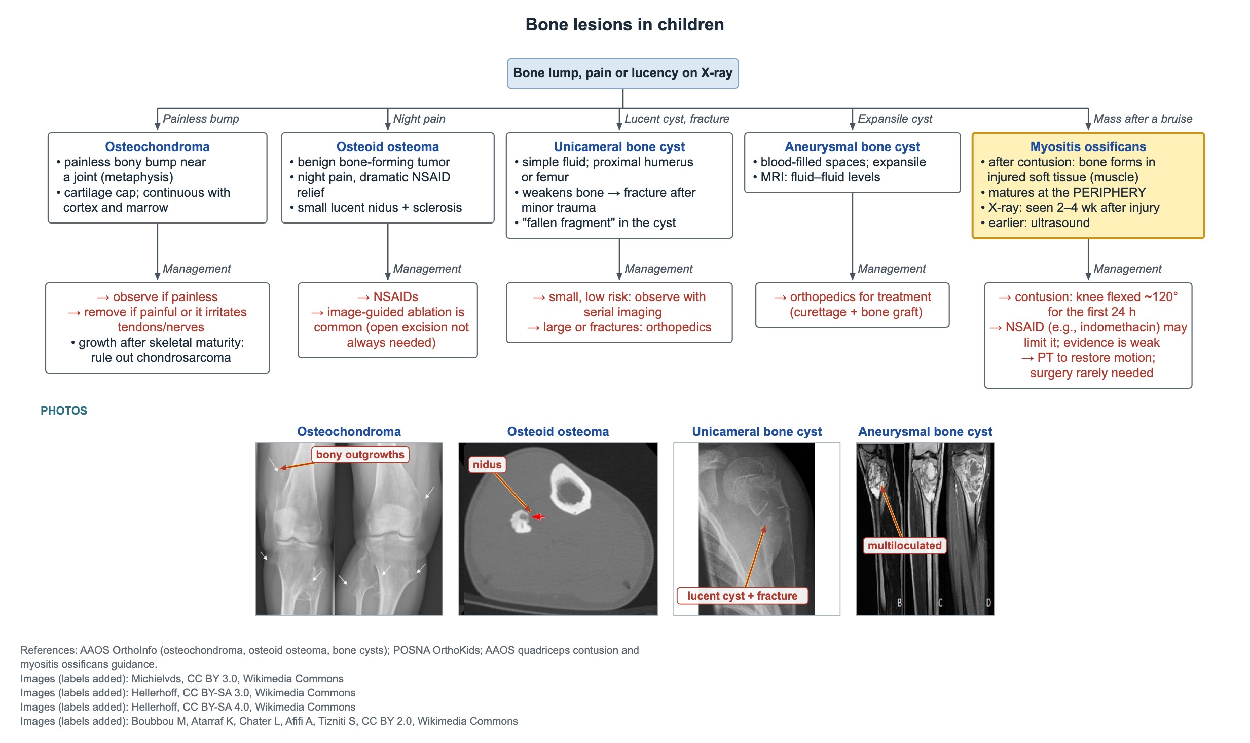
Traumatic myositis ossificans is bone forming inside injured soft tissue, most often the quadriceps after a hard blow. Think of it when a firm, enlarging thigh mass and loss of knee motion appear 2–4 weeks after a contusion.
The first test is a plain X-ray of the femur. Calcification usually shows 2–4 weeks after the injury; ultrasound can show it earlier while the X-ray is still normal. The bone matures at the periphery, with a less mature center, which is the opposite of osteosarcoma.
- The mass sits in the muscle and is separate from the bone.
- Avoid early biopsy: immature lesions can look like sarcoma under the microscope.
Preventing and treating myositis ossificans
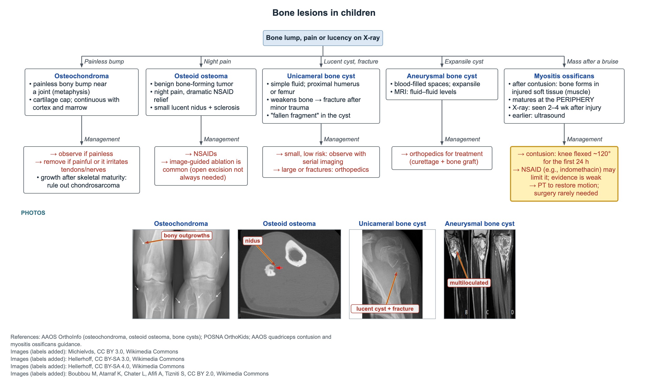
- Right after a quadriceps contusion: hold the knee flexed to about 120° for the first 24 hours, with ice.
- An NSAID such as indomethacin given early may limit heterotopic bone; the evidence is weak.
- Once it forms: physical therapy to restore knee motion; gentle, pain-free stretching.
- Surgery is rarely needed: excise only if still symptomatic after the bone has matured, about 6–12 months.
Common questions
Why does aspirin or ibuprofen relieve osteoid osteoma pain so well?
The nidus makes large amounts of prostaglandins. NSAIDs block prostaglandin production, so night pain eases dramatically.
How soon does myositis ossificans show on X-ray?
Usually 2–4 weeks after the injury. Ultrasound can detect it earlier.
Do unicameral bone cysts need surgery?
Not always. Small cysts with low fracture risk are watched with serial X-rays. Large cysts, cysts in the proximal femur and cysts that fracture are referred to orthopedics.
Study this topic
Sources
- Rosenblatt J, Koder A. Understanding unicameral and aneurysmal bone cysts (Pediatrics in Review, 2019)
- Wolf M. Knee pain in children, part III: stress injuries, benign bone tumors, growing pains (Pediatrics in Review, 2016)
- Dilek I et al. CT-guided radiofrequency ablation of osteoid osteoma in children and adults (Skeletal Radiology, 2025)
- AAOS OrthoInfo: Osteoid osteoma
Written for Pedemy in our own words from the sources above; checked November 2026. For education only, not medical advice; check doses and local protocols before treating a patient. Pedemy is independent and not affiliated with or endorsed by the organizations cited.