

Main decision tree
Limp or hip pain in a child: first, the approach
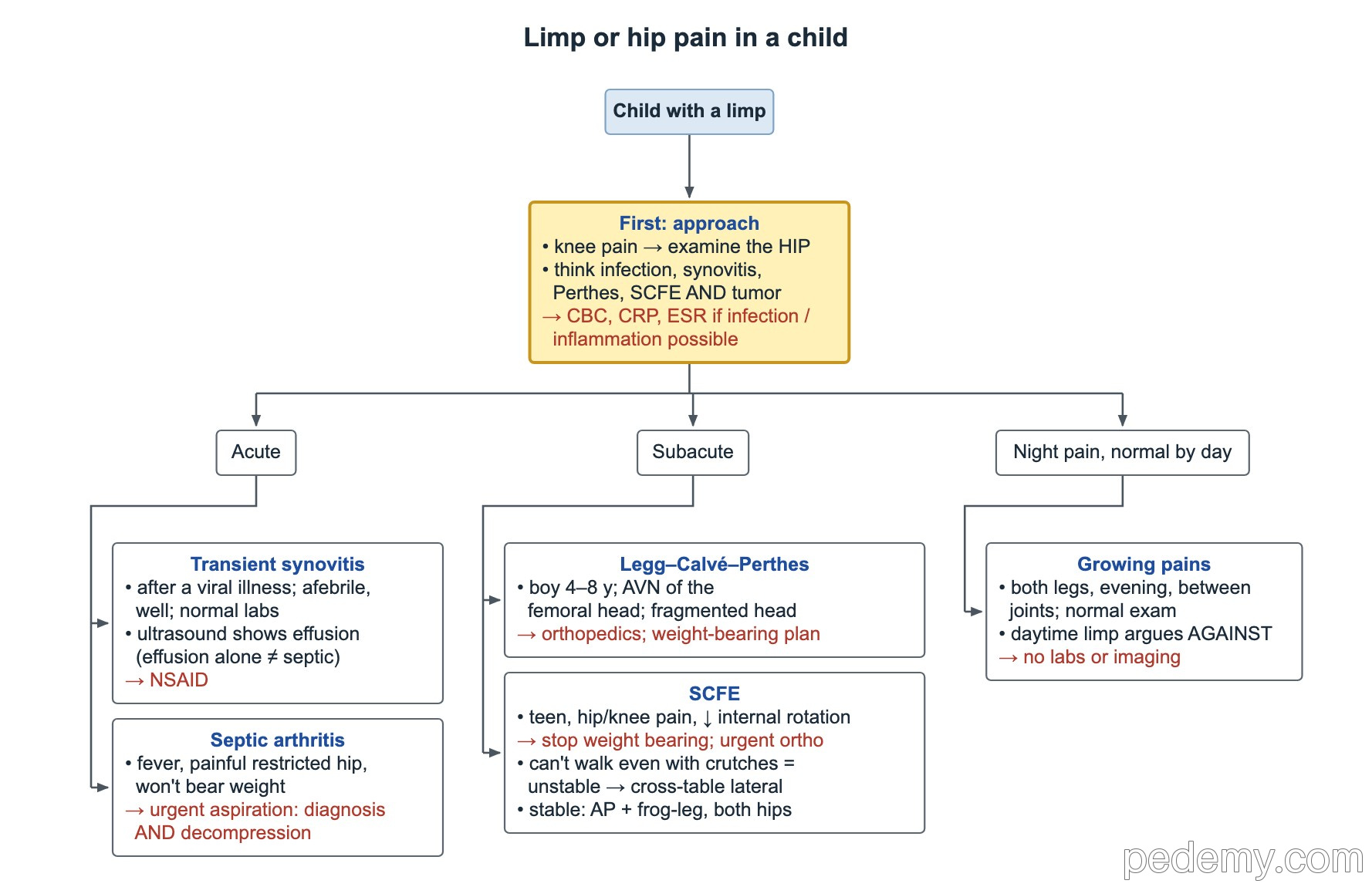
Key points
- Knee pain and limp: examine the hip; hip disease often refers pain to the knee.
- Kocher criteria for septic hip: fever >38.5 °C (101.3 °F), refusal to bear weight, ESR >40 mm/h, WBC >12,000/µL; CRP >2 mg/dL adds risk.
- Suspected septic joint → urgent aspiration for diagnosis and decompression; joint fluid WBC usually >50,000/µL with neutrophils.
- Kingella kingae is the commonest cause of septic arthritis at 6 months–4 years; culture is often negative, so use PCR.
- SCFE: teen, hip or knee pain, lost internal rotation → stop weight bearing, urgent orthopedics; AP and frog-leg X-rays of both hips.
- Perthes disease: avascular necrosis of the femoral head, usually boys 4–8 years.
- DDH: ultrasound before about 4–6 months, AP pelvis X-ray after; positive Ortolani or Barlow → orthopedic referral.
Approach to the limping child
Start with age, fever, trauma and whether the child will bear weight. Examine the whole limb, the spine and the abdomen, and always the hip when the complaint is knee pain.
Childhood hip pain covers infection, transient synovitis, Perthes disease, SCFE and tumor. When infection or inflammation is possible, check a CBC, CRP and ESR, and get a hip ultrasound to look for effusion.
| Age | Think of |
|---|---|
| Infant | DDH, septic arthritis, osteomyelitis |
| 1–3 years | Toddler fracture, septic arthritis, Kingella infection |
| 3–8 years | Transient synovitis, septic arthritis, Perthes disease |
| Adolescent | SCFE, apophyseal avulsion, overuse injury, tumor |
Septic arthritis vs transient synovitis
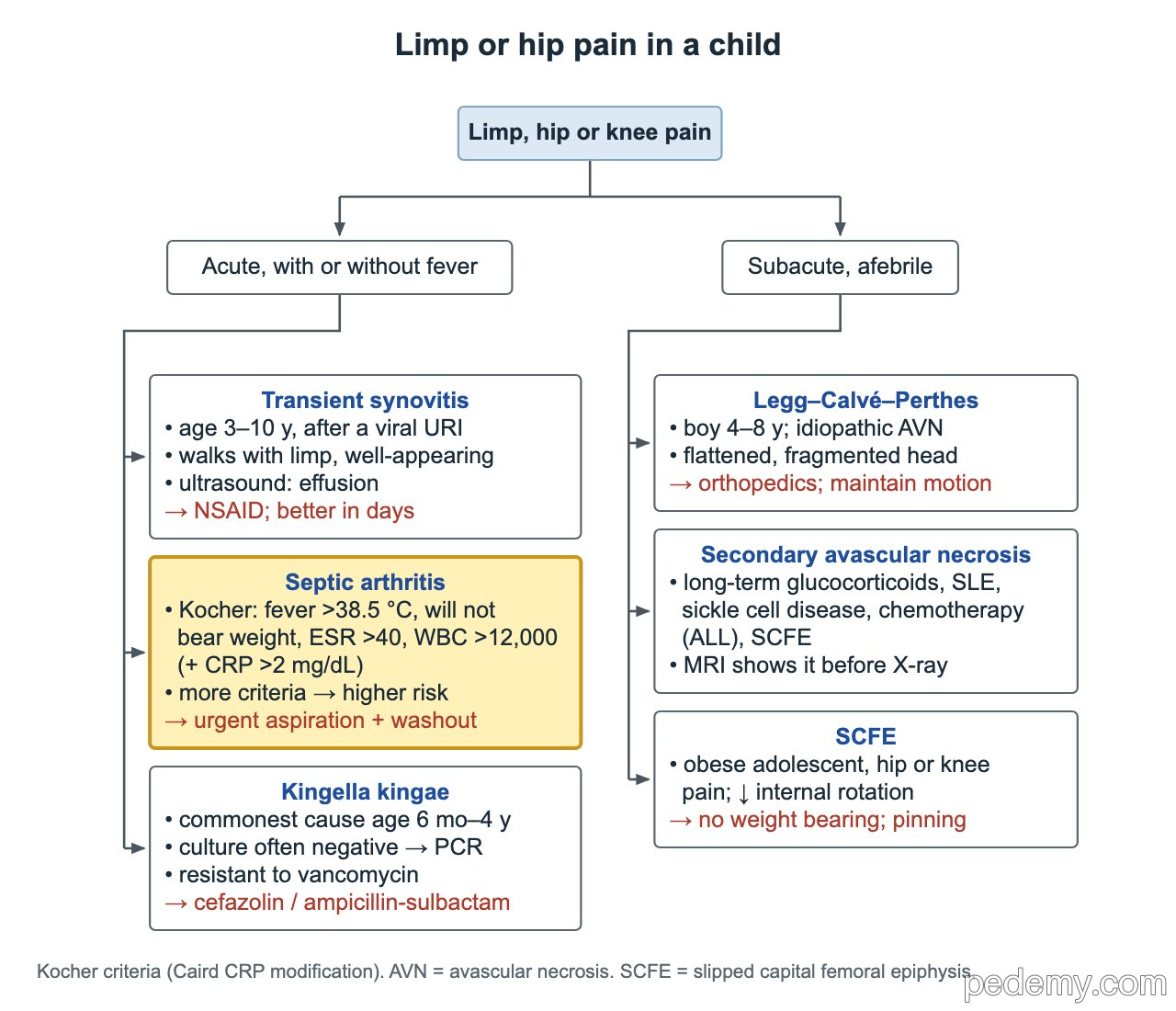
Fever, a painful hip with restricted motion and refusal to bear weight mean septic arthritis until proven otherwise. The Kocher predictors are fever above 38.5 °C (101.3 °F), refusal to bear weight, ESR above 40 mm/h and WBC above 12,000/µL. The more present, the higher the risk: with 3 the probability is about 90%, with all 4 it is near 100%. CRP above 2 mg/dL adds risk.
An effusion on ultrasound does not prove infection: transient synovitis also causes one. Combine exam, labs and aspiration.
- Confirm with urgent joint aspiration: joint fluid WBC usually above 50,000/µL with neutrophil predominance, plus Gram stain and culture.
- Urgent drainage also decompresses the joint to protect the femoral head's blood supply.
- Transient synovitis: afebrile, well child, often after a viral illness, normal or near-normal labs → NSAID and follow-up; it settles within days.
Kingella kingae and culture-negative arthritis
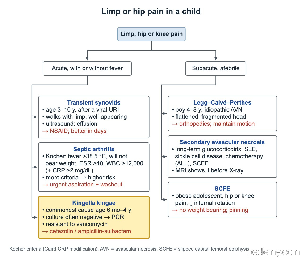
Kingella kingae is the commonest cause of septic arthritis in children 6 months to 4 years. Children often look well, with modest fever and labs. The organism grows poorly, so culture is often negative.
Detect it with PCR of joint fluid, or by inoculating joint fluid into blood culture bottles. Kingella is resistant to vancomycin; cefazolin or ampicillin-sulbactam covers it.
- IDSA/PIDS 2023: give empiric antibiotics that cover Staphylococcus aureus and, in young children, Kingella.
- Most children can switch to oral antibiotics after a short IV course once they improve.
Slipped capital femoral epiphysis (SCFE)
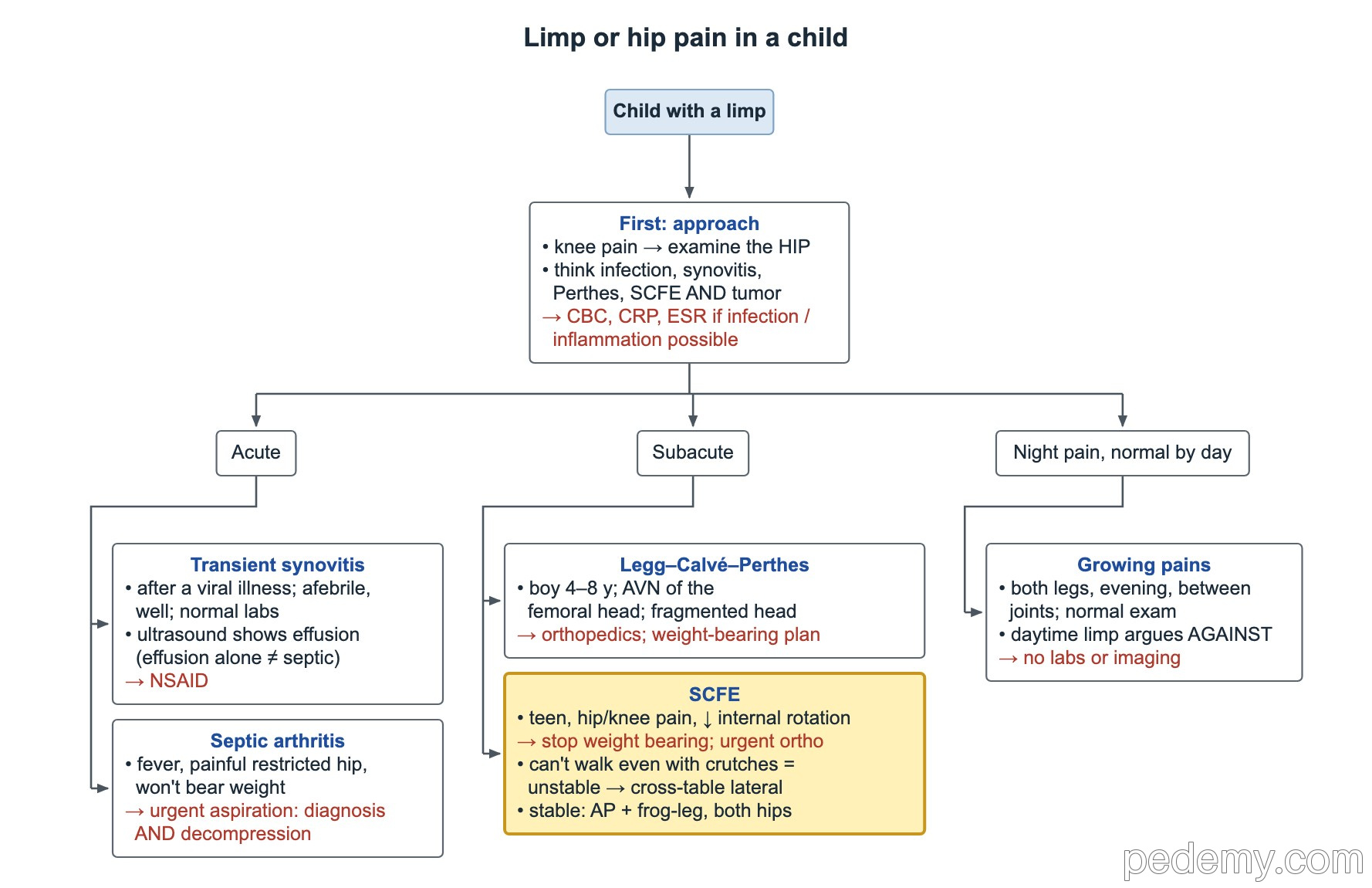
SCFE is a slip of the femoral head through the physis, usually in a young adolescent with obesity during the growth spurt. Pain may be in the hip, thigh or knee. On exam the hip has reduced internal rotation and rolls into external rotation when flexed.
Suspected SCFE: stop weight bearing and arrange urgent orthopedic assessment. Treatment is surgical fixation in situ.
- Stable SCFE (can walk, with or without crutches): AP and frog-leg lateral X-rays of both hips.
- Unstable SCFE (cannot walk even with crutches): use a cross-table lateral, not a forced frog-leg view; avascular necrosis risk is much higher.
- Check both hips: the other side often slips too.
- SCFE before 10 years or in a thin child: look for hypothyroidism or another endocrine cause.
Legg-Calvé-Perthes disease and avascular necrosis
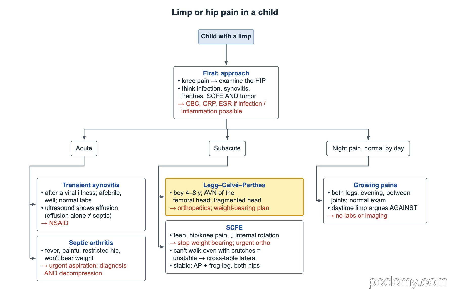
Perthes disease is idiopathic avascular necrosis of the femoral head. The typical patient is a boy aged 4–8 years with an atraumatic limp and mild hip or knee pain. X-rays show a dense, flattened and later fragmented femoral head.
Refer to orthopedics for an activity and weight-bearing plan; younger children usually do better. The quick contrast: younger child with femoral-head necrosis = Perthes; adolescent with a physeal slip = SCFE.
- Secondary avascular necrosis in a teen: long-term glucocorticoids, systemic lupus erythematosus, sickle cell disease, chemotherapy for ALL, or after SCFE.
- MRI shows avascular necrosis before X-ray does.
Developmental dysplasia of the hip (DDH)
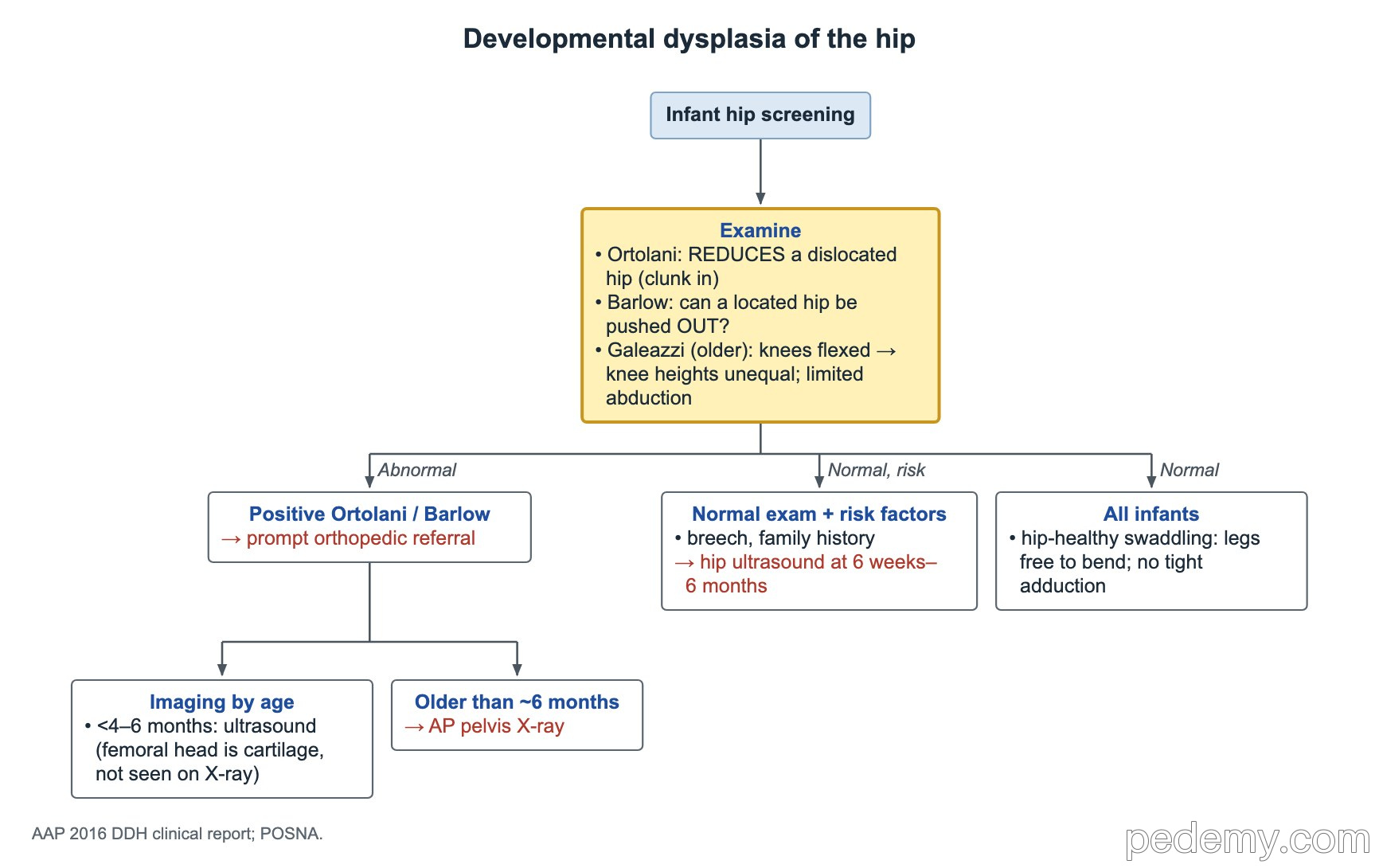
- Ortolani: reduces an already dislocated hip (clunk in).
- Barlow: tests whether a located hip can be pushed out (displaced) of the acetabulum.
- After about 3 months: limited abduction and a positive Galeazzi sign (hips and knees flexed, one knee lower).
- Positive Ortolani or Barlow in a newborn → referral to pediatric orthopedics.
- Breech in the third trimester or a family history, with a normal exam → hip ultrasound at about 6 weeks (or X-ray at about 4 months).
- Imaging by age: ultrasound before about 4–6 months, because the femoral head is still cartilage; AP pelvis X-ray once it ossifies.
- Hip-healthy swaddling: legs free to bend at the hips; avoid legs held straight and pressed together.
- Congenital muscular torticollis: DDH coexists in up to about 1 in 5; examine the hips, with or without ultrasound.
Growing pains and apophyseal avulsions
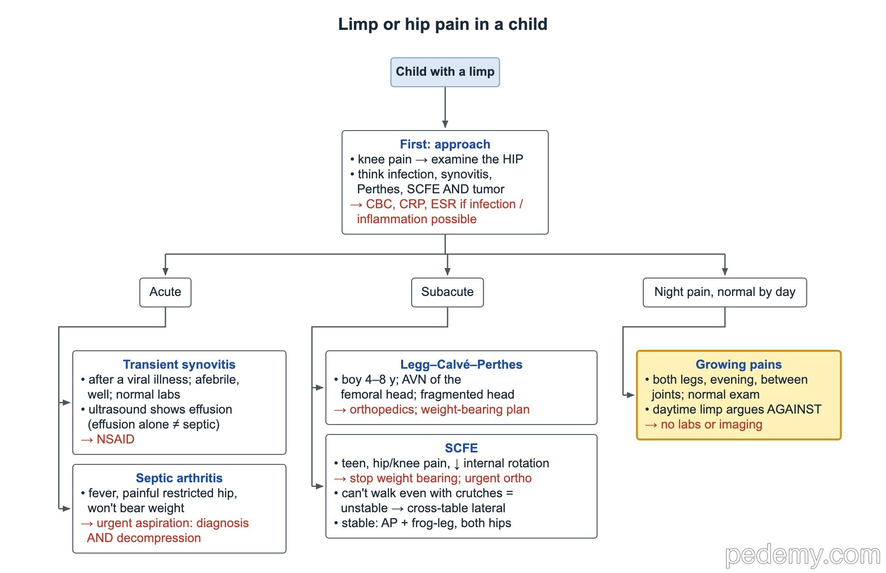
Growing pains are bilateral evening or night pains in the calves or thighs, between the joints, in a child who walks normally by day and has a normal exam. Routine labs and imaging are not needed. Daytime limp, joint swelling, one-sided pain or systemic symptoms argue against growing pains.
In adolescent athletes, a sudden forceful muscle contraction (sprinting, kicking, jumping) can pull off an apophysis: an apophyseal avulsion fracture. Common sites are the anterior superior and anterior inferior iliac spines and the ischial tuberosity. Confirm with a pelvis or hip X-ray; most heal with rest, crutches and rehab.
Common questions
How do you tell septic arthritis from transient synovitis?
Use the Kocher criteria: fever >38.5 °C, refusal to bear weight, ESR >40 mm/h and WBC >12,000/µL, plus CRP >2 mg/dL. More criteria mean higher risk. When septic arthritis is likely, aspirate the joint urgently.
What imaging is used for SCFE?
AP and frog-leg lateral X-rays of both hips for a stable slip. If the child cannot walk, use a cross-table lateral instead of a frog-leg view.
When is hip ultrasound used for DDH?
Before about 4–6 months of age, for an abnormal exam or for risk factors such as breech position, usually at about 6 weeks. After that age an AP pelvis X-ray is used.
Study this topic
Sources
- AAP: Evaluation and referral for developmental dysplasia of the hip in infants (Pediatrics, 2016)
- PIDS/IDSA 2023 guideline on acute bacterial arthritis in pediatrics (J Pediatric Infect Dis Soc)
- Karkenny AJ et al. Pediatric hip disorders: SCFE and Legg-Calvé-Perthes disease (Pediatrics in Review, 2018)
Written for Pedemy in our own words from the sources above; checked November 2026. For education only, not medical advice; check doses and local protocols before treating a patient. Pedemy is independent and not affiliated with or endorsed by the organizations cited.