

Main decision tree
Hydronephrosis and urinary obstruction
Key points
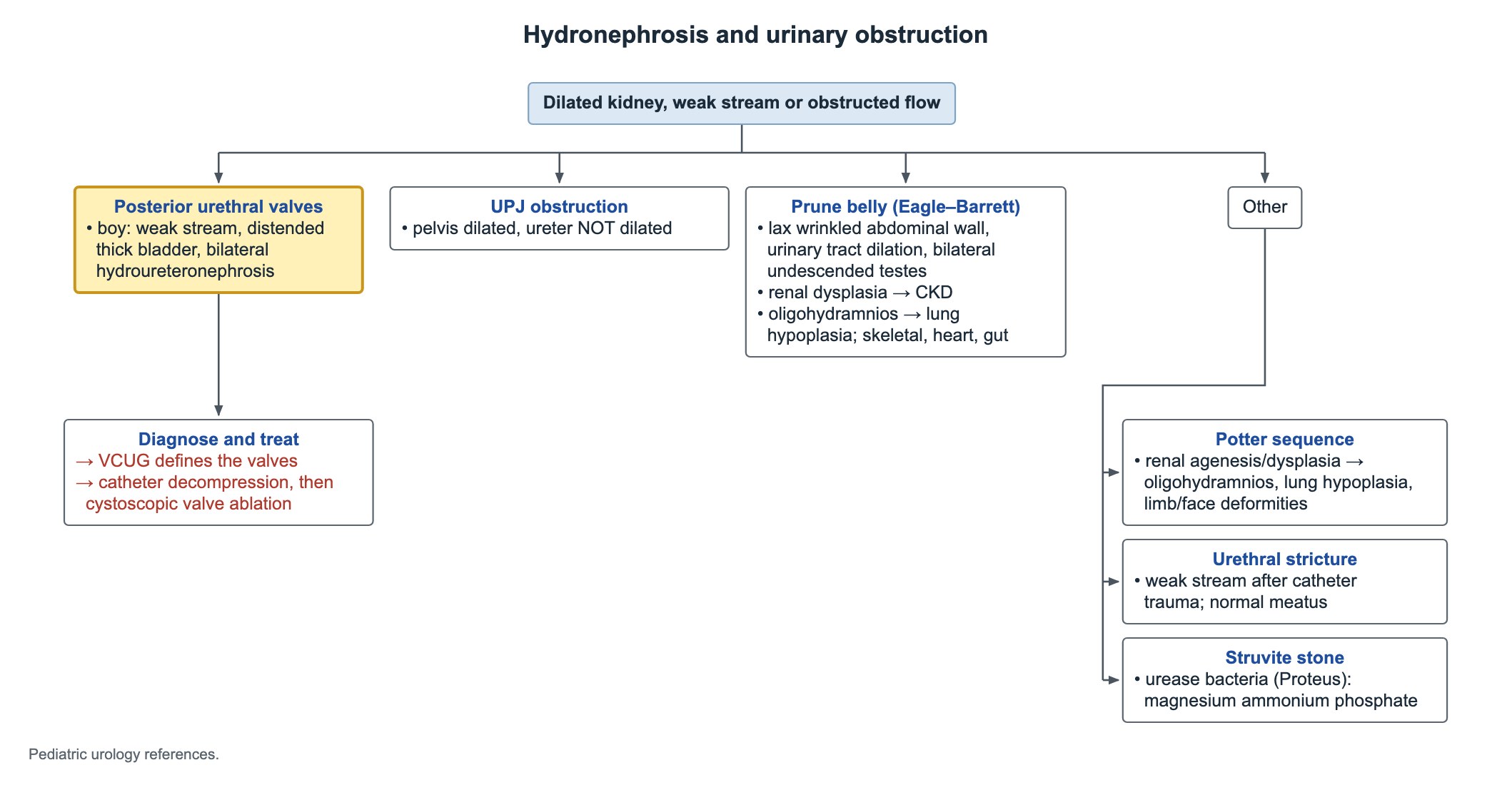
- Boy with a weak stream, a distended thick-walled bladder and bilateral hydroureteronephrosis: posterior urethral valves until proven otherwise.
- A VCUG defines the valves; drain the bladder with a catheter first, then ablate the valves endoscopically.
- Renal pelvis dilated but the ureter is not: think ureteropelvic junction (UPJ) obstruction.
- Prune belly (Eagle-Barrett) triad: deficient abdominal wall muscles, urinary tract dilation and bilateral undescended testes.
- Bilateral renal agenesis → no amniotic fluid → Potter sequence: pulmonary hypoplasia, flat face and limb deformities.
- Struvite (magnesium ammonium phosphate) stones form with urease-producing bacteria, classically Proteus.
- Postnatal ultrasound for prenatal dilation is best done after 48 hours of life, sooner if valves or severe bilateral dilation are suspected.
Posterior urethral valves: recognize them
Posterior urethral valves (PUV) are folds of tissue in the posterior urethra of boys that block bladder emptying. They are the most common cause of lower urinary tract obstruction in male infants.
The block raises pressure in the bladder, which thickens, and the pressure passes up both ureters to both kidneys. The kidneys may be dysplastic from early fetal life.
- Prenatal: bilateral hydronephrosis, a dilated thick bladder with a dilated posterior urethra (keyhole sign), and low amniotic fluid in severe cases.
- Newborn: weak or dribbling stream, palpable bladder, poor feeding, urinary ascites or respiratory distress from small lungs.
- Older boy: UTI, daytime wetting or a poor stream in milder valves.
- Only boys: a girl with bilateral hydroureteronephrosis needs another explanation.
Posterior urethral valves: diagnose and treat
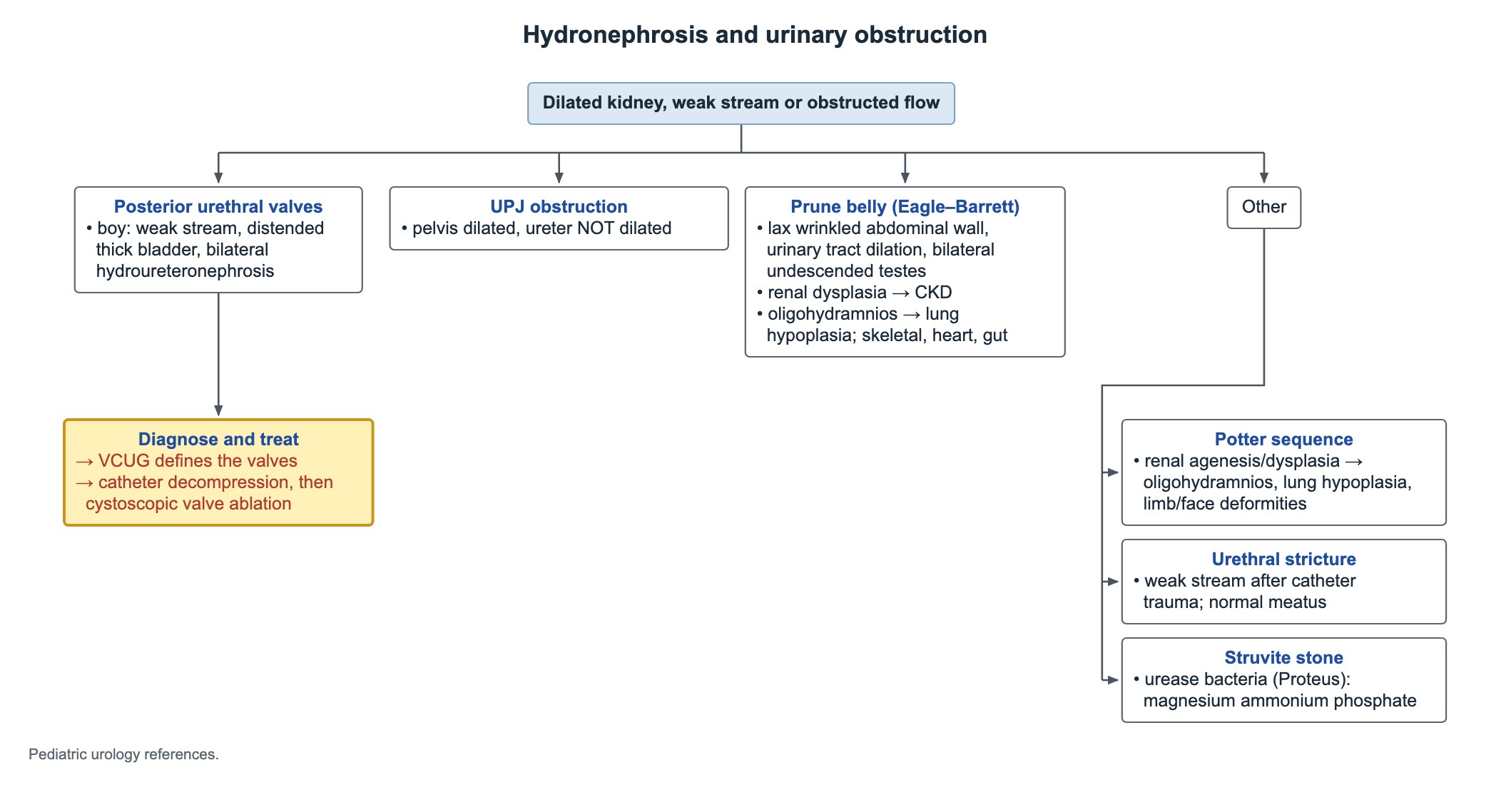
- First, decompress: place a small urethral catheter (feeding tube) to drain the bladder, and follow creatinine, electrolytes and urine output.
- Expect post-obstructive diuresis with salt and water loss after drainage.
- Diagnose: the voiding cystourethrogram (VCUG) shows the dilated posterior urethra and the valve; renal and bladder ultrasound shows the upper tract damage.
- Definitive treatment: endoscopic (cystoscopic) valve ablation once the infant is stable; a vesicostomy is used if the urethra is too small.
- Long term: many boys develop chronic kidney disease, and bladder dysfunction can persist after ablation, so nephrology and urology follow-up continue for life.
UPJ obstruction
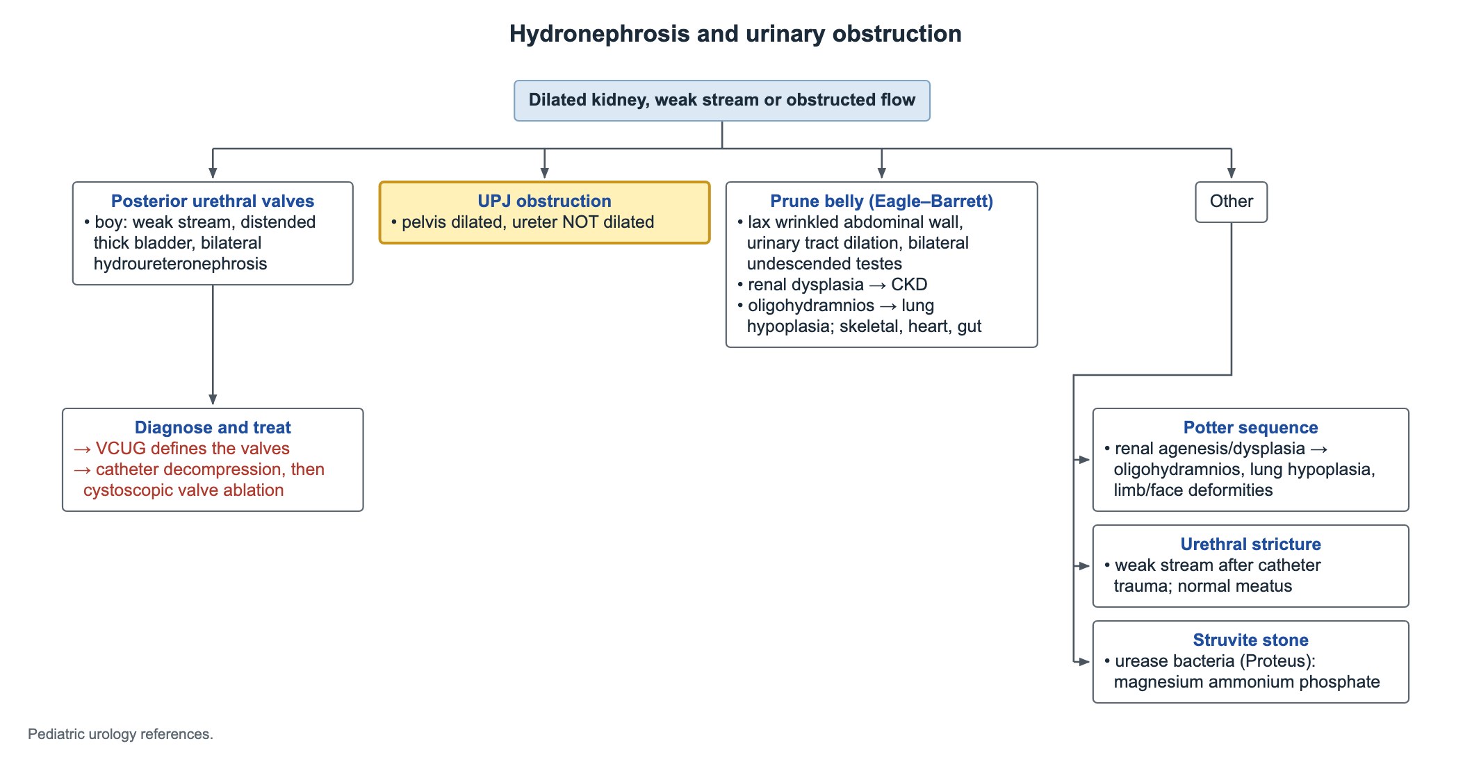
Ureteropelvic junction obstruction is the most common cause of significant hydronephrosis found on prenatal ultrasound. The block sits where the renal pelvis meets the ureter, so the pelvis and calyces dilate but the ureter below stays normal.
- Ultrasound: renal pelvis and calyceal dilation with a normal-caliber ureter and a normal bladder.
- A dilated ureter points lower: ureterovesical junction obstruction, vesicoureteral reflux or valves.
- Older children may have episodic flank or abdominal pain with vomiting, often after a large fluid intake.
- A diuretic renal scan (MAG3) measures split function and drainage.
- Pyeloplasty is offered for falling function, worsening dilation, pain, stones or infection; many infants are only watched.
Prune belly (Eagle-Barrett) syndrome
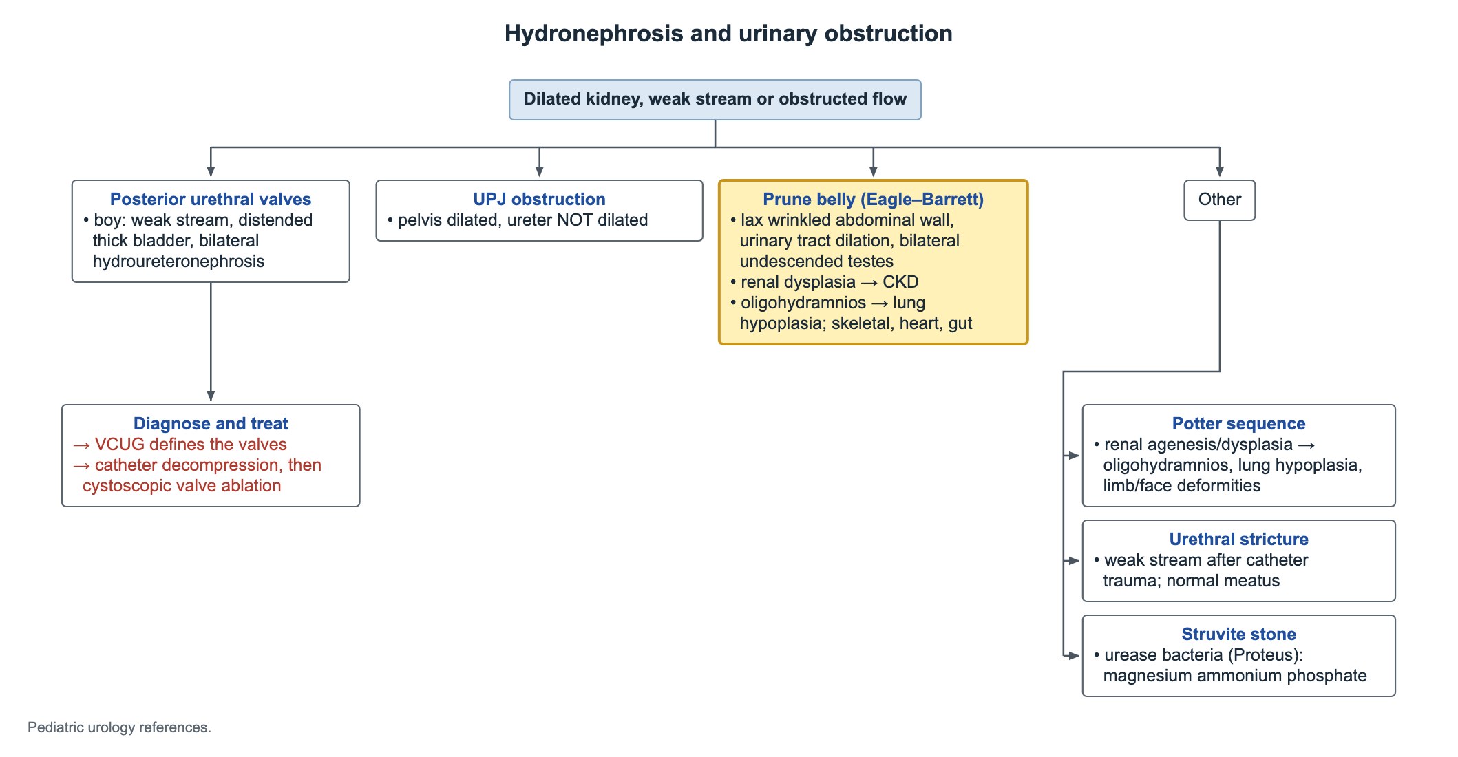
Prune belly syndrome occurs almost only in boys. The classic triad is a lax, wrinkled abdominal wall from deficient muscles, a dilated urinary tract (large bladder, tortuous ureters, often dysplastic kidneys) and bilateral undescended testes.
- Lungs: low amniotic fluid can cause pulmonary hypoplasia, the main threat at birth.
- Kidneys: renal dysplasia leads to progressive chronic kidney disease, sometimes kidney failure, so kidney function is followed for life.
- Other systems: skeleton (clubfoot, hip dislocation, scoliosis), heart defects and gut (malrotation, constipation).
- Testes are intra-abdominal and need orchiopexy.
Potter sequence
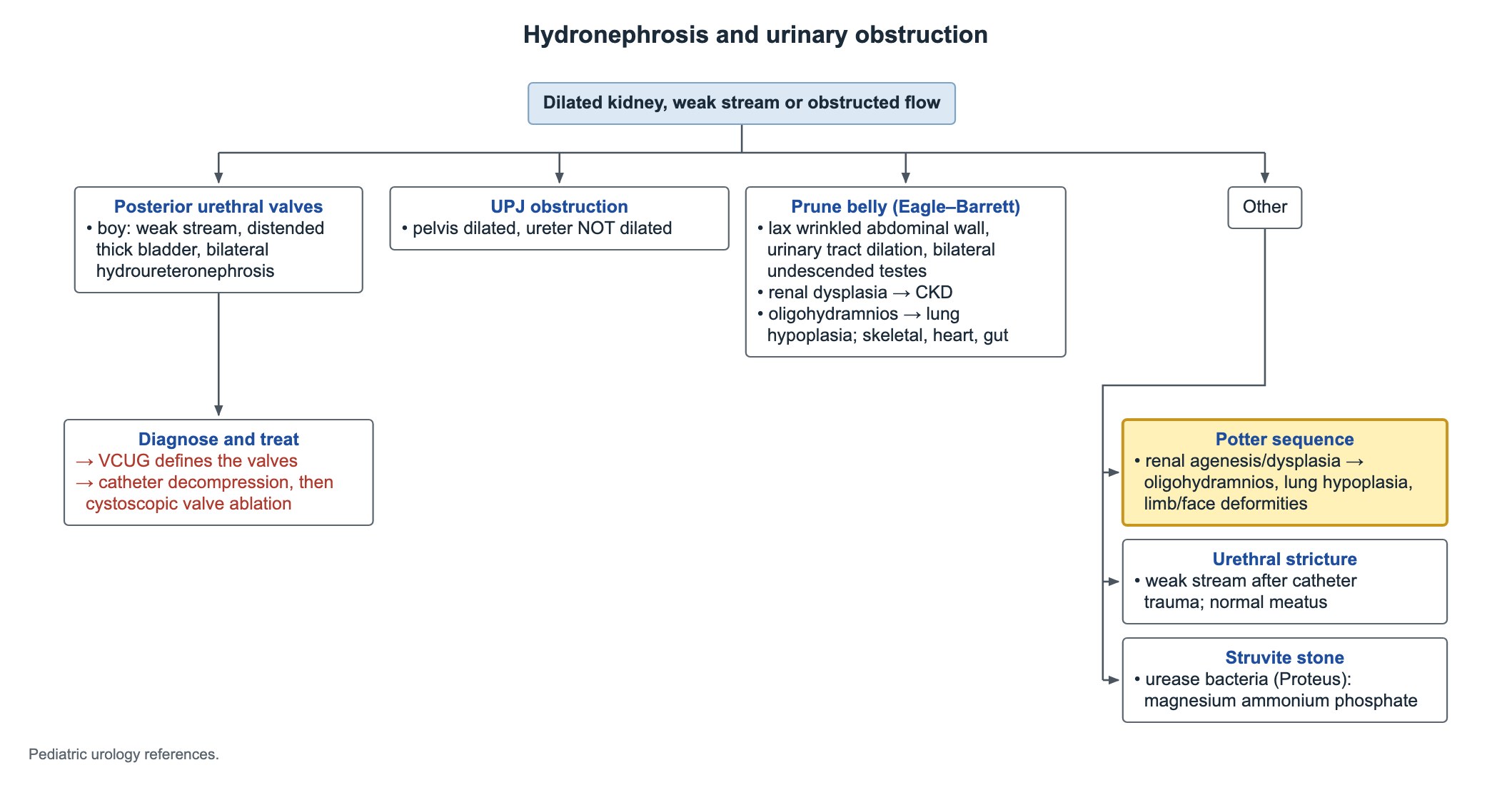
Potter sequence is the chain of findings caused by too little amniotic fluid from early in pregnancy. The classic cause is bilateral renal agenesis; severe bilateral dysplasia or obstruction can do the same.
Without fetal urine there is little amniotic fluid. The fetus is compressed and the lungs cannot grow.
- Pulmonary hypoplasia: the cause of death in bilateral renal agenesis.
- Face: flat face, flattened nose, receding chin, low-set ears and prominent skin folds below the eyes.
- Limbs: clubfeet, joint contractures and hip dislocation.
Struvite stones
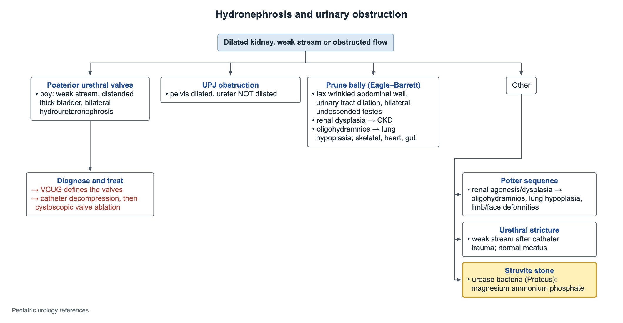
Struvite stones are made of magnesium ammonium phosphate. Urease-producing bacteria, classically Proteus, split urea into ammonia, which makes the urine alkaline and lets struvite crystals form.
They can grow fast into staghorn stones that fill the collecting system. Children with urinary stasis, such as an obstructed or neurogenic bladder, are at higher risk. Treatment needs both complete stone removal and treatment of the infection, because bacteria live inside the stone.
| Stone | Key association |
|---|---|
| Calcium oxalate | The most common stone type in children |
| Struvite | Urease-producing infection (Proteus); alkaline urine; staghorn shape |
| Cystine | Cystinuria; hexagonal crystals |
| Uric acid | Acidic urine, high cell turnover; seen poorly on plain x-ray |
Common questions
How are posterior urethral valves diagnosed?
A voiding cystourethrogram (VCUG) shows the dilated posterior urethra and the valve. Ultrasound shows the thick bladder and dilated ureters and kidneys.
What is the treatment for posterior urethral valves?
Drain the bladder with a catheter first, then remove the valves by endoscopic ablation. Long-term kidney and bladder follow-up is needed.
What are the three features of prune belly syndrome?
Deficient abdominal wall muscles, dilation of the urinary tract and bilateral undescended testes.
Study this topic
Sources
- Multidisciplinary consensus on prenatal and postnatal urinary tract dilation (UTD classification), J Pediatr Urol 2014
- EAU/ESPU Guidelines on Paediatric Urology
Written for Pedemy in our own words from the sources above; checked November 2026. For education only, not medical advice; check doses and local protocols before treating a patient. Pedemy is independent and not affiliated with or endorsed by the organizations cited.